

What a Nerve Conduction Study Shows Before Cubital Tunnel Surgery and Why It Matters

Key Takeaways
- The key measurement in a cubital tunnel nerve conduction study is how much velocity drops in the elbow segment compared with the forearm segment, which localizes the compression to the elbow.
- Falling response amplitude signals actual nerve fiber loss, while slowing alone signals insulation damage, and the two carry different recovery implications.
- Practice parameters call for testing with the elbow bent, typically 70 to 90 degrees, because a straight elbow can hide the slowing entirely.
- A normal study does not exclude early cubital tunnel syndrome, since the test samples only the large fast fibers that are often spared at first.
- Needle EMG adds a second layer by showing whether muscles supplied by other nerves from the same neck root are also affected, separating the elbow from the spine.
- Compression of the ulnar nerve at the elbow is the second most common arm entrapment after carpal tunnel syndrome, and the opposite elbow is routinely tested because bilateral involvement is common.
A nerve conduction study before cubital tunnel surgery measures how fast and how strongly electrical signals travel along the ulnar nerve as it passes behind the elbow. It helps confirm that the nerve is compressed at the elbow rather than the neck or wrist, grades how severe the damage is, and gives a baseline for judging recovery. Results guide, but do not replace, the surgeon's clinical judgment.
The ring and little fingers are the usual messengers. They go numb on a long phone call, tingle when an elbow rests on the car door, and feel clumsy with buttons by evening. By the time someone sits across from an orthopedic or hand surgeon, they often expect to talk about an operation. Instead, the surgeon asks for a test first: the nerve conduction study cubital tunnel specialists order before they commit to surgery.
That request can feel like a delay. It is closer to a map. The ulnar nerve runs a long route from the neck to the fingertips, and symptoms in the hand cannot tell you where along that route the trouble sits. A study of the nerve’s electrical behavior can.
This explainer walks through what the test records, how it feels, how to read the numbers on the report, and why a thoughtful surgical team treats those numbers as evidence rather than a verdict.
What is cubital tunnel syndrome, and why does the elbow pinch the ulnar nerve?
Cubital tunnel syndrome is irritation or compression of the ulnar nerve where it passes behind the inner bump of the elbow, the medial epicondyle. The ulnar nerve is the one you strike when you hit your “funny bone”; the jolt down the forearm is the nerve itself complaining. According to Johns Hopkins Medicine, compression of this nerve at the elbow is the second most common entrapment neuropathy in the arm, behind only carpal tunnel syndrome.
The anatomy explains the vulnerability. The nerve sits in a shallow groove covered by a thin band of tissue, with little padding between it and the skin. Every time the elbow bends, the tunnel narrows and the nerve stretches a few millimeters. Hold that position for an hour, reading in bed or sleeping with arms folded, and the nerve’s blood supply drops while pressure inside the tunnel rises. Repeated often enough, the lining of the nerve thickens, signals slow, and the muscles it supplies begin to weaken.
Those muscles matter more than most people realize. The ulnar nerve powers most of the small muscles that spread the fingers, pinch firmly between thumb and index finger, and keep the ring and little fingers from curling. It also carries sensation from the little finger, half of the ring finger, and the outer edge of the hand. That pattern is why early cubital tunnel syndrome feels like “two sleepy fingers” long before the hand looks different.
Not every case needs an operation. The Cleveland Clinic notes that many people improve with activity changes, keeping the elbow straighter at night, and padding the elbow during the day. The nerve conduction study helps sort the people who are likely to settle with those measures from the people whose nerve is already struggling.
What a nerve conduction study for cubital tunnel syndrome actually measures
A nerve conduction study is a test that delivers small electrical pulses to a nerve at one point and records the response at another, timing how long the signal takes to arrive. Divide the distance by the time and you get conduction velocity, reported in meters per second. Healthy, well-insulated nerve fibers in the arm conduct quickly; damaged ones conduct slowly or lose part of the signal altogether.
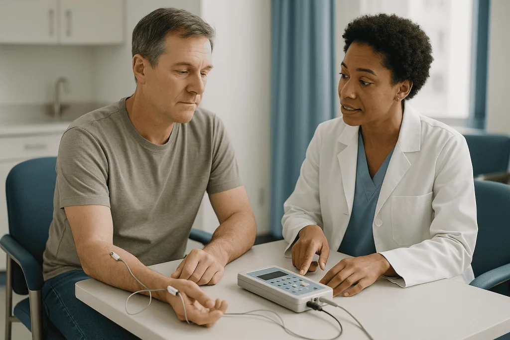
For the ulnar nerve, the examiner stimulates at the wrist, below the elbow, and above the elbow, recording from a small hand muscle at the base of the little finger. This produces three separate segments: forearm, across the elbow, and upper arm. The crucial comparison is between the elbow segment and the forearm segment. If the nerve conducts normally through the forearm but slows sharply across the elbow, the problem is almost certainly at the elbow. Published electrodiagnostic practice parameters treat a drop of roughly 10 meters per second across the elbow compared with the forearm segment as a meaningful sign of focal slowing.
Two other numbers appear on most reports. Amplitude describes the size of the response, which reflects how many nerve fibers are still conducting; a falling amplitude points toward actual fiber loss rather than simple insulation damage. Latency is the delay at a fixed distance, used mainly for the wrist-to-hand segment.
The examiner may also test sensory fibers separately, recording from the little finger, and may stimulate at several short intervals across the elbow, a technique called inching, to pinpoint the exact spot where the signal slows. According to MedlinePlus, the entire procedure is usually painless apart from the brief pulses, and no anesthesia is needed. What the test cannot do is show pain directly; it measures electrical function, not how the arm feels.
What actually happens in the testing room, step by step
The appointment begins with a conversation, not a wire. The technologist or physician asks where the symptoms are, which hand is dominant, and whether there is diabetes, thyroid disease, prior neck problems, or a pacemaker, since these change how the results are interpreted. Skin temperature is checked; a cool arm conducts more slowly and can produce a falsely abnormal result, so a cold hand is often warmed with a pack before testing starts.
Next come the stickers. Small adhesive recording electrodes go on the hand muscle and on the little finger. A handheld stimulator, about the size of a thick marker, is pressed against the skin at the wrist. The examiner delivers a series of brief pulses, each lasting a fraction of a millisecond, gradually increasing until the response stops growing. That ceiling is called a supramaximal response, and it ensures every available fiber has been counted.
The stimulator then moves to the forearm just below the elbow and to the upper arm just above it. Elbow position matters here: practice parameters recommend testing with the elbow bent, typically between 70 and 90 degrees, because a straight elbow lets the nerve slacken and can hide slowing. The examiner measures the skin distance between stimulation points with a tape; accurate distances produce accurate velocities.
Many laboratories test the median nerve in the same arm as a comparison and may test the opposite ulnar nerve, even without symptoms there. Then the needle portion, if ordered, follows. Mayo Clinic describes the combined session as typically lasting 30 to 60 minutes. Patients can drive afterward, eat normally, and return to work the same day. The report is written by a physician who reviews the raw waveforms, which is why results usually reach the surgeon a few days later rather than at the door.
Is an EMG test painful? An honest answer about the two parts
People searching “how painful is an EMG nerve test” are usually asking about two different procedures that happen in one visit. The nerve conduction study uses surface electrodes and electrical pulses. The electromyogram, or EMG, uses a fine needle placed into muscle to listen to its electrical activity. They feel different, and it helps to know which is which.
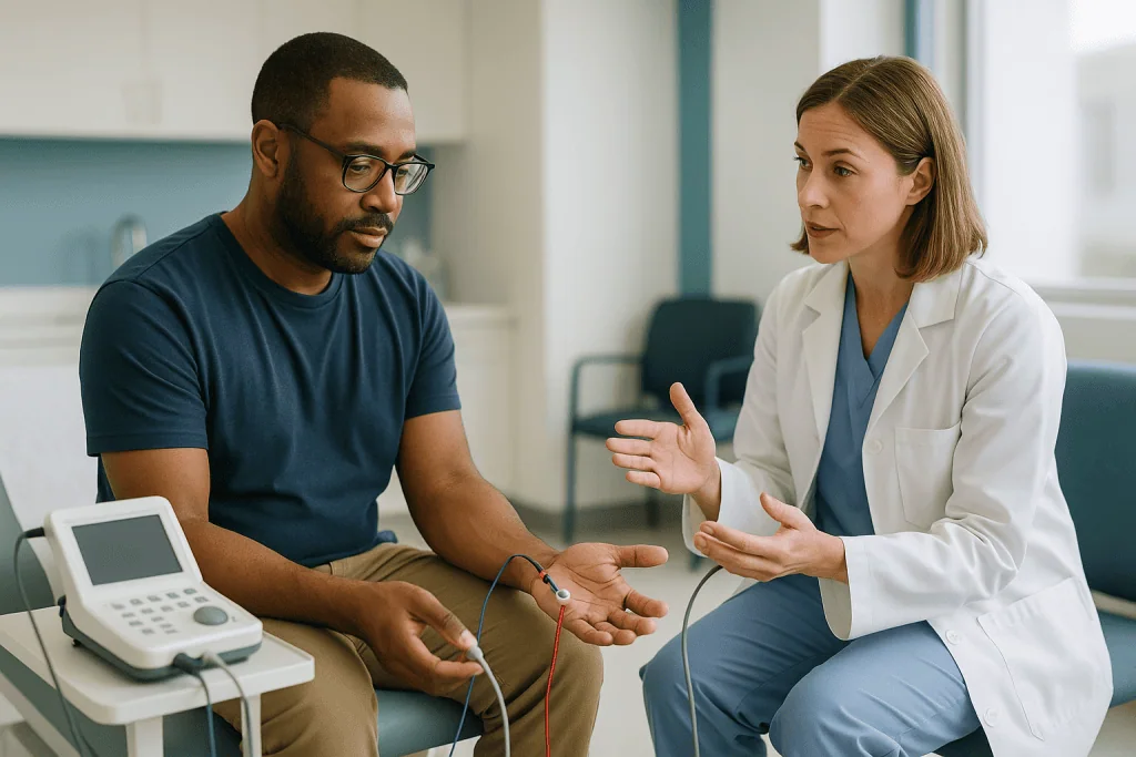
The conduction pulses are startling more than painful. Each one feels like a quick static shock or a rubber band snap, and the muscle twitches on its own, which surprises people the first time. The sensation ends the instant the pulse ends; nothing lingers. The pulses near the elbow, where the ulnar nerve is close to the surface, tend to feel sharpest, and a tingling may shoot into the ring and little fingers. That is expected and is in fact useful information for the examiner.
The needle EMG is a different experience. The needle is thinner than those used to draw blood and carries no injection; it records only. Insertion feels like a brief pinch, followed by a dull ache while the examiner asks you to gently tighten and relax the muscle. Mayo Clinic notes that some people feel mild soreness or see a small bruise for a day or two afterward. Most describe the whole session as uncomfortable rather than painful, and nobody needs sedation.
A few practical points reduce the discomfort. Arrive warm, because cold skin makes pulses feel sharper and slows the nerve. Skip lotion on the arm so electrodes stick without extra pressing. Say so if a particular site is especially sensitive; examiners can usually adjust technique or order. Finally, remember that the number of muscles sampled is tailored to the question being asked, and for a focused ulnar study it is often small.
EMG test for ulnar nerve: why the needle portion is added
An electromyogram, or EMG, records the electrical signals produced by muscle fibers at rest and during contraction. For an EMG test for ulnar nerve problems, the examiner samples a handful of muscles chosen for a reason: some are supplied by the ulnar nerve below the elbow, some above the elbow, and some by entirely different nerves that share the same root in the neck.
The logic is a chain of elimination. If the small hand muscles supplied by the ulnar nerve show abnormal activity but the forearm muscle supplied by the same nerve just below the elbow is normal, the lesion probably sits at or above the forearm branch. If muscles supplied by other nerves from the C8 and T1 spinal roots are also abnormal, the problem may be in the neck or the brachial plexus rather than the elbow. The pattern of which muscles are affected tells the story that symptoms alone cannot.
Needle EMG also detects something nerve conduction can miss: active nerve fiber loss. When a nerve fiber dies, the muscle fibers it supplied begin to fire spontaneously, producing tiny signals called fibrillation potentials and positive sharp waves. Their presence suggests ongoing damage, a finding Johns Hopkins describes as a marker of more severe compression. Over months, surviving nerve fibers sprout to adopt orphaned muscle fibers, creating larger, longer motor unit potentials that signal chronic injury with some repair.
Surgeons value this layer because it separates a nerve that is merely slow from one that is losing fibers. Slow nerves often recover well once pressure is relieved. Nerves showing dense spontaneous activity and few remaining motor units may improve more slowly and less completely, which changes what a surgeon can honestly say about expectations. Needle findings are therefore less about whether to operate and more about how to counsel.
Nerve conduction study results in cubital tunnel: how to read your report
Reports are written for the referring physician, so the language is dense. Three threads run through almost all of them: localization (where), type of injury (insulation versus fiber loss), and severity (mild, moderate, severe). The table below translates common phrases into what they usually imply. Interpretation always belongs to the physician who performed the study and the surgeon who reads it alongside the examination.
| Report phrase | What was measured | What it usually suggests |
|---|---|---|
| Focal slowing across the elbow | Conduction velocity drops in the elbow segment relative to the forearm | Insulation (myelin) damage localized to the elbow; typical of early or moderate cubital tunnel syndrome |
| Conduction block | Response amplitude falls sharply when stimulating above versus below the elbow | Fibers still alive but signals not getting through the compressed zone; often reversible |
| Reduced motor amplitude at all sites | Small response even from the wrist | Loss of nerve fibers (axonal injury); points toward more advanced disease |
| Absent sensory response from the little finger | No measurable sensory signal | Significant sensory fiber loss; also seen in some neck or plexus problems, so context matters |
| Fibrillations in ulnar-innervated muscles | Spontaneous activity on needle EMG | Ongoing fiber loss; grades severity higher |
| Normal study | No abnormality detected | Does not rule out mild compression; symptoms and examination carry more weight |
Severity grading combines these elements. Mild disease usually shows only slowing or a sensory change. Moderate disease adds reduced amplitude or early needle changes. Severe disease shows low amplitudes, absent sensory responses, and clear spontaneous activity, often alongside visible wasting of the hand muscles on examination. Where a laboratory reports exact velocities, remember that thresholds vary slightly between laboratories and depend on temperature and technique. A report is a snapshot taken with a particular method on a particular day; the surgeon’s job is to place it in the frame of the whole clinical picture.
Why surgeons want the study before cubital tunnel surgery
Four reasons sit behind the request, and each protects the patient from a different kind of mistake.
The first is localization. Numb ring and little fingers can come from the elbow, the wrist, the neck, or the network of nerves above the collarbone. An operation at the elbow does nothing for a pinched root in the neck. The elbow-versus-forearm velocity comparison is the single most useful tool for proving the elbow is the culprit, which is why MedlinePlus lists nerve conduction testing among the core investigations for ulnar nerve dysfunction.
The second is severity. A nerve that is only slow behaves differently from one that is losing fibers. Cleveland Clinic guidance notes that muscle weakness or wasting shifts the conversation toward surgery, because fiber loss that continues is harder to recover from. The study quantifies what the examination suggests.
The third is a baseline. If surgery goes ahead and symptoms persist or return, a repeat study can be compared with the original. Without a “before” picture, no one can tell whether the nerve is healing slowly, has been compressed again, or was never the main problem.
The fourth is honesty in counseling. Surgeons cannot promise outcomes, and the study does not let them. What it does is let them describe realistic trajectories: nerves with preserved amplitude tend to recover function more fully, while nerves with marked fiber loss may regain protective sensation and some strength over a long period but not always full fine control. That conversation, grounded in data, is the foundation of informed consent.
Not every surgeon orders the test in every case. A patient with classic symptoms, clear signs, and visible wasting may proceed on clinical grounds in some practices. The choice of whether to test, and when, remains with the treating team.
Ulnar nerve entrapment at the elbow versus its mimics
A good electrodiagnostic study is as much about what it rules out as what it confirms. Several conditions produce symptoms that overlap with ulnar nerve entrapment at the elbow, and each leaves a different electrical fingerprint.
Compression at the wrist, in a passage called Guyon’s canal, affects the same fingers but spares the sensation on the back of the hand, because the branch supplying that skin leaves the nerve above the wrist. On testing, the forearm and elbow segments are normal while the wrist-to-hand latency is prolonged.
A pinched nerve root in the lower neck, typically C8, can numb the little finger and weaken the hand. The giveaway is that muscles supplied by other nerves from the same root, such as some thumb muscles supplied by the median nerve, also show needle changes, while sensory responses from the finger remain normal because the injury sits before the sensory cell bodies.
Thoracic outlet involvement, where the lower part of the brachial plexus is compressed above the collarbone, often reduces the sensory response from the inner forearm rather than the little finger and affects a broader group of hand muscles.
Generalized nerve disease, such as diabetic polyneuropathy, slows many nerves at once. Testing the median nerve and the opposite arm reveals whether the ulnar slowing is a local event or part of a wider pattern.
Carpal tunnel syndrome deserves a mention because people often ask about both tests together. It involves a different nerve, the median, which supplies the thumb, index, and middle fingers. The NHS describes nerve conduction testing as the standard way to confirm it when the diagnosis is unclear, using the same principles at the wrist that the ulnar study applies at the elbow. The two conditions can coexist, which is one more reason examiners test both nerves in the same session.
Can the nerve conduction study be normal and still be cubital tunnel syndrome?
Yes, and this is the most misunderstood result of all. Electrodiagnostic testing samples the large, fast, heavily insulated nerve fibers because they produce the clearest signals. Early compression frequently irritates the nerve without yet slowing those fibers in a measurable way. Someone can have genuine, daily symptoms and a report that reads “no electrodiagnostic evidence of ulnar neuropathy at the elbow.”
Several technical factors widen that gap. If the elbow is tested straight rather than bent, the nerve lies relaxed and slowing can vanish. If the skin over the elbow is cool, velocities drift. If the measured distance across the elbow is short, small timing errors become large velocity errors, which is why practice parameters favor a standardized distance across the elbow segment. Symptoms that come only with the elbow bent for long periods, such as during sleep, may simply not be present in a laboratory where the arm is tested after a few minutes at rest.
What should happen next depends on the clinical picture. A normal study with mild, intermittent symptoms usually supports a period of conservative management: avoiding prolonged elbow flexion, padding, night positioning, and a review in a few months. A normal study with persistent symptoms and convincing physical signs, such as a positive elbow flexion test or reproducible tingling when the nerve is tapped, may prompt a repeat study with inching technique, an ultrasound to look at nerve size and position, or a frank conversation about proceeding on clinical grounds.
The reverse also happens. Mild slowing across the elbow turns up in people with no symptoms, particularly those who lean on their elbows or have had old elbow injuries. A borderline abnormality in an asymptomatic arm is not, on its own, a reason to operate. Numbers inform the decision; they do not make it.
Who is usually offered surgery, and who is usually asked to wait
There is no single threshold on a nerve conduction report that triggers an operation. Guidance from Johns Hopkins and the Cleveland Clinic converges on a practical framework in which the test, the examination, and the patient’s own experience are weighed together.
Surgery is more often discussed when there is measurable weakness or visible thinning of the hand muscles, when sensory loss is constant rather than intermittent, when the study shows reduced amplitudes or active fiber loss on needle testing, or when symptoms have persisted despite a genuine trial of conservative care. The reasoning is time-sensitive: once nerve fibers die, recovery depends on slow regrowth, and the small hand muscles may not wait indefinitely.
A period of watching and modifying is more often advised when symptoms are intermittent and provoked by posture, when the examination is normal apart from tingling on tapping, when the study is normal or shows only mild slowing with preserved amplitude, and when the person has not yet changed the habits that load the nerve. Many people in this group improve over weeks to months, and the surgical option remains open if they do not.
Some situations call for a pause for different reasons. Uncontrolled diabetes, active infection, a recent elbow injury still healing, or a study suggesting the neck rather than the elbow all redirect the plan. People taking blood thinners need coordination rather than exclusion.
The operation itself varies. Simple decompression opens the roof of the tunnel. Transposition moves the nerve to the front of the elbow so it no longer stretches on bending. A medial epicondylectomy removes part of the bony bump. Which approach, if any, suits a particular nerve is a judgment for the treating surgeon, informed by anatomy at the time of surgery as much as by the study beforehand.
Cubital tunnel syndrome in both arms: what the study often shows
Having cubital tunnel syndrome in both arms is common enough that experienced examiners routinely test the asymptomatic side. The ulnar nerve faces the same anatomy on each side, and the habits that compress it, leaning on elbows at a desk, sleeping with arms tightly folded, holding a phone for long calls, tend to be bilateral too. MedlinePlus lists repeated elbow pressure and prolonged bending as leading contributors, and neither respects handedness.
Testing both sides answers three questions. First, it provides a personal comparison: velocities vary between people, so an individual’s “good” arm is the best reference for the “bad” one. Second, it uncovers silent disease. Mild slowing on the symptom-free side is a frequent incidental finding, and while it rarely needs treatment on its own, it tells the patient that the habits driving the symptomatic side are already affecting the other. Third, bilateral abnormalities raise the question of a generalized nerve problem, prompting the examiner to look at the median and leg nerves as well.
When both arms are symptomatic and both studies are abnormal, decisions become sequential rather than simultaneous. Surgeons generally address the more severe or more disabling side first, partly so the patient has one functioning arm during recovery and partly to see how the first nerve responds before planning the second. The study helps rank the two sides objectively; the arm that feels worse is not always the one with the lower amplitude.
People in this situation often ask whether something systemic is wrong. Sometimes it is, which is exactly why the broader testing is done. More often the answer is mechanical, and the most effective intervention for the milder side is the least dramatic: taking the elbows off the desk and straightening them at night.
What the days and weeks around the test and after surgery usually look like
The testing visit itself needs almost no preparation. Mayo Clinic advises skipping lotions and creams on the day, wearing clothing that lets the arm be bared to the shoulder, and mentioning any implanted electrical device or blood-thinning medicine in advance. Afterward, the stimulation sites may feel faintly tender, and needle sites can show a small bruise for a day or two. Normal activity resumes at once.
Results typically take days rather than minutes, because a physician reviews the waveforms and writes a formal interpretation. The surgical consultation that follows is where the numbers meet the examination. Expect a discussion of severity grade, where the compression sits, what conservative measures remain untried, and what the operation would and would not aim to do.
If surgery proceeds, the early period is about protecting the wound and keeping fingers moving. Many patients wear a soft dressing or splint briefly, then begin gentle elbow motion within the first couple of weeks, with timing set by the surgeon and the type of procedure. Grip strength and comfort improve over weeks.
Nerve recovery runs on a slower clock. Tingling and intermittent numbness, which reflect insulation damage, frequently ease early. Constant numbness and muscle weakness, which reflect fiber loss, depend on regrowth from the elbow toward the hand. Johns Hopkins and the Cleveland Clinic both describe this as a process of months, and in severe cases some deficits may not fully return. Odd sensations such as buzzing or shooting tingles during recovery are often a sign of fibers reconnecting rather than a setback.
A repeat nerve conduction study is not routine but may be ordered if progress stalls or symptoms recur, giving an objective comparison against the preoperative baseline. The schedule for follow-up, splinting, therapy, and return to driving or work belongs to the treating team.
What people often get wrong about nerve testing and cubital tunnel
“A normal test means nothing is wrong.” It means the large fast fibers conducted normally on that day in that position. Early compression often escapes detection, which is why examiners and surgeons give the history and examination real weight.
“An abnormal test means I need surgery.” Mild slowing with preserved amplitude is compatible with full recovery through posture changes and night positioning. The test grades the nerve; it does not issue instructions.
“The test is dangerous or involves high voltage.” The pulses are brief and low in energy, delivered through the skin, with no lasting effect on the nerve. MedlinePlus describes the procedure as safe for nearly everyone, with precautions mainly around implanted devices.
“EMG and nerve conduction are the same thing.” They are two complementary tests. One measures nerves with surface electrodes; the other listens to muscles with a fine needle. Many visits include both, but a focused question can sometimes be answered with one.
“The numbers are exact.” Velocity depends on temperature, elbow angle, measured distance, and technique. Laboratories standardize these, yet small differences between laboratories are normal. Comparing a new report with an old one from a different laboratory needs care.
“Surgery will fix the numbness right away.” Pressure is relieved in the operating room; nerve fibers regrow slowly afterward. Expecting instant sensation sets people up to feel the operation failed when it is simply early.
“It only happens to people who use their hands for work.” Sleeping posture and resting elbows on hard surfaces compress the nerve as effectively as any job. Office workers and students are well represented in every clinic.
“If it is in one arm, the other is safe.” The same habits usually load both elbows, which is why the other side is tested and why habit changes should apply to both.
Questions to ask your care team before and after the study
A nerve conduction report is only useful if it changes a conversation. These questions help turn a dense page of numbers into a plan you understand.
Before the test, ask who will interpret it and roughly when results will reach the surgeon, whether the needle EMG portion is planned and why, whether both arms and the median nerve will be tested for comparison, and whether anything you take or any device you have needs to be flagged.
After the test, ask these:
- Does the study confirm that the compression is at the elbow rather than the neck or wrist?
- How would you grade the severity, and which finding drove that grade?
- Is there evidence of nerve fiber loss, or only slowing?
- If the study is normal, what weight are you giving my symptoms and examination?
- Which conservative measures have I not yet tried properly, and for how long should I try them?
- If surgery is recommended, which procedure, and what did the study contribute to that choice?
- What is a realistic recovery picture for a nerve in my grade of severity?
- Would you repeat the study if symptoms persist, and how would you compare it with this one?
- What signs would make you want to see me sooner?
Bring someone with you if you can; the detail is easier to absorb with a second set of ears. Ask for a copy of the report for your own records, because a future clinician may want the baseline. Above all, ask the surgeon what they would do if the numbers and their examination disagreed. The answer tells you how they think, which matters more than any single value on the page.
When to call your doctor: red-flag signs that should not wait for the next appointment
Most cubital tunnel symptoms evolve slowly and can be reviewed at a scheduled visit. A smaller set of changes deserves a prompt call, whether you are waiting for a nerve conduction study, waiting for a surgical date, or recovering from an operation.
Contact your care team promptly if you notice:
- New or rapidly worsening weakness in the hand, such as dropping objects, inability to spread the fingers, or the ring and little fingers beginning to curl into a claw position
- Visible hollowing or thinning of the muscle between the thumb and index finger or along the outer edge of the hand
- Numbness that has become constant after previously coming and going
- Symptoms spreading beyond the ring and little fingers, into the whole hand, the forearm, or the other arm, or new neck pain with arm symptoms
- Weakness or numbness in a leg, trouble with balance, or changes in bladder or bowel control, which point away from the elbow and need urgent assessment
After surgery, call the same day for fever, spreading redness or warmth around the wound, pus or persistent bleeding, calf pain or swelling, chest pain, or shortness of breath. Sudden severe pain, a hand that turns pale or blue, or fingers that feel cold and will not warm are emergencies.
The Cleveland Clinic and Johns Hopkins both emphasize that earlier evaluation of progressive weakness protects the small muscles of the hand, which recover least well once wasted. Calling early is never an overreaction; the worst outcome of an unnecessary call is reassurance. Every decision about testing, timing, and treatment rests with the clinicians who can examine the arm in person.
Frequently asked questions
What is cubital tunnel syndrome?
Cubital tunnel syndrome is compression or irritation of the ulnar nerve where it passes behind the inner bump of the elbow. The nerve supplies sensation to the little finger and half of the ring finger and powers many small hand muscles. Early symptoms are tingling and numbness in those fingers, especially with the elbow bent; later stages can bring weakness and visible muscle thinning in the hand.
Is an EMG test painful?
Most people describe it as uncomfortable rather than painful. The nerve conduction pulses feel like brief static shocks that end instantly. The needle EMG involves a fine recording needle inserted into a few muscles, which feels like a pinch followed by a dull ache. Mayo Clinic notes mild soreness or a small bruise may last a day or two. No sedation is needed and normal activity resumes immediately.
What is a nerve conduction study used for in carpal tunnel syndrome diagnosis?
In carpal tunnel syndrome it measures the median nerve as it crosses the wrist, looking for delayed signals compared with a nearby nerve. The NHS describes it as the standard confirmatory test when the diagnosis is uncertain. The principle is identical to the ulnar nerve study at the elbow, and examiners often test both nerves in one session because the two conditions can coexist.
Is it common to have cubital tunnel syndrome in both arms?
Yes, bilateral involvement is common, because the habits that compress the nerve, leaning on elbows and sleeping with arms folded, usually affect both sides. Examiners routinely test the symptom-free arm as a personal comparison and to detect silent slowing. When both sides need treatment, surgeons generally address the more severe side first and reassess before planning the other.
How long does an EMG test for ulnar nerve problems take?
Mayo Clinic describes a combined nerve conduction and EMG session as typically taking 30 to 60 minutes, depending on how many nerves and muscles are examined. A focused ulnar study with a comparison nerve and the opposite arm sits within that range. Results usually follow within days, after a physician reviews the recorded waveforms and writes a formal interpretation.
Can I have ulnar nerve entrapment at the elbow with a normal nerve conduction study?
Yes. Early compression frequently irritates the nerve without measurably slowing the large fibers the test records, and factors like a straight elbow or cool skin can mask slowing. A normal result with convincing symptoms and examination findings may lead to repeat testing with inching technique, an ultrasound, or a decision based on clinical grounds. The surgeon weighs all of these together.
What number on the report means the nerve is damaged?
No single number decides it. Examiners look at the velocity drop across the elbow relative to the forearm, published practice parameters treating roughly 10 meters per second as meaningful, together with response amplitude and needle findings. Thresholds vary slightly between laboratories and depend on temperature and technique, so the interpreting physician’s written conclusion matters more than any isolated value.
Does an abnormal study mean I need surgery?
Not on its own. Mild slowing with preserved amplitude is often managed by avoiding prolonged elbow bending, padding, and night positioning, with review after a period of weeks to months. Surgery is more often discussed when there is weakness, muscle wasting, constant numbness, reduced amplitudes, or active fiber loss on EMG. The decision rests with the treating surgeon and the patient together.
Why does the examiner bend my elbow during the test?
Bending the elbow puts the ulnar nerve under the same stretch and pressure that provoke symptoms, so slowing across the elbow becomes measurable. Practice parameters recommend an angle between roughly 70 and 90 degrees. With the elbow straight the nerve slackens, measured distances become unreliable, and genuine compression can produce a falsely normal result.
Will a repeat nerve conduction study be needed after surgery?
Not routinely. A repeat study is usually reserved for situations where recovery stalls or symptoms return, when an objective comparison with the preoperative baseline helps distinguish slow healing from recurrent compression or a different cause. Because nerve fibers regrow slowly, surgeons typically allow months before judging recovery and would time any repeat study accordingly.
References
- Cleveland Clinic: Cubital Tunnel Syndrome
- MedlinePlus: Nerve conduction velocity
- MedlinePlus: Ulnar nerve dysfunction
- NHS: Carpal tunnel syndrome
This article is for general information only and is not a substitute for professional medical advice. Please consult a qualified doctor about your individual situation.

More from the Blog

How Closed Reduction Works: Realigning a Broken Bone Without an Incision
A closed reduction procedure realigns a displaced broken bone by hand, without cutting the skin. After pain control, usually a local anesthetic, sedation or…

Knuckle and Joint Cracking: The Physics and Whether It Harms Joints
Knuckle cracking is the sound of a gas cavity forming in the lubricating fluid of a joint when the joint surfaces are pulled apart.…

Hip Arthritis Pain: Where It Is Felt, How It Differs from Back Pain and What Helps
Hip arthritis pain is most often felt deep in the groin or the front of the thigh, and it can travel to the buttock…

How Do You Know You Need a Hip Replacement? The Functional Signs
You may need a hip replacement when arthritis pain in the groin or thigh persists despite several months of nonsurgical care and begins limiting…

Foot Surgery When You Have Diabetes: Wound Healing, Blood Sugar and Extra Precautions
Foot surgery with diabetes is often appropriate, but it carries higher risks of slow wound healing, infection and nerve-related complications. Surgeons usually ask for…

Preparing for Finger or Toe Surgery: Blood Thinners, Imaging and Arranging Help
Preparing for finger or toe surgery usually involves a medication review with the team that prescribes your blood thinners, any imaging the surgeon needs…