

What Do Feet Usually Look Like After Clubfoot Treatment? Realistic Long-Term Expectations

Key Takeaways
- Clubfoot affects about 1 in 1,000 babies, roughly half of them in both feet, and boys about twice as often as girls, according to the NHS and Mayo Clinic.
- The Ponseti method corrects the foot with weekly casts over about 5 to 8 weeks, usually followed by a minor Achilles tenotomy, and then holds the correction with boots and a bar for years.
- Full-time bracing lasts about three months, then night-time bracing continues until roughly age four or five in the NHS schedule, and stopping early is the leading cause of relapse.
- A treated foot is commonly slightly smaller than the other, with a thinner calf and a little less ankle movement, and these differences do not predict pain or disability.
- Relapse is a known fork in the road, usually managed with repeat casting or a tendon transfer from around age three, after which the foot generally looks much like one that never relapsed.
- Adults treated with extensive release surgery decades ago face more stiffness and arthritis than children on today's casting-based pathway, so older outcomes do not predict current ones.
After successful clubfoot treatment, most feet look close to typical: the sole sits flat on the floor, the foot points forward, and ordinary shoes fit. The treated foot is often slightly smaller than the other, the calf thinner, and ankle movement a little reduced. Most children walk, run and play sports, although relapse can happen, especially if bracing stops early, so follow-up continues through childhood.
The first thing many parents do in the delivery room is count fingers and toes. Then they notice the toes are pointing the wrong way. A newborn’s clubfoot turns inward and downward so firmly that the sole can face sideways, and the question that follows is rarely about the cast or the boots. It is about the far future: will this foot ever look ordinary, and will anyone be able to tell?
Honest answers about clubfoot treatment results are harder to find than you would expect. Some sources promise feet that are indistinguishable from any other. Others, usually older ones, describe stiff, scarred feet from an era of extensive surgery. The truth lives between those two pictures, and it depends more on the bracing years than on the casting weeks.
This explainer walks through what the foot usually looks like at each stage, which small differences tend to stay, and what the long-term evidence genuinely supports.
What do clubfoot treatment results usually look like years later?
Picture a ten-year-old at a swimming pool. If that child was born with clubfoot and treated with the Ponseti method, the most likely observation is that nobody notices anything at all. The foot rests flat, the heel sits under the leg rather than tucked inward, and the toes point forward in the direction of travel. That is the standard the treating team is aiming for, and the NHS describes most children treated this way as able to walk, run and take part in normal activities without a noticeable difference.
Look more closely, as a parent inevitably does, and small asymmetries often show up. The treated foot is commonly a little shorter and narrower than the other, sometimes by half a shoe size or more. The calf on that side is usually thinner, because the muscle never fully develops to match the other leg. The ankle may not bend upward quite as far. Mayo Clinic lists all three as expected features even after a good result, and they are differences of degree rather than signs that something went wrong.
Two points matter most. First, the appearance of the foot at the end of casting is not the final result; the shape is held and protected by years of night-time bracing, and it is the bracing that determines whether the foot stays corrected. Second, “corrected” in orthopedic terms means plantigrade, a foot that can be placed flat on the ground and loaded through the heel and sole. It does not mean identical to a foot that was never affected, and expecting perfect symmetry sets families up for disappointment over details that have no bearing on how well the child moves.
How the Ponseti method works, step by step
Clubfoot, known clinically as congenital talipes equinovarus, is a condition present at birth in which the foot is turned inward and downward because the ligaments and tendons on the inner side of the foot are short and tight. The Ponseti method, now the mainstream first-line approach in most of the world, treats this without major surgery by taking advantage of how soft and stretchable a newborn’s connective tissue is.

The method has three parts. In the first, a clinician gently manipulates the foot a few degrees toward the correct position and holds it there with a plaster cast that runs from the toes to the top of the thigh. The NHS describes the cast being changed roughly every week over about 5 to 8 weeks, with each change moving the foot a little further. The order of correction matters: the front of the foot is turned outward first, around the ankle bone, before the heel is addressed.
The second part deals with the Achilles tendon, the cord at the back of the heel that pulls the foot downward. A tenotomy is a small cut in that tendon, made under local or brief general anesthesia, that lets the heel drop into position. Both the NHS and Mayo Clinic note that most babies need this step. The tendon heals longer than before during a final cast of around three weeks.
The third part is the one families underestimate: a brace made of two boots joined by a bar, worn full time at first and then during sleep for years. Correction happens in the casts; it is kept in the brace. The team’s decisions about timing and progression rest on the individual foot, and the method is adjusted rather than applied by rote.
Who the Ponseti method is usually for, and who may be asked to wait
Roughly 1 in every 1,000 babies is born with clubfoot, according to the NHS and Mayo Clinic, and about half have both feet affected. Boys are affected around twice as often as girls. For the large majority, the clubfoot is “idiopathic,” meaning it occurs on its own in an otherwise healthy baby, and that group is the one the Ponseti method was designed for.
Treatment usually begins in the first week or two of life, when the tissues are most pliable, though the method still works in older infants and has been adapted for children whose treatment was delayed. Mayo Clinic notes that the same casting principles apply to a toddler, with more casts and a higher likelihood of needing additional procedures.
Some babies are asked to wait, or follow a modified pathway. A premature or unwell newborn may need to be medically stable before serial casting begins. Clubfoot that is part of a broader condition, such as spina bifida or arthrogryposis (a condition in which several joints are stiff from birth), tends to be stiffer and more prone to relapse; Mayo Clinic flags these “syndromic” or “non-idiopathic” cases as harder to treat. The treating team may still use casting, but with different expectations and a lower threshold for surgery.
Finally, a foot that merely looks turned in at birth is not always clubfoot. Positional foot deformities caused by how the baby lay in the womb are flexible and usually resolve with gentle stretching. Distinguishing the two is a clinical judgment made by examining the foot, sometimes with ultrasound or X-rays, and that call belongs to the pediatric orthopedic team rather than to a photograph on a phone.
Is there surgery for clubfoot?
Yes, but what “surgery” means has changed dramatically. For much of the twentieth century, the standard operation was a posteromedial release: an extensive procedure that opened the back and inner side of the foot and lengthened or divided many tendons and ligaments at once. It produced a foot that looked corrected, but Mayo Clinic now cautions that extensive surgery tends to leave the foot stiff and more likely to develop arthritis in adulthood. That history explains why older adults with clubfoot often have a different experience from today’s children.
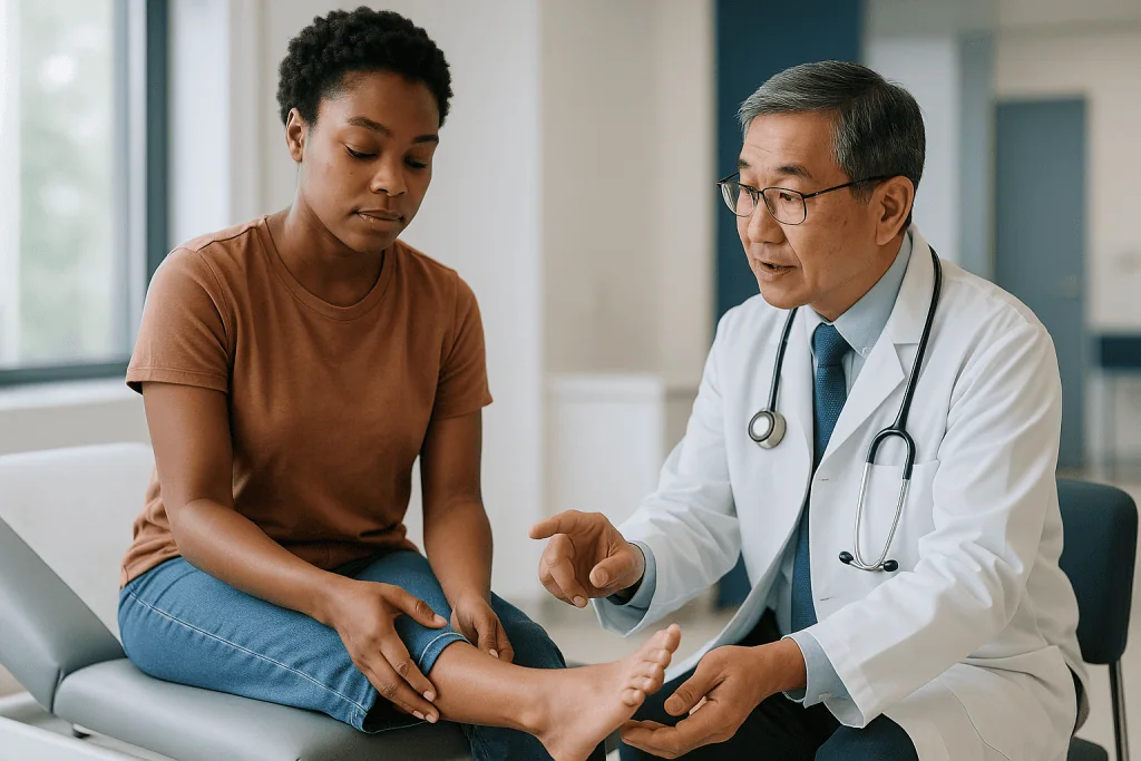
Current surgery is smaller and more targeted. The Achilles tenotomy at the end of casting is technically an operation, yet it takes minutes, leaves a tiny scar and does not open the joints. Beyond that, the most common procedure is a tibialis anterior tendon transfer, in which a tendon on the top of the foot is moved toward the outer side so that it stops pulling the foot inward. It is typically considered when a foot that was corrected begins to drift back during the preschool years and the child is old enough for the bone it attaches to to have formed, usually from around age three.
More extensive release surgery still has a place, mainly for very stiff syndromic feet or for older children whose clubfoot was never treated. In those situations the team weighs a straighter, more functional foot against the trade-off of long-term stiffness.
The practical takeaway for parents is that “needing surgery” is no longer a sign of failure. A tenotomy is part of the standard pathway, a tendon transfer is a planned response to a common pattern, and the decision about any of them rests with the treating team after examining the child.
What the first weeks of clubfoot treatment usually look like
The casting phase is intense but short, and knowing the rhythm helps. In a typical week, the family brings the baby to clinic, the old cast is removed, the foot is examined and gently stretched, and a new cast is applied in a slightly better position. The whole visit may take an hour. Between visits, the cast stays dry and the parents check that the toes remain pink and warm and that the cast has not slipped down the leg.
Babies generally tolerate the casts well. They feed, sleep and kick much as before, and the plaster adds surprisingly little weight. Parents often find the hardest part is practical: cast-friendly clothing, car seats, and the emotional weight of watching a newborn in plaster up to the thigh.
Across the 5 to 8 casts the NHS describes, the change is visible from one week to the next. The sole, which may have faced sideways at the start, rotates toward the floor. By the final cast the forefoot is often over-corrected into an outward position that looks alarming but is intentional, because it leaves room for the natural drift back toward neutral as the child grows.
The tenotomy, when needed, comes near the end. The foot is placed in a final cast afterward for about three weeks while the tendon heals. When that cast comes off, the boots and bar go on the same day. Mayo Clinic describes the full-time bracing phase as around three months, and the NHS as 23 hours a day for about three months, before the brace becomes a sleep-only routine. Those first days in the brace are the moment the real work begins.
Why Ponseti method results depend so heavily on the boots and bar
If one message belongs in bold, it is this: the single strongest predictor of a good long-term result is whether the brace is worn as prescribed. Mayo Clinic states plainly that relapse is more likely when the brace is not used consistently, and the NHS gives the same warning.
The reason is mechanical. The tissues that caused the clubfoot retain a tendency to tighten again as the child grows, particularly in the first few years when growth is fastest. The boots hold each foot turned outward and the bar holds them a shoulder-width apart, so every night of sleep becomes several hours of gentle stretch. Nothing about the brace changes the shape of the foot; its job is to stop the shape from changing back.
The schedule the NHS describes is full-time wear, about 23 hours a day, for roughly three months, followed by wear during naps and overnight until the child is around four or five years old. Mayo Clinic gives a slightly shorter range for the night-time phase, which reflects genuine variation in practice; the treating team sets the plan for each child.
Families struggle most at two points. The first is the transition from cast to boots, when a baby who has been immobile is suddenly able to kick against a bar; a few nights of unsettled sleep are common and usually pass. The second is the toddler years, when a child who walks confidently all day resists being strapped into boots at bedtime. Teams with experience in this phase often suggest making the brace part of an unchanging bedtime ritual rather than a negotiation. Skin checks, correct heel seating in the boot and prompt replacement when the boots are outgrown all protect the result as surely as the schedule itself.
Clubfoot treatment results by pathway: a comparison table
Different starting points lead to different endings, and families benefit from seeing the range laid out rather than a single best case. The table summarizes what the mainstream sources cited in this article describe for each pathway. None of these outcomes is a promise; each is the typical pattern the evidence supports.
| Pathway | What it involves | What the foot usually looks like later | Points to know |
|---|---|---|---|
| Ponseti method, started in infancy, brace worn as prescribed | Weekly casts, usually a tenotomy, then boots and bar through early childhood | Flat, forward-pointing, flexible foot; often slightly smaller with a thinner calf | The reference standard today; follow-up continues until growth is complete |
| Ponseti method with relapse treated by tendon transfer | Repeat casting, then a tendon moved to the outer side of the foot, typically from around age three | Usually returns to a flat, functional foot; small scar on the top of the foot | Common and planned for; not a sign of failed treatment |
| Extensive release surgery (historical standard, or very stiff syndromic feet) | Opening the joints and lengthening multiple tendons and ligaments | Straighter but stiffer foot; more scarring; higher risk of arthritis in adulthood | Reserved today for feet that do not respond to casting |
| Delayed or untreated clubfoot | Casting adapted for an older child, often with additional procedures | Depends on age at treatment; improvement is usual but residual stiffness is more likely | Walking on the side or top of the foot causes skin and joint problems if untreated |
Two things stand out from the table. The gap between the first two rows is small, which is why a relapse managed promptly rarely changes the long-term picture. The gap between the first and third rows is large, which is why current practice tries so hard to avoid extensive surgery and why the experiences of adults treated decades ago do not predict what today’s children should expect.
A smaller foot and thinner calf: the differences that usually stay
Parents often first notice the calf. A child born with clubfoot on one side will usually have a visibly slimmer calf on that side, even after an excellent correction, and no amount of exercise closes the gap. Mayo Clinic lists a smaller calf muscle as an expected feature of a treated clubfoot. The difference is not weakness in the usual sense; it reflects how the muscle developed before birth, and the leg still functions well.
The foot itself tends to be shorter and a little narrower. Buying shoes can mean two different sizes, or accepting a slightly loose fit on the smaller foot. Some families use an insole on that side; the treating team can advise, and this is a matter of comfort rather than medical necessity.
Ankle movement is the third difference. Dorsiflexion, the ability to pull the top of the foot up toward the shin, is commonly a little reduced. Most children never notice. It can show up in activities that demand a deep squat with heels down, where the treated side may lift slightly earlier.
The leg may be marginally shorter as well, usually by a small amount that does not affect walking. Cleveland Clinic describes these size differences as common and generally mild, and routine follow-up includes checking that any difference is not growing in a way that would need attention.
None of these features predicts pain or disability, and that is the point worth holding onto. A foot that is a little smaller, attached to a calf that is a little thinner, with an ankle that bends a little less, is a foot that has been treated well and can carry a child through an ordinary active life.
Walking, running and sport after clubfoot
Children treated with the Ponseti method usually begin walking at the same age as their peers, and the brace, worn only at night by then, does not interfere. The NHS describes most children treated this way as able to walk, run and join in sport normally, and Mayo Clinic says the same, noting that children generally wear ordinary shoes and take part in activities without restriction.
What families sometimes observe is a subtle difference in gait, such as a tendency to turn the foot slightly inward when tired, or a less powerful push-off on the treated side because the calf is smaller. These differences tend to be invisible to anyone not looking for them and do not prevent participation in running, football, dance, gymnastics or swimming.
Sport can matter for a different reason. A clinician watching a child run is observing whether the heel strikes the ground, whether the foot stays flat during stance, and whether the forefoot swings inward. A change in any of these during the growing years is one of the things the team looks for at follow-up, which is why clinic visits continue through childhood even when all seems well.
Fatigue is sometimes reported in adolescence on long walks or hikes, and ankle flexibility may matter in activities such as weightlifting or sprinting that depend on it. Physical therapy focused on calf strength and ankle range can help some adolescents, and the treating team decides when that is useful. The honest summary is that sport is expected, not an exception, and that the differences that remain are usually matters of fine detail rather than barriers.
Clubfoot relapse signs the care team watches for
Relapse means that a foot which had been corrected begins to drift back toward its original position. It is common enough that every treatment plan assumes it might happen, and both Mayo Clinic and the NHS identify inconsistent brace use as the leading reason. Relapse is most likely in the first years after casting, when growth is fastest, and becomes less likely with each year the brace is worn through.
At clinic visits, the team examines the foot for a set of changes rather than relying on a single measurement. They check whether the heel can still be brought up toward neutral, whether the front of the foot has started to curve inward, whether the sole stays flat when the child stands, and whether the child walks on the outer border of the foot. Parents are usually the first to notice a change in the way shoes wear down or the way the child runs, and reporting that observation is far more useful than trying to interpret it at home.
What happens next depends on the degree of relapse and the child’s age. A mild drift in a young child is often managed with a short return to casting, sometimes followed by a renewed emphasis on bracing. In a child old enough, the tendon transfer described earlier is the common surgical response, and it usually restores the foot to a flat, functional position. More extensive surgery is unusual for a relapsed Ponseti foot.
Relapse feels like a setback to families, and that reaction is understandable. In clinical terms it is a known fork in the road with a well-mapped route onward, and the long-term appearance of the foot after a promptly treated relapse is generally similar to a foot that never relapsed.
Adult clubfoot long term effects: what the evidence shows
The long-term picture for adults depends heavily on which era of treatment they belong to, and this is where honesty matters most. Adults treated decades ago with extensive release surgery are the group most often studied, and Mayo Clinic notes that this kind of surgery is associated with stiffness and a higher likelihood of arthritis in later life. Many of those adults report aching after long days on their feet, limited ankle motion and difficulty with uneven ground. Their experience shaped the reputation of clubfoot as a lifelong problem.
Adults who were treated with the Ponseti method as infants are a younger group, because widespread adoption of the method is relatively recent, so the evidence on them into middle age is still accumulating. What is available suggests a more flexible foot with fewer symptoms than the surgical generation, which is the reason the method became the standard of care. Residual features in adulthood mirror those in childhood: a smaller foot, a thinner calf, somewhat reduced ankle motion and, for some, mild stiffness or tiredness with heavy use.
A few points deserve stating plainly. Clubfoot itself is not a progressive disease; the joints and tissues do not deteriorate simply because the condition was present at birth. Arthritis, when it develops, is linked to joint stiffness and abnormal loading rather than to the diagnosis. Footwear that fits each foot, calf and ankle conditioning, and attention to any change in pain or shape are the ordinary tools adults use.
An adult with a treated clubfoot who develops new pain, swelling or difficulty walking should be assessed by a clinician, because the foot can have ordinary problems like any other foot, and because a few may benefit from targeted treatment later in life.
What people often get wrong about clubfoot treatment
Several beliefs about clubfoot persist long after the evidence has moved on, and correcting them changes how families experience the early years.
“The foot will be fixed when the casts come off.” The casts correct the shape; the brace keeps it. The NHS and Mayo Clinic both describe bracing as continuing for years, and both identify stopping early as the main reason feet drift back.
“Clubfoot is caused by something the mother did.” In most cases there is no identifiable cause. Mayo Clinic lists family history, male sex, certain congenital conditions and smoking during pregnancy as factors that raise the likelihood, but the majority of babies with clubfoot are born to parents with none of these.
“Treatment is a surgery with a long recovery.” The modern pathway is mostly casting and bracing. The tenotomy is minor, and extensive surgery is now the exception rather than the rule.
“If the child needs another procedure, the first treatment failed.” A tendon transfer for relapse is a planned option, not a failure, and the foot afterward usually looks much like one that never relapsed.
“The child will not be able to play sports.” Mainstream sources describe most children treated with the Ponseti method as taking part in normal activities, including sport.
“The treated foot should look exactly like the other one.” It usually does not, and does not need to. A slightly smaller foot and thinner calf are expected after a good result.
“Adults with clubfoot are destined for pain.” Much of that reputation comes from older surgical techniques. Today’s children follow a different path, and the long-term outlook with the current approach appears meaningfully better, even though evidence into later adulthood is still being gathered.
Questions to ask your care team
Good questions do two things: they fill the gaps that worry you, and they tell the team what you have understood. These are the ones that tend to be most useful across the stages of treatment, and the answers belong to the clinicians examining your child.
- Is this an idiopathic clubfoot, or is it linked to another condition, and how does that affect what you expect?
- How many casts do you anticipate, and what does each visit involve for us?
- Will my baby need a tenotomy, and how will the anesthesia and the day be handled?
- What exactly is the bracing schedule you want us to follow, and for how many years?
- How will we know when the boots need replacing, and how quickly can that happen?
- What should we look for at home that would make you want to see the foot sooner than the next appointment?
- How often will follow-up visits continue once the brace is only worn at night, and until what age?
- If the foot relapses, what are the likely options at my child’s age?
- Will the treated foot be a different shoe size, and how do you suggest we manage that?
- Is physical therapy part of the plan at any stage?
- Who is our contact for a problem with the cast or brace between appointments?
Bring a notebook or use your phone, because answers given while a newborn is being casted are easy to lose. Ask the team to show you how the heel should sit in the boot before you leave the clinic the first day; seeing it done once prevents most fitting problems later. If a plan changes, ask why; the reasons are usually straightforward and knowing them makes the long bracing years easier to sustain.
When to call your doctor
Most of clubfoot treatment runs on a schedule, but a few situations call for a prompt call to the treating team rather than waiting for the next appointment. The guidance here reflects what mainstream sources such as the NHS and Mayo Clinic advise for casts and braces in general; your own team’s instructions take priority.
During the casting phase, contact the clinic the same day if the toes become pale, blue, cold or swollen, if the baby seems inconsolable in a way that is not explained by feeding or tiredness, if there is a bad smell or discharge from the cast, or if the cast slips so that the toes disappear inside it or the knee becomes visible. A cast that is cracked, soft from getting wet or loose enough to move should also be reported, because a loose cast cannot hold the correction.
During the bracing years, call if you see red or broken skin on the heel, ankle or sole that does not settle after adjusting the boot, if the child cannot be comforted in the brace after the first week or two of adjustment, or if you notice the foot turning inward again, the heel lifting off the ground or the child walking on the outside edge of the foot. A persistent change in the way shoes wear down is worth mentioning too.
At any age, new pain, swelling, warmth or redness in the foot, a limp that lasts more than a day or two without an obvious cause, or a fever alongside any of these should be assessed. None of these signs means something has gone wrong; they are the points at which the team would rather examine the foot than guess, and every decision about what to do next rests with them.
Frequently asked questions
What does a clubfoot after treatment look like compared with the other foot?
It usually looks close to typical: flat on the floor, pointing forward, fitting ordinary shoes. On close inspection the treated foot is often a little shorter and narrower, the calf thinner and the ankle slightly less flexible. Mayo Clinic describes these as expected features of a well-treated clubfoot rather than signs of a problem.
Is there surgery for clubfoot, and will my child need it?
Most children need a minor Achilles tenotomy at the end of casting, which is a small cut to lengthen the tendon. Larger operations are now uncommon. A tendon transfer may be used if the foot relapses in the preschool years, and extensive release surgery is reserved for very stiff feet. The treating team decides based on examining the foot.
What is the Ponseti method used for correcting clubfoot?
It is the mainstream non-surgical approach in which the foot is gently manipulated and held in a series of weekly casts, usually followed by a small tenotomy, then braced with boots and a bar during sleep for several years. The NHS describes it as the main treatment for clubfoot and notes that most children treated this way walk and run normally.
What are the chances of clubfoot?
About 1 in 1,000 babies is born with clubfoot, according to the NHS and Mayo Clinic. The chance is higher with a family history, in boys, with certain conditions such as spina bifida, and with smoking during pregnancy, but most affected babies have none of these factors and no cause is identified.
What are the main clubfoot relapse signs parents report?
Parents usually notice a change in walking first, such as the foot turning inward, the heel lifting off the ground or the child walking on the outer edge of the foot, along with uneven shoe wear. These observations should be reported to the treating team, who examine the foot to decide whether it has relapsed and what to do.
Will the treated foot be a different shoe size?
Often, yes. The treated foot is commonly slightly shorter and narrower than the other, sometimes by around half a size or more, and the difference persists into adulthood. Many families buy for the larger foot and use an insole on the smaller side. This is a comfort issue rather than a medical one, and the care team can advise.
Can children play sports after clubfoot treatment?
Yes. Both the NHS and Mayo Clinic describe most children treated with the Ponseti method as able to walk, run and take part in sport without restriction. Some notice a less powerful push-off on the treated side or slightly reduced ankle flexibility, but these rarely limit participation in running, football, dance, gymnastics or swimming.
What are the adult clubfoot long term effects?
They depend on how the foot was treated. Adults who had extensive release surgery decades ago are more likely to have stiffness and arthritis, which Mayo Clinic links to that kind of surgery. Adults treated with casting and bracing as infants generally keep a more flexible foot, with a smaller foot and calf and some reduced ankle motion, though long-term evidence is still accumulating.
Who are some famous people with clubfoot?
Many athletes, actors and public figures have spoken openly about being born with clubfoot, and their stories are easy to find. We do not name individuals here, partly out of respect for privacy and partly because a single person’s outcome, whether exceptional or difficult, says little about what any child should expect. The evidence-based picture matters more.
How long does clubfoot treatment take from start to finish?
Casting usually takes about 5 to 8 weeks, followed by a final cast of roughly three weeks after the tenotomy. Full-time bracing then lasts about three months, and night-time bracing continues until around age four or five in the NHS schedule. Follow-up visits continue through childhood to check the foot as it grows.
References
This article is for general information only and is not a substitute for professional medical advice. Please consult a qualified doctor about your individual situation.

More from the Blog

Recovery After Trauma Surgery: Why Physiotherapy Starts Early and How Milestones Are Set
Trauma surgery recovery usually begins within a day of the operation, when a physiotherapist helps the patient sit, breathe deeply and, where the repair…

Total vs Partial Shoulder Replacement: Which Joint Surfaces Are Replaced and Why
A total shoulder replacement resurfaces both sides of the ball-and-socket joint: the rounded top of the upper arm bone and the shallow socket on…

Who Is a Candidate for Robotic Knee Replacement? What Orthopedic Surgeons Weigh
A robotic knee replacement candidate is generally an adult with advanced knee arthritis whose pain and stiffness limit daily life despite non-surgical care such…

Hip Precautions After Hip Replacement: Which Movements Are Limited Early and for How Long
Hip precautions after hip replacement are movement limits that protect the new joint while the surrounding soft tissue heals. After a posterior approach they…
Does Foot Pain Always Need Surgery? Orthotics, Injections and When an Operation Makes Sense
No. Most foot pain is managed without an operation. Conditions such as plantar fasciitis, bunions, Morton's neuroma and many fractures usually respond to footwear…

Flying Home After Hand Surgery: Splints, Swelling and Timing Your Follow-Up Abroad
Most people can fly after minor hand surgery once the anesthetic has fully worn off, pain is controlled with the medicines they were prescribed,…