

When Radiotherapy Is Added to Lymphoma Treatment and When Chemotherapy Alone Is Enough

Key Takeaways
- Consolidation radiotherapy is given after chemotherapy to destroy any lymphoma cells left at the original site, and reaching that stage usually means the pathway is on track rather than off it.
- The four facts that drive the decision are subtype, stage, whether any mass was classed as bulky at diagnosis, and the Deauville score on the end-of-treatment PET scan.
- For PET-negative early-stage classical Hodgkin lymphoma, guidelines endorse both chemotherapy plus radiotherapy and chemotherapy alone, trading a small extra relapse risk against fewer late effects.
- Advanced-stage Hodgkin lymphoma and most advanced diffuse large B-cell lymphoma that reach a complete metabolic response are generally treated with chemotherapy alone.
- Lymphoma radiotherapy is a short course of weekday sessions over a few weeks, each lasting roughly 10 to 30 minutes with the beam itself on for only a minute or two, and it does not make a person radioactive.
- Fatigue and skin reactions typically peak around the final week and the weeks just after, and the late risks that matter most, to heart, lungs, thyroid and breast tissue, depend on exactly which part of the body is in the field.
Radiotherapy is usually added after chemotherapy for lymphoma when disease was localized or bulky at diagnosis, or when a post-treatment PET scan still shows active tissue in one area that a targeted beam can reach. Chemotherapy alone is usually enough for advanced-stage disease that reaches a complete metabolic response, and increasingly for early-stage disease that becomes PET-negative. The treating team decides case by case.
The scan report is folded in half on the clinic desk. Six cycles of chemotherapy are done, the port is still in, and the first words from the oncologist are not ‘you are finished’ but ‘we need to talk about radiotherapy.’ For many people with lymphoma that sentence lands like a demotion. Usually it is not one. Radiation after chemotherapy for lymphoma is a planned, targeted step in a pathway that was sketched out at the first staging scan, not a sign that the drugs failed.
What makes the conversation confusing is that the person in the next chair, with what sounds like the same diagnosis, was told chemotherapy alone was enough. Both can be right. The decision turns on subtype, stage, how large the biggest mass was at the start and, above all, what the PET scan shows once the drugs have done their work.
This explainer walks through that logic the way a tumor board does, then covers what the sessions, the tiredness and the skin actually feel like.
What does radiation after chemotherapy for lymphoma actually mean?
Oncologists call it consolidation radiotherapy: a short course of radiation aimed at the places where lymphoma was sitting, given once chemotherapy has shrunk or cleared the disease, with the aim of destroying any cells the drugs left behind. The word consolidation carries the whole idea. The radiation is there to lock in a response that has already happened, not to rescue a response that never came.
The technique used today is involved-site radiotherapy, which means the beam is shaped to the original site of disease plus a small safety margin. Older approaches treated entire regions of the body, such as the whole neck and chest together, and much of what people fear about lymphoma radiotherapy comes from that era. Modern fields are smaller, planned in three dimensions on a CT scan, and avoid as much normal tissue as the anatomy allows.
Lymphoma is one of the most radiation-sensitive cancers. Lymphoma cells are damaged at exposures lower than many solid tumors need, so the course is short. The NHS describes lymphoma radiotherapy as brief daily sessions, usually on weekdays, spread over a few weeks rather than months. Each visit involves lying still on a treatment couch while a machine called a linear accelerator delivers invisible, painless beams from several angles.
Nothing about the treatment makes a person radioactive. The energy passes through the body and is gone in seconds; what remains is damage to the DNA of cells in the beam path, which rapidly dividing lymphoma cells cannot repair as well as healthy tissue can. That difference in repair is the entire basis of the treatment, and it is also why side effects are confined to the treated area and build gradually rather than hitting all at once.
Why chemotherapy and radiotherapy do different jobs
Chemotherapy is systemic. The drugs travel in the bloodstream and reach lymphoma cells wherever they are hiding, including places no scan can see. Radiotherapy is local. It treats only the tissue inside the planned field and nothing beyond it. Those two facts explain almost every decision in this article.
Lymphoma spreads through the lymphatic system, the network of vessels and nodes that carries immune cells around the body. In some people it stays in one or two neighboring regions; in others it is scattered through nodes above and below the diaphragm, or into bone marrow, liver or spleen. Widespread disease needs a systemic treatment. A local one cannot chase it.
Large masses are the exception that keeps radiotherapy in the picture. A bulky node has a poorly supplied core where blood flow is sluggish, oxygen is low and fewer cells are actively dividing. Chemotherapy drugs reach that core less reliably, and cells that are not dividing are less vulnerable to them. When lymphoma comes back after chemotherapy alone, it tends to return at the sites that were bulkiest at the start. A beam aimed at exactly those sites addresses the weak point of the drugs.
The trade-off is geography. A field in the groin touches very different normal tissue than a field in the middle of the chest, where the heart, lungs, breast tissue and thyroid sit close together. The benefit of consolidation is roughly similar wherever the disease was; the long-term cost depends heavily on what is nearby. Teams therefore weigh the same question differently for a mass behind the breastbone than for one in the armpit, and a younger person with decades ahead is weighed differently again.
How PET scans changed the decision
A PET scan uses a mildly radioactive sugar tracer that collects in cells burning energy quickly, so active lymphoma lights up while scar tissue stays dark. Before PET, a residual lump on a CT scan after chemotherapy was a genuine puzzle: live disease or a fibrous shell? Radiotherapy was often given simply because nobody could tell. PET answered the question and, in doing so, rewrote the rules.
Two scans matter. An interim PET, taken after the first couple of chemotherapy cycles, shows how quickly the disease is responding. An end-of-treatment PET shows what is left. Radiologists grade the result on the Deauville scale, a five-point score that compares tracer uptake in any remaining tissue against the liver and the blood in the large chest vessels. Low scores are read as a complete metabolic response, which means no detectable active disease; high scores mean something is still burning.
Large trials in early-stage Hodgkin lymphoma tested a simple idea: if the interim PET is negative, skip the radiotherapy. The results were honest rather than tidy. Leaving radiation out carried a modest extra risk of relapse, yet most people who relapsed could be treated again, and overall outcomes between the two approaches were close. Guidelines now describe both options for PET-negative early-stage disease, which is why the same scan can lead to different recommendations in different clinics, each defensible.
PET has limits. Inflammation, infection, recent chemotherapy and the thymus gland regrowing in young adults can all create false-positive signals. Timing the scan a few weeks after the last cycle reduces this, and a positive result in a single spot usually prompts discussion rather than automatic action. Sometimes the answer is a biopsy; sometimes it is radiotherapy to that one site; occasionally it is a repeat scan.
Who is usually offered radiation after chemotherapy for lymphoma?
Several situations recur in guidelines across Europe and North America, and the NHS lists most of them in its treatment pages for Hodgkin and non-Hodgkin lymphoma.
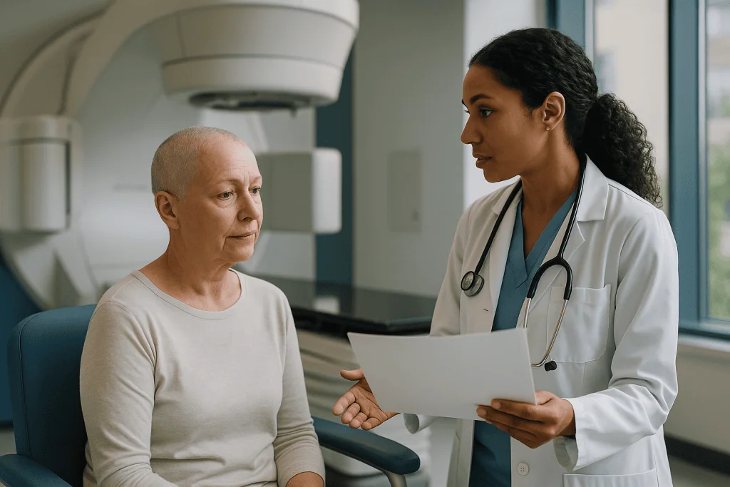
Early-stage classical Hodgkin lymphoma is the clearest example. Stage I or II disease, meaning nodes on one side of the diaphragm, is commonly treated with a short course of chemotherapy followed by involved-site radiotherapy, an approach known as combined-modality treatment. The short chemotherapy course is only considered sufficient because radiation finishes the job locally.
Bulky disease at diagnosis is the second. Bulky means a mass larger than a threshold set in the staging guideline, most often measured in the chest. Because relapses cluster at bulky sites, radiotherapy to that site after chemotherapy is frequently recommended even when the end-of-treatment PET is clean, particularly in Hodgkin lymphoma.
A PET-positive residual area confined to one region after full chemotherapy is the third. Here radiation is doing something closer to active treatment than consolidation, and the team may also consider further drug therapy or a biopsy first.
Limited-stage diffuse large B-cell lymphoma, the most common aggressive non-Hodgkin subtype, is a fourth. Radiotherapy may allow fewer chemotherapy cycles, or may be added for bulk, bone involvement or certain extranodal sites such as the testis or stomach.
Nodular lymphocyte-predominant Hodgkin lymphoma and stage I follicular lymphoma round out the list; in both, radiotherapy alone may be the entire treatment rather than an add-on. Finally, people who cannot tolerate a full chemotherapy course because of heart, lung or kidney problems may be offered radiation to make up the shortfall. None of these is automatic; each is a starting position for the tumor board discussion.
When is chemotherapy alone enough for lymphoma?
The short answer is: when the disease was too widespread for a beam to help, or when the scans say the drugs have already finished the work.
Advanced-stage classical Hodgkin lymphoma, stage III or IV, is treated with a full course of chemotherapy, and radiotherapy is generally omitted when the end-of-treatment PET shows a complete metabolic response. Some teams still consider radiation to a site that was bulky at the outset or that remains PET-positive, but routine consolidation of every original site is no longer standard. The NHS notes that advanced Hodgkin lymphoma is usually managed with chemotherapy alone.
Advanced diffuse large B-cell lymphoma follows the same logic. Immunochemotherapy, which pairs chemotherapy with an antibody that attaches to a protein on the surface of B cells and flags them for destruction, is the backbone, and a complete response ends treatment for most people. Residual PET-positive sites are handled individually.
PET-negative early-stage Hodgkin lymphoma is the gray zone described earlier. Chemotherapy alone is a guideline-endorsed option, and it is chosen more readily when the field would include breast tissue in a young woman, the heart in a person with existing cardiac risk, or the growing tissues of a child or adolescent. Pediatric protocols are built specifically to avoid radiation whenever the response allows, because young tissue is the most vulnerable to late effects.
Indolent lymphomas that are widespread at diagnosis rarely receive consolidation radiotherapy; when a beam is used in these cases it is to relieve a symptom, such as a painful or obstructing node, not to lock in a remission.
Omitting radiotherapy is not the same as doing less. It is a deliberate trade: a small added relapse risk, usually treatable, in exchange for a lower lifetime burden of late effects. Reasonable experts land on different sides of that trade for the same patient, which is exactly why the recommendation should be explained rather than simply announced.
Radiotherapy for Hodgkin lymphoma versus non-Hodgkin subtypes
Lymphoma is not one disease but dozens, and the role of radiation shifts with subtype. The table summarizes the typical starting positions described in mainstream treatment guidance; individual plans depart from them for good reasons.
| Situation | Usual approach | Reasoning |
|---|---|---|
| Early-stage classical Hodgkin, not bulky, PET-negative | Short chemotherapy plus radiotherapy, or chemotherapy alone | Both endorsed; choice weighs small relapse risk against late effects |
| Early-stage classical Hodgkin, bulky or PET-positive | Chemotherapy followed by involved-site radiotherapy | Relapse risk concentrates at bulky or still-active sites |
| Advanced classical Hodgkin, complete metabolic response | Chemotherapy alone | Disease too widespread for local treatment to add much |
| Limited-stage diffuse large B-cell lymphoma | Immunochemotherapy, sometimes shortened, with or without radiotherapy | Radiation can replace cycles or cover bulk and extranodal sites |
| Advanced diffuse large B-cell lymphoma, residual PET-positive site | Individualized: biopsy, radiotherapy or further drug therapy | Single residual site may be reachable; false positives possible |
| Stage I follicular lymphoma | Radiotherapy alone is a standard option | Highly radiation-sensitive, localized disease |
Two patterns stand out. In Hodgkin lymphoma, radiotherapy is tied tightly to stage and PET response; the scan effectively votes. In aggressive non-Hodgkin lymphoma the drugs carry more of the load and radiation is used more selectively, for bulk, for specific anatomical sites or for a stubborn residual area. In indolent lymphoma the beam can be the whole treatment when disease is confined, and almost irrelevant when it is not.
The subtype label on the pathology report, the stage, the bulk measurement and the Deauville score are the four pieces of information that drive the column on the right. Asking the care team to walk through each of those four, in that order, usually makes the recommendation feel less arbitrary.
What happens during involved-site radiotherapy, step by step
The first appointment is planning, not treatment. A dedicated CT scan is taken in the exact position used for every later session, often with the arms raised or the chin extended. For neck and chest fields a thermoplastic mask is molded while warm and sets in a few minutes; it keeps the head and shoulders in the same place each day and is removed the moment the beam is off. Tiny permanent skin marks, usually three or four dots the size of a freckle, or non-permanent markings, allow the lasers in the treatment room to line up the body precisely.
Radiation oncologists then outline the target on the planning scan, drawing around where the disease was at diagnosis, adjusted for how the anatomy has shifted after the mass shrank. Physicists calculate beam angles that cover that target while sparing heart, lungs, spinal cord, thyroid, salivary glands or breast tissue as far as possible. Techniques such as intensity-modulated radiotherapy, which varies the beam strength across the field, and deep-inspiration breath hold, where the patient holds a full breath to pull the heart down and away from a chest field, are common in lymphoma planning.
Treatment itself is uneventful. The Mayo Clinic describes a typical external-beam session as lasting around 10 to 30 minutes, most of it spent on positioning and imaging checks rather than beam-on time, which is usually a minute or two. Nothing is felt. Nothing is heard beyond the hum and clicks of the machine moving. People drive themselves, go to work and pick up children afterward.
A radiographer or nurse reviews side effects at least weekly, and the radiation oncologist sees the patient through the course. Any new symptom, including ones that seem unrelated, belongs in that conversation.
How long after chemo does radiotherapy start, and what the weeks look like
There is a deliberate pause between the last chemotherapy cycle and the first radiation session. It serves three purposes: blood counts recover, the end-of-treatment PET is taken late enough to avoid false positives from chemotherapy-related inflammation, and the planning scan can be based on the body’s new shape once the mass has shrunk. In practice that pause is measured in weeks. Teams aim to keep it short, since a long gap gives any surviving cells time to regrow, and they will say if a delay is becoming a concern.
Here is the usual shape of the period, drawn from NHS and Mayo Clinic descriptions of radiotherapy pathways:
- Weeks after final chemotherapy: blood tests, end-of-treatment PET, tumor board review, decision discussion.
- One to two weeks before treatment: planning CT, mask if needed, skin marks, written information on skin care and side effects.
- Treatment course: daily weekday sessions over a few weeks; side effects are mild early and build toward the final week.
- First weeks after the last session: skin reaction and fatigue often peak before they begin to settle, because the biological effect lags behind the beam.
- A few months after treatment: response assessment, usually with PET, timed to let treatment-related inflammation fade.
During the course, most people are asked to keep a loose diary of energy, appetite, swallowing (for neck or chest fields), bowel habit (for abdominal or pelvic fields) and skin. Patterns in that diary guide the weekly review better than memory does.
The follow-up rhythm after the response scan is set by the hematology team and typically spaces out over several years, with earlier reviews of thyroid function, heart health and, for women who had chest radiation young, breast screening arrangements. Survivorship care is part of the treatment, not an afterthought to it.
What does radiation fatigue feel like?
People reach for the same words: heavy, as if the limbs were full of wet sand; a wall around mid-afternoon; waking after eight hours and feeling as though it were four. Radiation fatigue is not ordinary tiredness and sleep does not fully reset it. The NHS lists it as the most common side effect of radiotherapy, notes that it tends to build through the course, and says it can continue for weeks or months after treatment finishes before gradually lifting.
The mechanism is partly repair. The body is clearing damaged cells and rebuilding tissue inside the field every day, and that is metabolically expensive. Layered on top are the leftovers of chemotherapy: a blood count that has not fully recovered, muscle lost over months of treatment, disrupted sleep, the daily commute to the department and the quiet strain of being a patient for so long. Low mood and anxiety add to the load and are worth naming out loud.
Fatigue after lymphoma treatment is also a prompt to check a few things that are easy to treat. Anemia, an underactive thyroid (especially after neck radiation, though this usually appears later), poor sleep and depression all produce the same heavy feeling and all have their own solutions. A persistent crash deserves a blood test, not only reassurance.
What helps, according to NHS guidance on radiotherapy side effects, is counterintuitive: gentle, regular activity such as daily walking tends to reduce fatigue more than prolonged rest does. Pacing matters, meaning spreading tasks across the day rather than saving them for the morning, short naps rather than long ones, protein with each meal and enough fluid. Most people can keep working through a short lymphoma course, often with adjusted hours; some cannot, and that is not a failure of will.
Skin care and the real do's and don'ts during radiation treatment
Skin changes happen only inside the treated area, and for lymphoma fields they are usually modest. The NHS describes a reaction that starts a week or two into the course, looks and feels like mild sunburn, may darken or peel, and typically settles within a few weeks of the final session. Neck and groin fields, where skin folds, tend to react more than flat areas.
The following practical points come from NHS and MedlinePlus skin-care guidance; the department’s own leaflet takes precedence where it differs.
- Wash the area daily with lukewarm water and a mild, unperfumed soap; pat dry, never rub.
- Apply the moisturizer the team suggests, from day one, and skip it in the hours just before a session only if told to.
- Choose soft, loose clothing; avoid tight collars, bra straps or waistbands that rub the field.
- Keep the area out of the sun during treatment and cover it in the months afterward, since treated skin burns more easily for a long time.
- Leave the skin marks alone; if they fade, the radiographers redraw them.
- Avoid heat pads, ice packs, adhesive tape and strong products such as perfumed lotions or exfoliants on the field.
- Ask before shaving the area; an electric razor is usually acceptable, a wet blade often is not.
- Report broken, weeping or increasingly painful skin promptly rather than waiting for the next review.
Beyond the skin, a few habits make the course easier. Swallowing can become sore with neck or upper chest fields, so softer, cooler foods and avoiding alcohol and very spicy meals help. Smoking slows tissue healing and raises the risk of later lung and heart problems after chest radiation; this is one of the best moments to get support to stop. Tell the team about every supplement, because some high-dose antioxidant products are thought to blunt the effect of radiation, and about any vaccine appointments, which may need rescheduling.
Radiotherapy side effects in lymphoma: the late effects that drive the decision
Short-term side effects of lymphoma radiotherapy are usually manageable. The late effects, which surface years or decades later, are the real reason teams argue over whether to add it at all. Honesty about them is the most useful thing this section can offer.
Chest fields may expose part of the heart, and radiation accelerates coronary artery disease and valve stiffening, with the risk rising alongside ordinary factors such as smoking, high blood pressure and cholesterol. Lung tissue in the field can become inflamed in the months after treatment (pneumonitis) and later scarred. Breast tissue exposed in adolescence or young adulthood carries a higher lifetime breast cancer risk, which is why survivorship guidelines bring screening forward for these women. Neck fields commonly lead, eventually, to an underactive thyroid, easily picked up on a blood test and treated. Salivary glands in the field may leave a drier mouth and more dental decay. Pelvic fields raise questions about fertility, which should be discussed before treatment starts.
Two things soften this picture. First, the risks above were largely measured in people treated with the large fields and higher exposures of previous decades; involved-site planning, breath-hold techniques and modulated beams now keep far more normal tissue out of the path, and the long-term effect of those changes is still being measured. Second, almost every late effect has a surveillance plan: thyroid function tests, blood pressure and cholesterol control, earlier breast screening, dental follow-up, and a low threshold for investigating chest symptoms.
Put plainly, the question a team asks is not ‘is radiotherapy safe’ but ‘for this person, with this anatomy and this scan, does the reduction in relapse risk outweigh the specific late risks of this field?’ A twenty-year-old woman with a residual mass over the heart and a sixty-five-year-old man with a bulky groin node are not the same calculation, and should not get the same answer.
What people often get wrong about radiation after chemotherapy for lymphoma
Misunderstandings cluster around a handful of points, and correcting them tends to lower the temperature of the whole conversation.
Radiotherapy means the chemotherapy failed. In most cases it means the opposite: the plan from the start was chemotherapy followed by consolidation, and reaching the radiotherapy stage means the pathway is on track. The exception, a PET-positive residual area, is a change of plan rather than a verdict, and the team will say so explicitly.
Skipping radiotherapy means the team is giving up. Omitting it after a complete metabolic response is a guideline-based choice to avoid late effects, backed by trials in which outcomes were close either way.
You become radioactive. External-beam radiotherapy leaves nothing behind. Holding a baby, sharing a bed or sitting next to a pregnant colleague is safe immediately after a session, as MedlinePlus and the NHS both state.
Radiation side effects feel like chemotherapy. They are local. Hair falls out only inside the field, nausea is unusual unless the abdomen is treated, and blood counts are affected far less than with drugs.
A lump that is still visible means the cancer is still there. Many residual masses on CT are scar tissue. The PET result, not the size on a CT, is what the team reads.
Fewer sessions means a weaker treatment. Lymphoma is highly radiation-sensitive, so courses are short by design, not by compromise.
Late effects are inevitable. They are risks, modified heavily by field size, technique, age and lifestyle factors such as smoking, and most are monitored for and treatable when they do appear.
None of these corrections should replace a direct conversation with the team about why a particular recommendation was made. They simply clear the ground so that the conversation is about the right things.
Questions to ask your care team
A good consultation about consolidation radiotherapy should leave a person able to explain, in their own words, why it is or is not being recommended. These questions tend to get there. Writing them down and bringing someone to take notes helps more than most people expect.
- What subtype and stage do I have, and was any site classed as bulky at diagnosis?
- What did my interim and end-of-treatment PET scans show, and what was the Deauville score?
- Is the radiotherapy you are suggesting consolidation after a complete response, or treatment of an area that is still active?
- What would happen if I chose chemotherapy alone, and how would relapse be detected and treated if it occurred?
- Exactly which part of my body would be in the field, and which organs are close to it?
- Which techniques, such as breath hold or modulated beams, will be used to protect my heart, lungs, breast tissue or thyroid?
- How many weeks will the course take, and how soon after chemotherapy should it begin?
- What short-term side effects should I expect for this particular field, and who do I contact if they become severe?
- What late effects apply to me specifically, and what monitoring will I have for them in the years ahead?
- Does this treatment affect fertility, and should I speak to a fertility specialist before starting?
- Will I need any vaccinations rescheduled, or any medicines or supplements paused?
- Is there a clinical trial relevant to my situation?
If the answers to the first three questions are not clear, it is reasonable to ask for the case to be summarized in a letter, or for a second opinion within the same health system. Teams expect this request and it does not delay care when raised early.
When to call your doctor
Most problems during and after lymphoma radiotherapy are mild and expected. A few are not, and they need same-day contact with the treating team or, where stated below, emergency care. Blood counts can remain low for weeks after chemotherapy, so infection remains the most urgent concern throughout the radiotherapy course.
- A temperature of 38°C (100.4°F) or higher, shivering, or feeling suddenly unwell, even without a fever: contact the emergency number given by the hematology team immediately, as this may be a serious infection while immunity is low.
- New or worsening breathlessness, chest pain, a dry cough or a racing heartbeat, during treatment or in the months after a chest field: seek urgent assessment, since pneumonitis, a clot or a heart problem must be ruled out.
- Swelling, pain or warmth in one leg, or sudden breathlessness: possible blood clot; seek care the same day.
- Swallowing so painful that fluids are being avoided, or weight falling quickly: the team can adjust pain relief and nutrition support.
- Skin in the field that is broken, weeping, spreading redness, or has an offensive smell: possible infection needing review.
- Persistent vomiting, severe diarrhea, or signs of dehydration such as dizziness and very dark urine.
- Unusual bleeding or bruising, or black stools.
- Fatigue so profound that standing causes faintness, or confusion or drowsiness that is new.
- A new lump, night sweats or unexplained fever after treatment has finished: not an emergency, but worth an early appointment rather than waiting for the scheduled review.
When in doubt, call. Radiotherapy departments and hematology units run on the assumption that patients will phone about things that turn out to be nothing, and they would far rather hear about a symptom on day one than day five. Every decision about pausing, adjusting or continuing treatment belongs to the treating team, who know the plan, the scans and the person.
Frequently asked questions
How long after chemo does radiotherapy start for lymphoma?
Usually within a matter of weeks after the final chemotherapy cycle. The gap lets blood counts recover, allows the end-of-treatment PET scan to be taken late enough to avoid false positives from inflammation, and gives time for a planning CT and mask. Teams try not to stretch the interval, because a long delay gives any remaining cells time to regrow, and they will explain if scheduling becomes a concern.
What is consolidation radiotherapy in lymphoma?
Consolidation radiotherapy is a short course of targeted radiation given to the original site of lymphoma after chemotherapy has already produced a response, with the aim of eliminating any cells the drugs left behind. It is planned from the outset in early-stage and bulky disease rather than added because treatment failed. Modern involved-site technique shapes the beam closely to where the disease was, sparing surrounding organs as far as the anatomy allows.
Is radiotherapy always used for Hodgkin lymphoma?
No. Radiotherapy is common in early-stage classical Hodgkin lymphoma, especially with bulky disease or a PET-positive residual area, but advanced-stage disease that reaches a complete metabolic response is generally treated with chemotherapy alone. For early-stage disease that becomes PET-negative, guidelines allow either approach, and teams weigh the small relapse-risk difference against late effects for the individual, particularly in young people and when the heart or breast tissue would sit in the field.
What are the side effects of radiotherapy for lymphoma?
Short-term effects are confined to the treated area: fatigue, skin redness like mild sunburn, hair loss only within the field, and a sore throat or swallowing discomfort with neck or chest fields. Nausea is uncommon unless the abdomen is treated. Late effects depend on location and may include thyroid underactivity after neck fields, and heart, lung or breast risks after chest fields, all of which have monitoring plans in survivorship care.
Does needing radiotherapy mean chemotherapy did not work?
In most cases, no. Combined-modality treatment, meaning chemotherapy followed by radiotherapy, is a standard plan for early-stage and bulky lymphoma decided at diagnosis, so reaching the radiation stage means the pathway is proceeding as intended. The exception is radiotherapy aimed at an area that remains PET-positive after a full chemotherapy course; that is a change of plan rather than a failure, and the team should say which situation applies.
What are the next steps after radiotherapy for lymphoma?
Side effects usually peak in the first week or two after the final session and then settle over several weeks. A response assessment, typically a PET scan, is scheduled a few months later so that treatment-related inflammation does not mimic disease. Follow-up then spaces out over years and includes thyroid blood tests after neck fields, heart risk-factor checks after chest fields, dental review where salivary glands were treated and earlier breast screening for women treated young.
What does radiation fatigue feel like and how long does it last?
People describe heavy limbs, an afternoon wall and waking unrefreshed despite a full night’s sleep. The NHS notes it builds through the course and can persist for weeks or months after treatment before gradually lifting. Light daily activity such as walking tends to help more than prolonged rest. Fatigue that is severe or not improving warrants blood tests for anemia and thyroid function and an honest conversation about sleep and mood.
Can I work or exercise during radiotherapy for lymphoma?
Many people continue to work through a short lymphoma course, often with adjusted hours around daily appointments, and gentle exercise is encouraged by NHS guidance because it reduces fatigue. Heavier training may need scaling back toward the end of the course. Swimming is usually discouraged while the skin in the field is reacting, and any activity that rubs the treated area should be avoided until the skin has settled.
Will I lose my hair with radiotherapy for lymphoma?
Only hair growing inside the treated field is affected. A neck field may thin the hair at the back of the scalp or the beard line; a chest field may affect chest hair; a groin field may affect pubic hair. Scalp hair is untouched unless the scalp itself is treated, which is rare in lymphoma. Hair in the field usually regrows over months, though it can return finer or a different shade.
Am I radioactive after a radiotherapy session?
No. External-beam radiotherapy delivers energy that passes through the body in seconds and leaves nothing behind, so there is no risk to children, pregnant partners or anyone else at home immediately after treatment. This differs from certain radioactive drug treatments and from the PET scan tracer, which carries brief precautions on the day of the scan. The department will state clearly if any precaution applies to a specific test.
References
- NHS: Hodgkin lymphoma, treatment
- NHS: Non-Hodgkin lymphoma, treatment
- NHS: Radiotherapy, side effects
- MedlinePlus: Radiation therapy
This article is for general information only and is not a substitute for professional medical advice. Please consult a qualified doctor about your individual situation.

More from the Blog

Surgical Oncology Preparation: Staging Scans, Blood Tests and the Anesthesia Assessment
Preparing for cancer surgery usually means three things happening in parallel: staging scans such as CT, MRI or PET-CT to map where the cancer…
What Neck Cancer Follow-Up Looks Like: Physical Exams, Imaging and Side-Effect Reviews
Follow-up after neck cancer treatment usually combines a hands-on physical exam of the neck, mouth and throat at each visit, a camera check of…
Follow-Up After Cervical Cancer Treatment: Pelvic Exams, Imaging and Recurrence Checks
Cervical cancer follow up after treatment usually combines a review of symptoms, a pelvic exam and, depending on the treatment given, cytology or HPV…
What a Chemotherapy Cycle Looks Like: Treatment Days, Low-Count Days and Recovery
A chemotherapy cycle is one treatment session (or a short run of sessions) followed by a planned rest period, commonly repeating every one to…
Living With Multiple Myeloma: Bone Protection, Kidney Care and Staying Active Safely
People living with multiple myeloma can protect their bones and kidneys through a combination of bone-strengthening medicines chosen by their hematology team, regular dental…

Before Parotidectomy Surgery: Imaging, Fine Needle Biopsy and the Anesthesia Evaluation
Before parotidectomy surgery, most people have three things: imaging (usually ultrasound first, then MRI or CT) to map the lump and its relation to…