

When to Get a Mole Checked: The Changes That Matter

Key Takeaways
- The "E" in ABCDE, evolution, outweighs the other letters: a mole changing in size, shape, or color over weeks to months is the strongest single reason to book an exam.
- Most melanomas begin on previously normal skin, so a brand-new dark spot in adulthood deserves the same scrutiny as a changing mole.
- Melanomas can be smaller than the classic 6-millimeter "pencil eraser" cutoff, so a small size should never be your reason to relax.
- The UK's 2-week rule means a GP who suspects melanoma gets you a specialist appointment within a fortnight: a useful urgency benchmark wherever you live.
- Nodular melanoma often breaks every ABCDE rule; watch instead for EFG: a bump that is elevated, firm, and growing over several weeks.
- In people with darker skin tones, melanoma more often appears on palms, soles, or as a dark streak under a nail, places sun exposure rarely explains.
Get a mole checked promptly if it changes in size, shape, or color over weeks to months; becomes asymmetrical; develops ragged borders or multiple shades; grows larger than about 6 millimeters; or starts to itch, bleed, or crust. A mole that simply looks different from all your others, or a brand-new dark spot appearing in adulthood, also deserves a doctor's exam.
It rarely happens in front of a mirror you planned to use. You’re toweling off after a shower, or a partner traces a finger across your shoulder and asks, “Has that always been there?” Suddenly a brown spot you’ve ignored for a decade has your full attention, and the internet’s worst-case answers are one search away.
Here’s the honest version: most moles are boring, and boring is exactly what you want. The average adult carries dozens of them, and nearly all will stay quietly unchanged for years. Melanoma, the skin cancer everyone worries about, is far less common than harmless moles, but it is also one of the few cancers you can literally watch for on your own skin.
The trick is knowing which changes are background noise and which ones are a signal. That difference is smaller, and more specific, than most articles let on.
Why moles change, and why most changes are harmless
Moles are not fixed features like fingerprints. They have a life cycle of their own. New ones appear through childhood and early adulthood, existing ones can slowly rise off the skin or lighten with age, and many quietly fade away after midlife. None of that is alarming: it’s biography, not pathology.
Hormones move moles around, too. During puberty and pregnancy, moles commonly darken slightly or grow a little, and the key detail is that they tend to do it together, evenly, across the whole spot. The NHS notes that this kind of uniform, hormone-driven change is normal and usually nothing to worry about.
What separates ordinary change from a warning sign is the pattern. A mole that darkens evenly along with its neighbors over years is telling one story. A single mole that changes on its own timetable, new colors appearing within it, one edge creeping outward, a texture shift over weeks or a few months, is telling a different one. Speed, asymmetry of change, and a mole behaving unlike the rest of your skin are the details that matter. Slow, symmetrical, universal change is the skin doing what skin does. Fast, lopsided, solo change is the reason skin checks exist.
What does a normal mole look like?
Before hunting for trouble, it helps to know what “fine” looks like. According to MedlinePlus, most people have between 10 and 40 moles, and they share a recognizable family resemblance:
- Shape: round or oval, roughly symmetrical, if you folded it in half in your mind, the sides would match.
- Border: a crisp, well-defined edge, like a coin pressed on the skin rather than an ink blot.
- Color: a single shade, tan, brown, pink, or skin-toned, spread evenly across the spot.
- Size: usually smaller than a pencil eraser, about 6 millimeters or a quarter inch across.
- Behavior: stable. Month after month, it looks the same.
Normal moles can be flat or raised, smooth or slightly bumpy, hairy or hairless. A hair growing from a mole is not a danger sign, despite the persistent myth, hair follicles and moles happily coexist. Some people also have “atypical” (dysplastic) moles that are larger or a bit irregular from the start; these deserve monitoring, but an odd-looking mole that has always looked that way is very different from a normal-looking mole that suddenly stops being normal. Your own baseline is the reference point everything else is measured against.
What are the five warning signs of melanoma? The ABCDE rule
When people search for “the five warning signs of melanoma,” they’re usually looking for the ABCDE rule: a checklist used across major medical guidance, including Mayo Clinic and the NHS. Each letter flags a feature that makes a mole more suspicious:
| Letter | What to look for | Everyday comparison |
|---|---|---|
| A: Asymmetry | One half doesn’t match the other in shape or color | A folded pancake that doesn’t line up |
| B: Border | Edges that are ragged, notched, scalloped, or blurred | A coastline instead of a coin |
| C: Color | Multiple shades in one spot, browns, black, or patches of red, white, or blue | A marbled surface rather than a solid tile |
| D: Diameter | Larger than about 6 millimeters (a quarter inch) | Bigger than a pencil eraser |
| E: Evolving | Any change in size, shape, color, height, or symptoms over weeks to months | A photo from last summer that no longer matches |
Two honest caveats. First, melanomas can be smaller than 6 millimeters, particularly early on, so size alone should never reassure you. Second, the letters aren’t equally weighted. In practice, E is the heavyweight. A stable, odd-looking mole is usually less concerning than an ordinary-looking one that is visibly changing. One strong feature is enough to warrant a professional look; you don’t need to collect all five.
What are three warning signs a mole is becoming cancerous?
If the five-point ABCDE list feels like a lot to hold in your head, three signs carry most of the weight, and they’re the ones clinicians consistently emphasize.
First: change you can track. A mole that grows, spreads, shifts color, or rises off the skin over weeks to a few months is the single most reliable warning. Mayo Clinic and the NHS both put evolution at the center of melanoma detection, because cancerous cells divide faster than normal mole cells, change is the visible fingerprint of that biology.
Second: a mole that stands out from the crowd. Your moles tend to look like siblings. One that is noticeably darker, larger, or shaped differently from every other spot on your body, the “ugly duckling”, deserves attention even if it doesn’t tick a single ABCDE box.
Third: symptoms in a mole that never had any. Itching that persists, tenderness, crusting, oozing, or bleeding without an injury are late-arriving signs, and they should shorten your timeline from “keep an eye on it” to “book an appointment this week.”
Notice what’s not on this list: ugliness, hairiness, or the mere fact of being raised. A mole can be unattractive and completely benign. What matters is behavior, not looks alone, and behavior means comparing today’s mole with last season’s.
The ugly duckling sign: the shortcut dermatologists actually use
Stand at a slight distance from a mirror and take in your skin as a whole rather than spot by spot. Most people’s moles cluster into a visual family, similar shades, similar sizes, similar shapes. The ugly duckling sign asks one question: does any single spot break the pattern?
It’s a deceptively powerful screen, and it solves a real problem with the ABCDE rule. Someone with many atypical moles might have twenty spots that each technically flag a letter or two, asymmetric here, multicolored there, yet all of them are stable and benign. Chasing every letter would mean endless biopsies. The ugly duckling reframes the question from “is this mole odd?” to “is this mole odd for this person?”
The duckling can be an outlier in any direction. On a body of small tan moles, a large dark one stands out. On a body of large dark moles, a small red or pink one might be the anomaly. Cleveland Clinic notes that melanomas can even lack pigment entirely, so “different” doesn’t always mean “darker.”
Practically, this sign pairs beautifully with photographs. A wide shot of your back or torso taken every few months makes the outlier, or the newcomer, jump out in a way that memory never will. Your comparison group is your own skin, which is a standard no generic checklist can match.
New mole or changing mole, which is more worrying?
Here’s a fact that surprises almost everyone: most melanomas don’t grow out of existing moles at all. Cleveland Clinic notes that the majority arise on previously normal-looking skin, appearing as a new dark spot rather than a transformation of an old friend. The cultural script, “watch your moles”, is only half the assignment. The other half is watching the blank spaces between them.
Timing shifts the odds. New moles appearing in childhood, the teenage years, and your twenties are routine; the skin is still building its collection. A genuinely new pigmented spot appearing in middle age or later is statistically more unusual, and it’s worth showing to a clinician even if it looks unremarkable. Not alarming, just worth a look.
Neither category outranks the other in urgency; they’re two doors into the same room. A new spot that grows steadily and a longstanding mole that starts changing both earn the same response: a proper exam, sooner rather than later.
What this means for self-checks is simple but underappreciated. Don’t just audit your known moles like inventory. Scan the whole territory, including places you rarely see, such as your back, scalp, the backs of your legs, and the soles of your feet. The newcomer you’ve never catalogued is exactly the spot a mole-by-mole checklist will miss.
What does a stage 1 melanoma mole look like?
People search for images of stage 1 melanoma hoping for a definitive picture, and the truthful answer is humbling: early melanoma often looks unimpressive. Staging is based mostly on depth, not surface appearance: Cleveland Clinic describes stage 1 tumors as thin, no more than about 2 millimeters deep, roughly the thickness of two stacked credit cards. On the surface, that can translate to a spot barely different from an ordinary mole.
That said, early melanomas do tend to share some tells. A typical picture is a flat or slightly raised patch, often between the size of a pencil eraser and a fingernail, with subtle irregularities: an edge that wanders rather than curves cleanly, or two or three shades of brown where a benign mole would have one. Some show a faint asymmetry you’d only notice by looking deliberately. Many cause no symptoms whatsoever, no itch, no bleeding, no pain.
This is precisely why the “it doesn’t bother me, so it’s fine” instinct fails. By the time a melanoma bleeds or hurts, it has usually moved beyond its earliest stage. Survival statistics are dramatically better for thin, early melanomas than for deep ones, which is the entire argument for acting on subtle visual change rather than waiting for the spot to announce itself. Early melanoma whispers. The job is to catch the whisper.
What is the 2-week rule for melanoma?
The “2-week rule” is a specific feature of the UK’s National Health Service, and it’s worth understanding wherever you live because of what it signals about urgency. Under NHS urgent suspected-cancer referral pathways, when a general practitioner sees a mole or skin lesion with features suggestive of melanoma, the patient is referred to a specialist to be seen within two weeks.
Two things about that rule deserve attention. First, it tells you how seriously health systems treat a suspicious mole: not as a someday concern, but as something a specialist should examine within a fortnight. Second, and this is the part people misread, the two-week clock starts after a doctor becomes suspicious, not after you first notice a change. It is not permission to watch a worrying mole for two weeks before doing anything.
If you’re outside the UK, there’s no formal equivalent, but the principle translates directly: when you notice a genuinely concerning change, aim to be examined within days to a couple of weeks, and say the words “changing mole” when you book. Many practices triage those appointments faster than routine skin concerns.
One reassurance to hold alongside the urgency: melanoma develops over weeks and months, not overnight. A prompt appointment is the goal; a same-hour emergency it is not. Speed matters, panic doesn’t.
The melanomas the ABCDE rule misses
An honest article has to say this plainly: the ABCDE rule doesn’t catch everything. A minority of melanomas break every letter of it, and knowing their disguises matters.
Nodular melanoma is the most important exception. Rather than spreading sideways as a flat, irregular patch, it grows downward and upward, fast. It often appears as a symmetric, single-colored bump, sometimes pink or red rather than brown. Clinicians use a different mnemonic for it: EFG: Elevated, Firm, and Growing steadily over several weeks. A firm new bump that keeps enlarging deserves an exam regardless of its color.
Amelanotic melanoma lacks pigment altogether. It can look like a stubborn pink patch, a pimple that never resolves, or a small sore. Cleveland Clinic notes these colorless melanomas are easy to dismiss precisely because they don’t look like “skin cancer” in anyone’s mental image.
Acral melanoma appears on palms, soles, and under nails, often as a new dark streak running the length of a fingernail or toenail. It’s rare overall, but it accounts for a disproportionate share of melanomas in people with darker skin tones, in whom sun-exposed-site melanoma is less common.
The unifying thread across all three: persistent, progressive growth. When a spot of any color keeps changing week over week, the mnemonics have done their job and it’s time for a professional opinion.
How to check your own skin without driving yourself anxious
A good self-exam takes about ten minutes, and guidance summarized by Cleveland Clinic suggests doing it roughly once a month, often enough to catch change, spaced enough to notice it. More frequent checking mostly breeds worry, because day-to-day skin looks slightly different in every light.
Do it after a shower, in bright light, with a full-length mirror and a hand mirror. Work systematically so you don’t skip territory:
- Face, ears, and neck, then part your hair in sections to scan the scalp, or ask a partner or hairdresser to look.
- Arms, underarms, palms, backs of hands, and between fingers.
- Torso front and back: the back is where unseen spots hide, so use the hand mirror or recruit help.
- Legs front and back, then the often-forgotten finale: soles of the feet, between the toes, and toenails.
Photographs turn a decent habit into a genuinely useful one. Take well-lit pictures of your back, torso, and any mole you’re keeping tabs on, with a coin beside it for scale. Human memory flattens gradual change; a photo from three months ago doesn’t. Comparing side by side answers the question “is it actually bigger?” with evidence instead of anxiety.
And a note on tone: the goal is familiarity, not vigilance-as-lifestyle. You’re learning your skin’s normal so that abnormal becomes obvious.
Who needs professional skin checks, and how often?
There’s no single schedule that fits everyone, and it would be dishonest to invent one, major guidelines don’t prescribe a universal screening interval for average-risk adults. What the evidence does support is matching vigilance to risk.
Mayo Clinic identifies several factors that raise melanoma risk, and having any of them is a reason to ask a clinician how often you personally should be examined:
- Fair skin that burns easily, red or blond hair, or light-colored eyes.
- A history of blistering sunburns, especially in childhood, or heavy cumulative sun exposure.
- More than 50 moles, or moles that are large and atypical.
- A personal history of melanoma or other skin cancer.
- Melanoma in a first-degree relative: a parent, sibling, or child.
- A weakened immune system, whether from a medical condition or from medicines that suppress immunity.
- Past use of tanning beds, which the NHS and CDC both flag as a source of skin-damaging ultraviolet exposure.
People who’ve already had a melanoma typically enter a structured follow-up schedule set by their care team. For everyone else, a sensible middle path is monthly self-checks plus a professional exam whenever anything on your skin earns a second glance, and a conversation with your doctor about whether your personal risk profile justifies routine full-skin exams. That conversation, tailored to your history, beats any one-size-fits-all rule.
What actually happens at a mole check?
Knowing what’s on the other side of the appointment removes one of the biggest reasons people delay booking it. A mole check is quick, painless, and far less dramatic than imagination suggests.
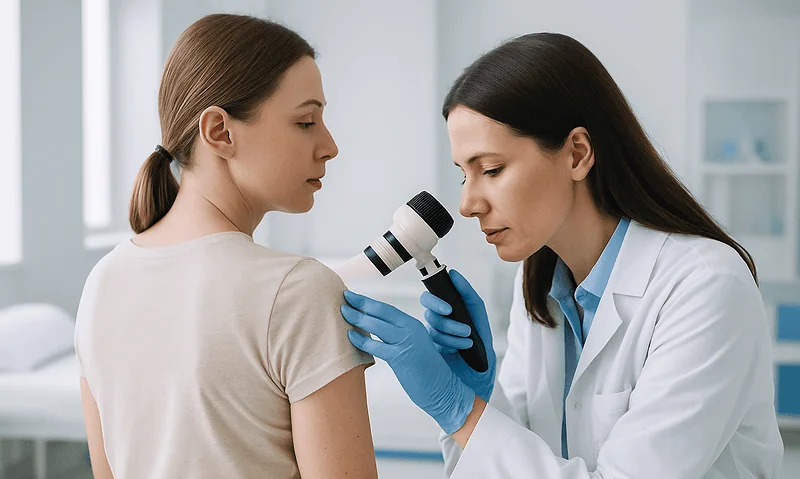
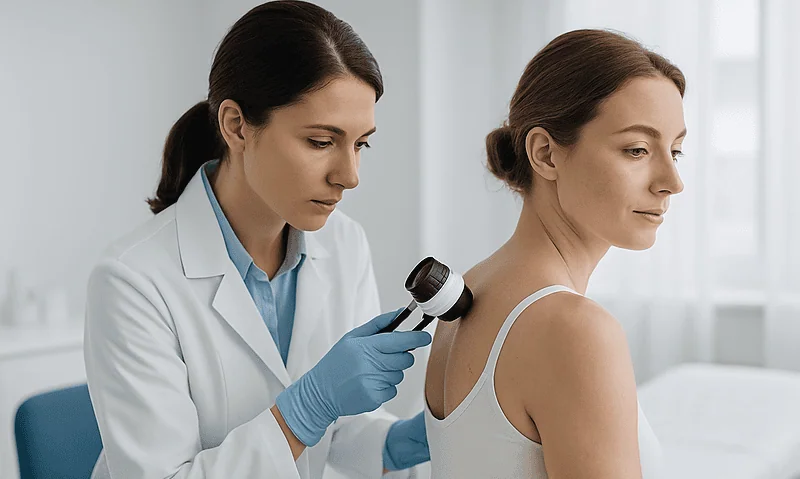
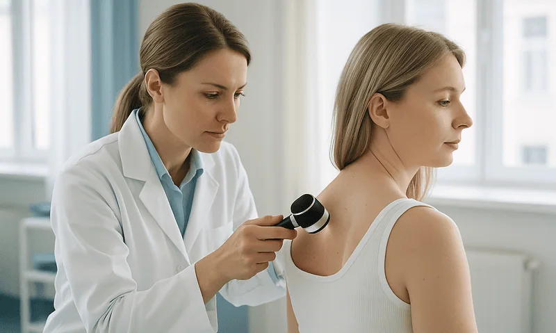
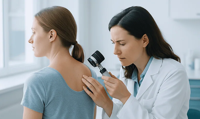
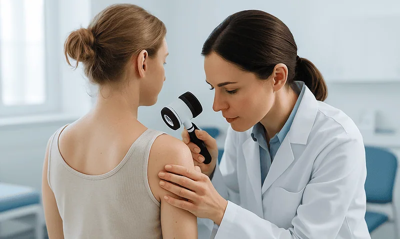
The clinician will ask when you noticed the spot and how it has changed, then examine it with a dermatoscope: a handheld magnifier with polarized light that reveals pigment patterns and structures invisible to the naked eye. In experienced hands, this one tool resolves most worries on the spot: the majority of checked moles turn out to be benign, and you’ll often hear that reassurance in the same visit. Many clinics also photograph moles or perform full-body “mole mapping” so future changes can be measured rather than guessed.
If a spot looks genuinely suspicious, the next step is a biopsy: the area is numbed with a local anesthetic, and the spot, or a sample of it, is removed for examination under a microscope. It typically takes minutes and leaves a small scar. Results usually arrive within days to a couple of weeks.
Two myths are worth retiring here. Biopsying or removing a mole does not “anger” it or cause cancer to spread: that’s folklore, contradicted by mainstream medical evidence. And a biopsy is not a verdict; it’s how doctors get an answer instead of a guess. Most answers are good news.
Is it even a mole? Spots that get mistaken for melanoma
A surprising share of skin-cancer worry attaches to spots that aren’t moles at all. Knowing the common look-alikes helps you triage, though when in doubt, the tie always goes to getting it examined.
Seborrheic keratoses are the great imposters of midlife skin: brown, black, or tan growths with a waxy, “stuck-on” appearance, as if pressed onto the skin like warm candle drippings. They multiply with age and are harmless, but their dark, irregular look sends many people to the dermatologist, reasonably so.
Freckles and sun spots are flat, uniform patches of pigment that darken with sun and often fade in winter. Uniformity and seasonality are their signatures. Cherry angiomas are small, bright-red dots made of blood vessels, common, benign, and unrelated to melanoma. Skin tags are soft, skin-colored flaps on stalks, favoring the neck, underarms, and eyelids.
Melanoma also isn’t the only skin cancer worth knowing. Basal cell carcinoma, the most common type, often shows up as a pearly or translucent bump, sometimes with visible tiny blood vessels, that may bleed and scab in cycles. Squamous cell carcinoma tends to appear as a rough, scaly, or crusted patch or sore that doesn’t heal. The NHS advises seeing a doctor about any skin sore or ulcer that persists rather than healing as an ordinary wound would. Different diseases, same doorway: a spot that won’t behave gets a professional look.
When to see a doctor: the changes that shouldn't wait
Everything above distills into one short list. Book a medical appointment promptly, within days, not months, if you notice any of the following, drawn from NHS and Mayo Clinic guidance:
- A mole that has changed in size, shape, or color over weeks to a few months.
- A mole with new asymmetry, a ragged or blurred border, or multiple shades within it.
- A mole that bleeds, oozes, or crusts without having been injured.
- Persistent itching, tenderness, or pain in a mole that never had symptoms.
- A firm bump of any color that keeps growing over several weeks.
- A new dark streak under a fingernail or toenail that wasn’t caused by an injury.
- A sore or ulcer on the skin that simply won’t heal.
- Any spot that stands out sharply from every other mark on your body.
One symptom from this list is enough: you don’t need a quorum. When you call, use the phrase “changing mole” or “possible skin cancer”; those words move appointments up the queue in most systems, and in the UK they can trigger the two-week urgent referral pathway.
A final calibration: none of these signs means you have cancer. Most checked moles are benign, and the point of going early isn’t that disaster is likely: it’s that early answers are cheap and late ones are expensive. This is one worry that’s genuinely worth outsourcing to a professional.
Sun habits that genuinely lower your risk
Watching your moles is surveillance; sun habits are prevention. Ultraviolet radiation is the major modifiable driver of melanoma risk, and the CDC’s core advice is refreshingly unglamorous: it’s the boring habits, repeated for decades, that shift the odds.
Shade is the most underrated tool. UV intensity peaks through the middle of the day, so structuring outdoor time toward mornings and late afternoons, or simply claiming the shady side of the street, reduces exposure without a single product. Clothing comes next: a wide-brimmed hat protects the ears, neck, and scalp, three places moles hide and mirrors miss, and tightly woven long sleeves outperform any lotion because they can’t be under-applied or forgotten.
Sunscreen matters, applied generously and reapplied after swimming or sweating; UK guidance recommends a broad-spectrum product with a sun protection factor of at least 30. Its most common failure mode isn’t the formula: it’s the thin, once-a-day application most of us actually do.
Two truths to end on. Tanning beds deliver concentrated UV and earn an unambiguous “avoid” from the CDC and NHS alike; there is no safe cosmetic tan from a machine. And prevention doesn’t retire surveillance: melanomas can arise on skin the sun rarely touches, from scalp to sole. The winning combination is modest sun discipline plus a monthly ten-minute look, small habits, compounding quietly, exactly the way risk does.
Frequently asked questions
What are three warning signs a mole is becoming cancerous?
The three signs that matter most are trackable change, standing out, and new symptoms. A mole that grows, shifts color, or changes shape over weeks to months is the strongest warning. A mole that looks clearly different from all your others, the ugly duckling, is the second. Third is a mole that starts itching, crusting, or bleeding without injury. Any one of these justifies a prompt medical exam.
What are the five warning signs of melanoma?
The five signs are summarized by the ABCDE rule: Asymmetry (halves don’t match), Border irregularity (ragged or blurred edges), Color variation (multiple shades in one spot), Diameter larger than about 6 millimeters, and Evolution (any change over time). Evolution is the most important of the five. One strong feature is enough to warrant a check: you don’t need all five to see a doctor.
What does a stage 1 melanoma mole look like?
Stage 1 melanoma is often subtle: a flat or slightly raised spot, frequently with a slightly irregular edge or two or three shades of brown, and typically no symptoms at all. Staging depends on depth rather than surface looks, stage 1 tumors are thin, about 2 millimeters or less. Because early melanoma can resemble an ordinary mole, visible change over time matters more than any single snapshot.
What is the 2-week rule for melanoma?
In the UK’s National Health Service, a GP who suspects melanoma refers the patient through an urgent pathway to be seen by a specialist within two weeks. The clock starts when the doctor becomes suspicious, not when you first notice a change, so it’s not a reason to wait two weeks before acting. Wherever you live, the takeaway is the same: a suspicious mole merits an exam within days to a couple of weeks.
Is a new mole in adulthood always a concern?
Not always, but it deserves more attention than a new mole in childhood. Most people accumulate moles through their twenties; a genuinely new pigmented spot appearing in middle age or later is less typical. Since most melanomas arise on previously normal skin rather than in existing moles, show any new adult-onset spot to a clinician, especially if it grows, darkens unevenly, or looks unlike your other moles.
Can a mole change and still be harmless?
Yes. Moles naturally darken slightly during puberty and pregnancy, can slowly rise off the skin over years, and often fade in older age. The reassuring pattern is slow, even change that affects your moles collectively. The concerning pattern is one mole changing alone, quickly, or unevenly, new colors within it, a spreading edge, or texture changes over weeks to months. When you can’t tell the difference, a quick exam settles it.
Does an itchy mole mean cancer?
Usually not. Dry skin, friction from clothing, eczema, and insect bites cause far more mole itching than melanoma does. The itch that matters is persistent, affects one specific mole, and arrives alongside other changes, growth, color shifts, crusting, or bleeding. A mole that itches once after a wool sweater is noise; a mole that has itched for weeks while visibly changing is a signal worth showing to a doctor.
How often should I check my own skin for mole changes?
About once a month is the interval commonly suggested in dermatology guidance, frequent enough to catch change, spaced enough to actually notice it. Use bright light, a full-length mirror, and a hand mirror, and cover hidden territory: scalp, back, buttocks, soles, between toes, and under nails. Photographs with a coin for scale make comparison objective. Checking daily tends to fuel anxiety without improving detection.
Does removing or biopsying a mole cause cancer to spread?
No. This is a persistent myth unsupported by medical evidence. A biopsy, removing all or part of a suspicious spot under local anesthetic for microscopic examination, is how doctors get a definitive answer, and it does not provoke or spread cancer. The procedure typically takes minutes, results arrive within days to a couple of weeks, and most biopsied moles turn out to be benign.
Can melanoma appear on skin that never gets sun, or on darker skin?
Yes. Melanoma can develop on the scalp, soles, palms, under nails, and other low-sun areas. In people with darker skin tones, it more often appears in exactly these places, commonly as a new dark streak in a nail or a changing spot on the palm or sole. Because these locations are easy to overlook, include them in every self-check regardless of your skin tone or sun habits.
References
This article is for general information only and is not a substitute for professional medical advice. Please consult a qualified doctor about your individual situation.

More from the Blog
Slugging: What Occlusive Skincare Does, and Who Should Skip It
Slugging means sealing your face overnight with a thin layer of an occlusive ointment, usually petroleum jelly, applied over your usual moisturizer. It reduces…
Why Photos of Your Flares Help: Preparing for an Atopic Dermatitis Visit When Skin Looks Calm
Atopic dermatitis tends to settle between flares, so the skin a clinician examines on appointment day often does not reflect your worst weeks. Dated,…
Folliculitis vs Acne: How Dermatologists Tell Them Apart and Why the Treatments Differ
Folliculitis and acne both produce red bumps and pustules, but they are different conditions. Acne involves clogged pores with blackheads or whiteheads, oil, and…
Preventing Cellulitis From Coming Back: Skin Care, Swelling Control and Foot Fungus Checks
Preventing recurrent cellulitis usually rests on three habits: caring for the skin barrier with daily moisturizer and prompt treatment of cuts, controlling chronic leg…

Is Rosacea Autoimmune, Does It Itch, and Does It Get Worse with Age?
Rosacea is not classified as an autoimmune disease. Current evidence describes it as a chronic inflammatory skin condition driven by an overactive innate immune…
Is It Eczema or Another Dermatitis? How Dermatologists Tell Atopic and Contact Types Apart
Eczema (atopic dermatitis) and contact dermatitis can look nearly identical, but they arise differently. Atopic eczema is a chronic, inherited tendency toward a leaky…