

When to Start Mammograms and How Often to Have Them
Key Takeaways
- The USPSTF now recommends a mammogram every two years from age 40 through 74: a full decade earlier start than its previous guidance.
- The NHS screens every three years between 50 and 71 because trial evidence shows most of the mortality benefit is preserved at that interval in postmenopausal women.
- There is no fixed stopping age in US guidance; after 74, the decision hinges on overall health and whether you'd likely benefit for another ten years.
- The radiation from a screening mammogram is a low dose, and the NCI rates the risk of harm as very low even with yearly exams over decades.
- After roughly ten annual mammograms, about half of women will have experienced at least one false alarm requiring extra imaging, most callbacks end in reassurance.
- About half of women over 40 have dense breast tissue, which can hide tumors on a mammogram, so a density notification is a cue to discuss supplemental options.
Most women at average risk can begin screening mammograms at age 40 and repeat them every one to two years, depending on the guideline their clinician follows. The US Preventive Services Task Force advises a mammogram every two years from 40 through 74, while the UK's NHS invites women every three years from 50 to 71. Women at higher risk may need to start earlier, sometimes with added MRI.
The reminder arrives quietly: a letter, a patient-portal ping, or a friend at lunch asking, “Wait, you haven’t had one yet?” And then the confusion starts. One cousin goes every year. A colleague in London goes every three. Your own mother wasn’t called in until 50, but your doctor mentioned 40 at your last visit.
None of them is wrong, exactly. Mammogram guidance has genuinely shifted in the past few years, and different expert groups read the same evidence with different priorities, some weighting early detection most heavily, others weighting false alarms and unnecessary biopsies. The result is a patchwork of start ages and intervals that can make a straightforward question feel like a debate.
Here is what the major guidelines actually say, why they differ, and how to land on a schedule that fits your risk, not your neighbor’s.
What age should you start getting mammograms?
For women at average risk in the United States, 40 is the new anchor. In 2024, the US Preventive Services Task Force (USPSTF) lowered its recommended starting age from 50 to 40, advising screening mammograms every other year from age 40 through 74, guidance the CDC now reflects in its screening information.
Why the change? Breast cancer diagnoses among women in their 40s have been rising, and the task force concluded that starting at 40 would prevent more deaths, with a particularly meaningful impact for Black women, who are more likely to die of breast cancer than white women. The American Cancer Society lands nearby: women should have the option to start at 40, and should begin yearly screening by 45.
“Average risk” is doing real work in that sentence. It means no known inherited mutation such as BRCA1 or BRCA2, no first-degree relative with such a mutation, no history of chest radiation therapy at a young age, and no prior high-risk biopsy finding. If any of those apply, the calendar changes, often substantially, and often before 40. A short risk conversation with your clinician, ideally by your late 20s, sorts you into the right lane before the first appointment is ever booked.
One thing every major guideline agrees on: waiting until symptoms appear is not a screening strategy. Mammography exists to find cancers years before they can be felt.
How often do you need mammograms, every year or every two?
Both schedules are defensible, and the honest answer is that expert groups split on this deliberately. The USPSTF recommends every two years for women 40 to 74, a position the CDC summarizes on its breast cancer screening pages. The American Cancer Society recommends yearly mammograms from 45 to 54, then a choice between yearly and every-other-year from 55 onward.
The trade-off is concrete. Annual screening catches some cancers a little earlier, which matters most for younger women, whose tumors tend to grow faster. But it also roughly doubles your lifetime exposure to false alarms, callbacks, extra views, sometimes biopsies for what turns out to be nothing. Biennial screening preserves most of the mortality benefit while cutting those harms substantially; that arithmetic is the core of the task force’s reasoning.
So how do you choose? A few practical tie-breakers:
- In your 40s and early 50s, the case for annual screening is strongest, because tumors in premenopausal women are more often fast-growing.
- After menopause, tumor biology typically slows, and every two years gives up very little.
- Dense breast tissue, family history, or simple peace of mind can all reasonably tip you toward yearly.
What matters far more than the interval is consistency. A mammogram every two years, kept faithfully for decades, protects you better than annual intentions that lapse after year three.
Why do the major guidelines disagree?
They are reading the same trials. What differs is how each group weighs a life saved against a false alarm endured, and how each defines an acceptable rate of overdiagnosis, cancers found and treated that would never have caused harm. The USPSTF is conservative about harms; the American Cancer Society leans toward maximizing detection; the NHS runs a taxpayer-funded population program and optimizes at the level of millions of women, not one exam room.
| Guideline | Start age | How often | Upper age |
|---|---|---|---|
| USPSTF (US) | 40 | Every 2 years | Through 74; evidence judged insufficient beyond |
| American Cancer Society (US) | 45 (option from 40) | Yearly 45–54, then every 1–2 years | Continue while in good health with roughly 10+ years of life expectancy |
| NHS (UK) | 50 (first invitation by the 53rd birthday) | Every 3 years | Invitations end after 71; women may self-refer afterward |
Notice what the table does not show: any guideline suggesting women skip screening entirely during their 50s and 60s. The decades where evidence for benefit is strongest, roughly 50 to 74, are common ground across every major body, including the WHO, which endorses organized mammography screening for women 50 to 69 in well-resourced health systems.
Disagreement at the edges, consensus at the core. That’s a more reassuring picture than the headlines usually paint.
Why are there three years between mammograms in the UK?
Americans are often startled by the NHS interval, three full years between screens, for women aged 50 to 71. It isn’t budget shorthand; it’s a design choice backed by trial data.
The UK program was built on randomized evidence showing that in women over 50, whose breast cancers are on average slower-growing, most of the mortality benefit of screening is preserved at a three-year interval. Stretching the gap sharply reduces the downsides: fewer false-positive recalls, fewer needle biopsies of harmless findings, and less overdiagnosis across a program that screens millions. The NHS is unusually transparent about this ledger: its patient information estimates that screening saves about one life from breast cancer for every 200 women screened regularly, while around three of those 200 will be diagnosed and treated for a cancer that would never have threatened them.
Context matters too. The NHS starts at 50, past the age range where fast-growing premenopausal tumors are most common, which makes the longer interval biologically safer than it would be for a 42-year-old. Women aren’t cut off after 71 either; routine invitations stop, but anyone older can request screening every three years by contacting their local service.
Neither system is careless. The US model prioritizes catching more cancers earlier and tolerates more false alarms; the UK model prioritizes efficiency and minimizing harm at population scale. Both, kept up consistently, reduce deaths from breast cancer.
What are the new mammogram guidelines for 2026?
Here is the plain truth behind that search: as of this writing, no major guideline body has announced a new mammogram recommendation taking effect in 2026. The most recent substantive change was the USPSTF’s 2024 update, which lowered the recommended starting age to 40 with screening every two years through 74. If a headline promises “new 2026 guidelines,” check it against the issuing organization directly, guideline bodies publish on multi-year review cycles, and secondhand summaries frequently misdate or overstate what changed.
That said, real questions are under active study, and future updates will likely address them:
- Screening beyond 74. The USPSTF currently says the evidence is insufficient to recommend for or against mammograms in women 75 and older: a gap researchers are working to close.
- Supplemental imaging for dense breasts. US facilities must now tell women their breast density, but whether adding ultrasound or MRI actually reduces deaths in this group is still being tested.
- Personalized intervals. Trials are exploring whether screening frequency should flex with individual risk scores rather than birthdays alone.
- 3D mammography. A large randomized trial is comparing 3D and standard 2D screening head-to-head on outcomes that matter, not just detection counts.
Until those results land, the current guidance stands, and following it consistently beats waiting for a hypothetical update.
Who should start screening before 40?
A meaningful minority of women should never wait for the standard start age, and many of them don’t know it. Guideline groups summarized by the CDC and Mayo Clinic flag several situations that shift a woman into high-risk screening:
- A known mutation in BRCA1, BRCA2, or certain other genes, or a first-degree relative (parent, sibling, child) who carries one, even if you haven’t been tested
- Radiation therapy to the chest between roughly ages 10 and 30, often for childhood lymphoma
- A strong family history: breast cancer in multiple close relatives, or in a relative diagnosed before 50
- A prior breast biopsy showing atypical cells or lobular carcinoma in situ
- An estimated lifetime breast cancer risk of about 20 percent or higher on a validated risk model
For these women, guidelines commonly recommend an annual breast MRI in addition to a yearly mammogram, often beginning around age 30: the exact starting point depends on the specific risk factor and should be set with a clinician, sometimes alongside genetic counseling.
The practical takeaway is timing. Risk assessment works best when it happens by the mid-to-late 20s, before the first screening decision arrives. If breast or ovarian cancer runs on either side of your family, fathers pass these genes just as readily as mothers, raise it at your next routine visit. A ten-minute family-history conversation can move your first screen up by a decade, exactly when it matters most.
What is the best age to stop mammograms?
There is no universal stop sign, and any article naming a single “best age to quit” is simplifying past the evidence. Here is what the major bodies actually say.
The USPSTF recommends screening through age 74, then declares the evidence insufficient to recommend for or against continuing, not because mammograms suddenly stop working at 75, but because the landmark trials enrolled few women that old. The American Cancer Society skips ages entirely and uses health instead: keep screening as long as you’re in good overall health and expect to live at least another ten years or so. The NHS stops sending invitations after 71 but explicitly allows older women to self-refer every three years.
The ten-year logic is worth understanding, because it’s genuinely useful. Screening finds cancers years before they would cause trouble; a slow-growing tumor detected at 88 in a woman with serious heart failure may never have affected her life, while the biopsy, surgery, and worry absolutely would. Conversely, a vigorous 78-year-old with a 15-year horizon still stands to benefit meaningfully from finding a cancer early.
So the honest framing is a question, not a birthday: would finding a cancer now change anything for the better? If you’re healthy, active, and would want treatment, continuing past 74 is entirely reasonable. If serious illness has shortened the horizon, stopping can be the wise and kind choice. Either way, it deserves an actual conversation with your clinician, not a silent lapse.
How frequently is it safe to have a mammogram?
Every schedule recommended by a major guideline, including annual screening for decades, is considered safe. The radiation question deserves a straight answer, though, because it stops some women from booking at all.
A mammogram uses a low-dose X-ray, and the dose per exam is small. The National Cancer Institute, part of the NIH, states plainly that the risk of harm from the radiation in screening mammography is very low, even with repeated yearly exams over many years. The theoretical risk of a radiation-induced cancer from a lifetime of screening is tiny compared with the demonstrated benefit of catching real cancers early, which is exactly why no guideline asks women to space out screens for radiation reasons. Modern machines are also regulated and quality-tested, with dose limits built into certification requirements.
A few practical notes:
- Extra views at a callback, or a diagnostic mammogram after a symptom, add only modestly to exposure and are worth it when a radiologist needs a better look.
- Tell the technologist if you are or might be pregnant. Routine screening is usually simply deferred; if imaging is truly needed, shielding and technique are adjusted.
- There is no benefit to screening more often than your guideline recommends. More frequent scans mostly buy more false alarms, not more protection.
The realistic risk of mammography isn’t radiation. It’s the callback that frightens you for a week and turns out to be nothing: a harm worth knowing about, and one covered a few sections down.
What actually happens at a screening appointment?
Twenty minutes, start to finish, and most of that is paperwork and changing. The imaging itself takes a few minutes per breast.
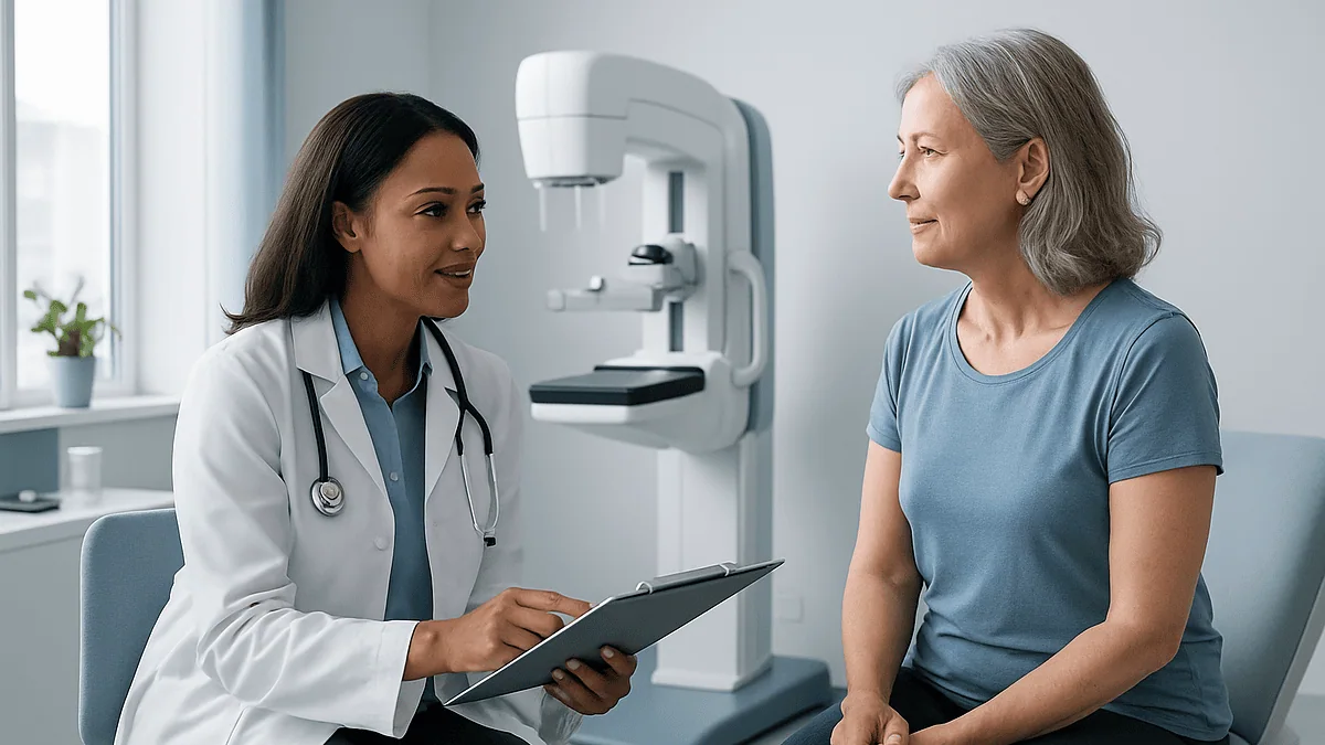
You’ll undress from the waist up and stand at the machine while a technologist positions one breast at a time on a flat plate. A second plate presses down for roughly ten to fifteen seconds per view, typically two views per breast. The compression is the part nobody enjoys, but it isn’t gratuitous: flattening the tissue spreads overlapping structures apart so small cancers can’t hide, holds the breast still to prevent blur, and lets the machine use a lower radiation dose. Firm pressure, briefly, in exchange for a sharper image.
A few things make the visit easier, echoed in patient guidance from MedlinePlus and Mayo Clinic:
- If you still menstruate, book for the week after your period, when breasts are least tender.
- Skip deodorant, antiperspirant, and powders that morning, aluminum particles can mimic calcifications on the image and trigger an unnecessary callback.
- Wear a two-piece outfit so you only undress from the waist up.
- Bring or arrange transfer of prior mammograms if you’ve switched facilities; comparison images prevent more false alarms than almost anything else.
Results follow by letter or portal: US facilities are required to notify you within 30 days, and most report much sooner; the NHS aims for results within about two weeks. No news is not good news here: if nothing arrives, call and ask.
Is a 3D mammogram better than a standard one?
Probably somewhat, for many women, and the field is honest that the final verdict is still being written.
A 3D mammogram, formally digital breast tomosynthesis, sweeps the X-ray tube in an arc and reconstructs the breast as a stack of thin image slices, rather than the two flat pictures of conventional 2D mammography. Radiologists can page through the tissue layer by layer, which helps in two ways: overlapping normal tissue is less likely to hide a real tumor, and less likely to masquerade as one. Studies summarized by the National Cancer Institute show that 3D screening finds modestly more cancers and generates fewer callbacks than 2D alone, with the advantage most pronounced in women with dense breasts.
What hasn’t been proven yet is the outcome that ultimately matters: whether 3D screening reduces deaths or advanced cancers compared with modern 2D. A large NIH-supported randomized trial is testing exactly that question, and guideline bodies have so far declined to declare 3D mandatory while it runs.
The practical guidance is refreshingly simple. If your screening facility offers 3D, most in the US now do, there’s no reason to decline it, and dense-breasted women may particularly benefit from fewer false alarms. If only 2D is available, or 3D would mean an out-of-pocket charge you can’t absorb, a standard mammogram remains an excellent, evidence-backed test. The best mammogram is the one you actually show up for, on schedule, year after year.
What does it mean if you have dense breasts?
If a results letter mentioned breast density, you’re in large company: roughly half of women over 40 have dense breast tissue, according to the CDC. Density has nothing to do with how breasts feel or look: it describes the ratio of glandular and fibrous tissue to fat, and only an X-ray can measure it.
Density matters for two separate reasons, and it’s worth keeping them distinct. First, the masking effect: dense tissue appears white on a mammogram, and so do tumors, making a cancer harder to spot: the classic comparison is searching for a snowball in a snowstorm. Second, density itself is associated with a modestly higher risk of developing breast cancer, independent of the imaging problem.
US mammography facilities are now required to tell every woman whether her breasts are dense, precisely so this conversation happens. If yours are, sensible questions for your clinician include:
- Would supplemental imaging, typically ultrasound, or MRI for higher-risk women, make sense for me?
- Does my overall risk profile (family history, prior biopsies) change the calculus?
- Is 3D mammography available, since it performs somewhat better in dense tissue?
Two honest caveats. Supplemental imaging finds additional cancers but also generates additional false positives, and researchers have not yet shown that adding it saves lives for average-risk women with dense breasts, which is why guidelines stop short of a blanket recommendation. Density also tends to decrease after menopause, so a “dense” designation at 45 may not follow you to 65.
How often do mammograms get it wrong?
More often than the pink-ribbon posters suggest, and knowing the numbers in advance is the best inoculation against panic.
False positives are the common error. According to the National Cancer Institute, over the course of about ten yearly mammograms, roughly half of women will experience at least one false alarm: a callback for extra views or ultrasound that turns out to be normal tissue, a cyst, or benign calcifications, and a smaller share will undergo a biopsy for something harmless. Callbacks are most frequent at a woman’s first mammogram, when radiologists have no prior images for comparison, and they become rarer with every screen thereafter. A callback is not a diagnosis; the large majority end with reassurance.
False negatives happen too. Mammograms miss roughly 20 percent of breast cancers present at the time of screening, per NCI, most often in dense tissue. This is why a new lump always deserves evaluation, even weeks after a normal mammogram. “The picture was clear” and “there is nothing there” are not the same sentence.
Then there’s overdiagnosis, the subtlest harm: some screen-detected cancers grow so slowly they would never have caused symptoms in a woman’s lifetime, yet once found, they’re treated. The NHS estimates that for every 200 women screened regularly, about three are treated for a cancer that would never have harmed them, alongside one life saved. Guidelines don’t hide this math: it’s precisely why start ages and intervals are debated so carefully.
Do regular mammograms really save lives?
Yes, with a benefit that is real, well-documented, and smaller per individual than most people assume. Both halves of that sentence deserve equal weight.
The scale of the disease frames the question. The WHO reports that breast cancer was diagnosed in about 2.3 million women worldwide in 2022, with roughly 670,000 deaths, making it the most common cancer in women in the large majority of countries. Randomized trials conducted across several decades, the evidence base cited by the National Cancer Institute and WHO alike, show that inviting women to regular mammography reduces breast cancer deaths, with the clearest and most consistent benefit in women aged 50 to 69. The mechanism is straightforward: cancers found at a small, node-negative stage are treated more successfully and often less aggressively than those found once they can be felt.
Translated to one person, the numbers turn modest. The NHS’s own figure, one life saved per 200 women screened regularly over the program, is a sober, credible estimate, not a slogan. Most women who attend every screen will never personally have their life saved by one, because most were never destined to die of breast cancer.
Is that worth twenty minutes every one to three years? For women in the core screening ages, every major medical body answers yes, and it’s a stronger yes for those who also know their family history, report changes promptly, and keep the schedule rather than treating one clear result as permanent permission to stop.
When should you see a doctor without waiting for a screening?
Screening is for people with no symptoms. The moment something changes, the calendar becomes irrelevant: a new finding needs diagnostic evaluation now, not a mammogram in eighteen months, and not reassurance from a normal result last spring.
Contact your clinician promptly if you notice any of the following, in either breast, at any age:
- A new lump or area of thickening in the breast or armpit, whether or not it hurts
- Nipple discharge you didn’t squeeze out, especially if it’s bloody or from one side only
- A nipple that has newly turned inward or changed direction
- Skin dimpling, puckering, or an orange-peel texture on the breast
- Redness, warmth, or swelling that doesn’t settle within a couple of weeks, particularly important because inflammatory breast cancer can mimic infection
- A persistent change in the size or shape of one breast, or focal pain that stays in one spot
Most of these turn out to be benign, cysts, fibroadenomas, hormonal changes, blocked ducts. But “usually benign” is a statistic, not an exam, and the NHS and Mayo Clinic are unambiguous that every new breast change warrants assessment. Men should take the same list seriously; about 1 in 100 US breast cancers occurs in men, per the CDC.
One more reason to book an appointment early: if breast or ovarian cancer clusters in your family and no one has ever formally assessed your risk, that conversation is itself a form of care, and it can reshape your entire screening timeline.
Frequently asked questions
Why are there three years between mammograms in the UK?
Because trial evidence showed that for women 50 to 71, whose breast cancers grow more slowly on average, a three-year interval preserves most of the life-saving benefit while substantially cutting false alarms and overdiagnosis. The NHS runs screening as a national program and optimizes that balance at population scale. Women over 71 can still self-refer for screening every three years after routine invitations stop.
What are the new mammogram guidelines for 2026?
No major guideline body has announced new mammogram recommendations taking effect in 2026. The most recent significant change was the 2024 USPSTF update, which lowered the recommended starting age to 40 with screening every two years through age 74. Guidelines are reviewed on multi-year cycles, so future updates may address screening after 74, dense-breast imaging, and 3D mammography, but for now, the 2024 guidance stands.
What is the best age to stop mammograms?
There is no single stop age. The USPSTF recommends screening through 74 and calls the evidence insufficient beyond that; the American Cancer Society advises continuing as long as you’re in good health with roughly ten or more years of life expectancy; the NHS stops routine invitations after 71 but allows self-referral. A healthy, active woman in her late 70s can reasonably keep screening: the decision belongs in a conversation with her clinician.
How frequently is it safe to have a mammogram?
Annual mammograms are considered safe, and no recommended schedule poses meaningful radiation risk. Each exam uses a low X-ray dose, and the National Cancer Institute states the risk of harm from screening mammography radiation is very low even with yearly repeats over many years. Extra diagnostic views after a callback are also safe. There’s no benefit, though, to screening more often than your guideline recommends: it mainly adds false alarms.
Should I get a mammogram at 35?
Not routinely, if you’re at average risk, no major guideline recommends screening mammograms before 40 for average-risk women. But higher-risk women are a different story: a BRCA mutation, chest radiation in youth, or a strong family history can justify starting around 30, often with annual MRI added. If cancer runs in your family, ask for a formal risk assessment now rather than waiting for a milestone birthday.
Do mammograms hurt?
Compression is uncomfortable for most women, firm pressure for roughly ten to fifteen seconds per view, but genuine pain is uncommon and brief. The squeeze is what makes the image sharp and keeps the radiation dose low. To minimize discomfort, schedule for the week after your period if you menstruate, and tell the technologist if positioning hurts; adjustments are usually possible. Most appointments are done within twenty minutes.
How long does it take to get mammogram results?
In the US, facilities are required to send you results within 30 days, and most patient portals post them within a few days; the NHS aims to mail results within about two weeks. If something needs a closer look, you’ll typically hear sooner, because callbacks are scheduled promptly. If no result arrives in the expected window, call the facility, silence should never be assumed to mean the mammogram was normal.
Do I need mammograms if breast cancer doesn't run in my family?
Yes. Most women diagnosed with breast cancer have no family history of the disease, which is exactly why screening is recommended for all average-risk women, not just those with affected relatives. Age is the biggest single risk factor. Family history changes when you start and whether MRI is added: it doesn’t determine whether screening is worthwhile in the first place.
Is a 3D mammogram worth it?
If it’s offered at no extra cost, take it. Studies summarized by the National Cancer Institute show 3D mammography (tomosynthesis) detects modestly more cancers and triggers fewer callbacks than standard 2D imaging, with the biggest advantage in dense breasts. Whether it ultimately saves more lives is still being tested in a large randomized trial. If only 2D is available to you, it remains an excellent, evidence-backed screening test.
Can I have a mammogram with breast implants?
Yes, and you should keep your regular schedule. Tell the scheduler and technologist about the implants in advance: facilities use special displacement views that gently move the implant back so more natural tissue is visible, which usually means a few extra images and a slightly longer appointment. Implants can obscure some tissue, so choose a facility experienced with augmented breasts, and report any new lumps or changes promptly.
References
- CDC: Screening for Breast Cancer
- NHS: Breast screening (mammogram)
- WHO: Breast cancer fact sheet
- MedlinePlus: Mammography
This article is for general information only and is not a substitute for professional medical advice. Please consult a qualified doctor about your individual situation.

More from the Blog

After Robotic Hysterectomy: Redness, Discharge or Fever and When to Contact Your Surgeon
After a robotic hysterectomy, mild pinkness at the small incisions, light brown or pink vaginal discharge for up to six weeks, and low-grade warmth…
What to Expect From PCOS Treatment: How Cycles, Skin and Weight Respond Over Months
PCOS treatment results arrive on different clocks. Hormonal treatments usually make bleeding predictable within one to three cycles, while skin and unwanted hair often…
Vaginal Infections During Pregnancy: Why Treatment Is Chosen Carefully and Follow-Up Matters
A vaginal infection during pregnancy is common and usually manageable, but clinicians choose treatment carefully because some medicines are preferred over others while a…

After Fibroid Surgery: Heavy Bleeding, Fever or Rising Pain and When to Call Your Team
After fibroid surgery, call your surgical team the same day if you soak through a sanitary pad every hour for two hours or more,…

How PCOS Is Tested and Diagnosed: Blood Tests, Ultrasound and the Rotterdam Criteria
PCOS is diagnosed by pattern rather than a single test. Clinicians use the Rotterdam criteria: at least two of three features, infrequent or absent…

How to Check for Fibroids, and What They Look Like
You cannot reliably check for fibroids at home. Fibroids are noncancerous growths in the muscular wall of the uterus, and they are usually confirmed…