

Which Dental Bone Graft Material Is Used? Your Own Bone, Donor, Animal-Derived and Synthetic
Key Takeaways
- All four graft types, autograft, allograft, xenograft and alloplast, work mainly as scaffolds that your own cells colonize and replace; only your own bone also contributes living cells and growth signals.
- Cleveland Clinic cites a usual healing window of about three to nine months between graft placement and implant surgery, with larger repairs such as sinus lifts often at the longer end.
- Animal-derived and dense synthetic minerals resorb very slowly, which preserves shape under the sinus or along the front of the mouth, but particles can remain embedded in new bone for years.
- Human donor bone is screened, stripped of cells, sterilized and regulated as tissue; declining it for personal reasons is legitimate, and a synthetic can nearly always be substituted.
- A healed graft becomes living bone with no separate lifespan; what keeps it is chewing load from an implant and freedom from gum inflammation, not the material it started as.
- Short, narrow, tilted or zygomatic implants, implant-supported bridges and conventional bridges or dentures are the main alternatives to grafting, each trading one risk for another.
Dental bone graft material comes in four families: autograft (your own bone, usually taken from the jaw), allograft (processed human donor bone), xenograft (processed animal bone) and alloplast (synthetic calcium-based minerals). Each works mainly as a scaffold that your body gradually replaces with new bone. The choice depends on the size and location of the defect, your general health and your preferences, and is made together with your dental team.
The consent form arrives on a clipboard, and somewhere on page two a box is already ticked: bovine-derived particulate. Nobody has explained what that means yet. You came in to ask about an implant for a molar lost two winters ago, and now you are being told the socket has sunk, the bone is too thin, and something has to be built back before a titanium post can go in.
This is where most people first meet the types of dental bone graft material, and the vocabulary can feel stranger than the surgery itself. Your own bone. Donor bone from a tissue bank. Mineral from a cow or pig. A powder made in a laboratory. The words sound like a menu of very different things.
They are closer than they sound. Every one of them is a scaffold, and your body does the actual building. What differs is where the scaffold comes from, how quickly it disappears, and what trade-offs come with it. Those differences are worth understanding before you sign.
What a dental bone graft actually does inside the jaw
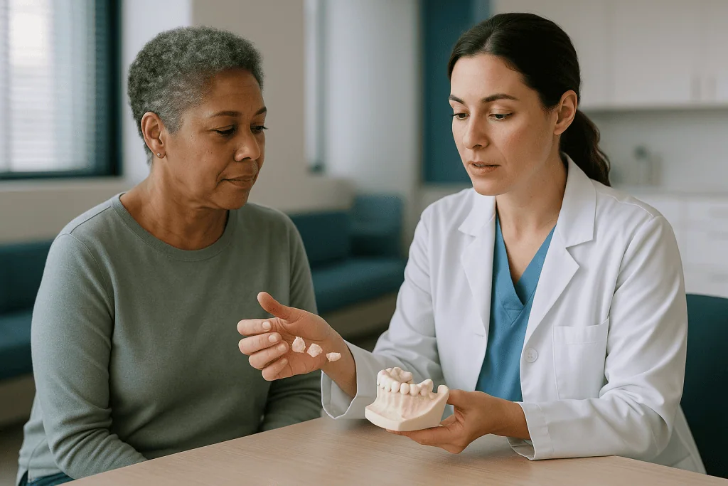
A dental bone graft is a small amount of bone or bone-like material placed where the jaw has thinned, so that there is enough solid bone to hold an implant. The jawbone that cradles the teeth is called the alveolar ridge, and it behaves a little like muscle: it stays strong only while it is being used. Once a tooth is pulled, the chewing forces that kept that stretch of ridge alive vanish, and the body starts to reabsorb it. Width is lost first, then height.
The graft interrupts that process. Surgeons describe three biological jobs a material can do. Osteoconduction means it acts as a scaffold that new bone can creep along. Osteoinduction means it carries protein signals that recruit bone-forming cells. Osteogenesis means it contains living bone cells of its own. Only fresh bone from your own body does all three; the other materials rely mostly on being good scaffolds.
Over the following months, blood vessels grow into the graft, cells lay down new mineral, and the scaffold is slowly broken down and replaced. Cleveland Clinic puts the usual healing window for a dental bone graft at roughly three to nine months before an implant can be placed, sometimes longer depending on the size of the repair.
The procedure itself is usually done under local anesthetic. The gum is lifted, the material is packed into the defect, often a thin membrane is laid over it, and the gum is stitched closed. Mayo Clinic lists the common settings: filling a fresh socket (socket preservation), rebuilding a collapsed ridge (ridge augmentation) and lifting the sinus floor in the upper back jaw (sinus lift). The material chosen often follows the setting.
Types of dental bone graft material: the four families at a glance
Dentists sort graft materials by origin, and the four categories cover almost everything you will see on a consent form. The table below is the version most people wish they had been handed in the chair.
| Material | Where it comes from | What it contributes biologically | Typical strengths | Typical trade-offs |
|---|---|---|---|---|
| Autograft | Your own bone (chin, back of the lower jaw, occasionally hip) | Scaffold, signaling proteins and living cells | Most complete biology; no foreign material | Second surgical site; limited volume; some donor sites resorb quickly |
| Allograft | Human donor bone from a regulated tissue bank, processed and sterilized | Scaffold; demineralized forms retain some proteins | No second wound; plentiful supply | No living cells; donor-source questions for some people |
| Xenograft | Animal bone, most often bovine or porcine, with organic matter removed | Mineral scaffold only | Very slow resorption keeps shape and volume | Particles can persist for years; religious or dietary concerns |
| Alloplast (synthetic) | Laboratory-made calcium phosphates, bioactive glass or calcium sulfate | Mineral scaffold only | No donor; consistent composition; resorption can be tuned | Purely a scaffold; less long-term data in large defects |
Two patterns are worth noticing. The further a material moves from your own body, the fewer biological signals it carries, and the more it depends on the health of the surrounding bone to do the work. At the same time, the materials that resorb slowest are often the ones chosen when holding a shape matters, such as under the sinus or along the visible front of the mouth.
Mixing is common. A surgeon may blend a little of your own bone with a slow-resorbing xenograft, or layer a synthetic over donor particles. The label on the form may name only one component, so it is reasonable to ask what else is in the mix.
Autograft: when your own bone is the material
An autograft is bone harvested from one part of your body and moved to another. In dentistry the donor site is usually inside the mouth: the chin, the thick bone behind the last lower molar, or shavings collected while preparing the implant site. For larger reconstructions, MedlinePlus notes that bone can be taken from the hip, though in routine implant work that is uncommon.
Biologically this is the material every other type is measured against. It carries living cells that start forming bone immediately, proteins that call in more cells, and a mineral framework for them to climb. Nothing has to be screened or sterilized, and there is no foreign substance for the body to react to.
The downsides are practical rather than biological. A second surgical site means a second area of swelling, bruising and tenderness, and a chin harvest can leave temporary numbness of the lower lip while small nerves recover. The amount available inside the mouth is limited, which matters when a large section of ridge has collapsed. Bone from the chin, which is relatively soft, also tends to shrink more during healing than denser block bone, so surgeons sometimes overbuild or combine it with a slower-resorbing particle.
In practice, pure autograft is reserved for situations where biology has to do heavy lifting: rebuilding a ridge in height, repairing a defect after trauma, or when a previous graft has not taken. For a single socket after a straightforward extraction, the second wound is rarely considered worth it, and a processed material does the job with less recovery. That is a judgment call for the treating team, informed by the shape of the defect on a three-dimensional scan.
Allograft: donor bone and how it is processed
An allograft is bone from another human being, obtained after death through a tissue bank. The word unsettles people, and it deserves a plain explanation rather than a reassuring wave.
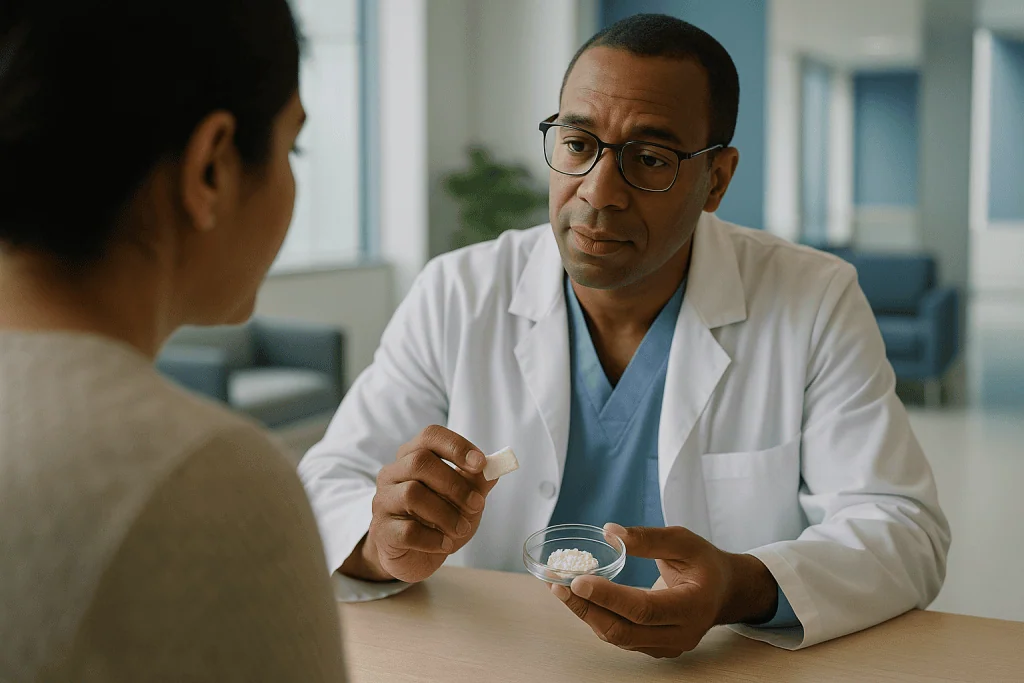
Donors are screened medically and tested for transmissible infections. The bone is then cleaned of blood, marrow and cells, and treated by freeze-drying, sometimes combined with a process that removes most of the mineral (demineralized bone matrix). It is sterilized and stored as granules, putty or small blocks. By the time it reaches a dental chair, there are no living donor cells left. What remains is a human mineral scaffold and, in demineralized forms, some of the natural proteins that encourage bone growth. In the United States, tissue banks operate under federal regulation for donor eligibility and processing.
Clinically, allograft occupies a middle ground. It avoids a second surgical site, it is available in whatever volume is needed, and its particles resorb at a moderate pace, usually faster than animal-derived mineral and slower than soft autograft. That makes it a frequent choice for socket preservation and for widening a ridge before an implant.
Honest limits apply. Because the living cells are gone, it cannot form bone on its own; it depends on healthy neighboring bone and good blood supply. Its processing also varies between tissue banks, which is one reason published comparisons are hard to pool. Some people decline human donor tissue on religious or personal grounds, and that preference is entirely legitimate; a surgeon can usually offer a synthetic alternative without changing the overall plan.
Xenograft: animal-derived material and why it resorbs so slowly
A xenograft is bone mineral from another species, most commonly cattle (bovine) or pigs (porcine), with equine sources used less often. The organic part of the bone, including cells and proteins, is removed with heat or chemical treatment. What is left is almost pure hydroxyapatite, the calcium phosphate crystal that makes up the hard part of all mammal bone, arranged in the porous honeycomb structure that cells find easy to colonize.
That structure is the appeal. Because the mineral is so similar to our own, bone grows readily onto and around it. Because the processing makes the crystals very stable, the body breaks them down extremely slowly. Particles can still be found in biopsies years after placement, embedded in new bone. For some procedures that is exactly what a surgeon wants. Under the sinus, where there is nothing above the graft to hold its shape, or along the front of the mouth, where even a millimeter of collapse shows as a shadow at the gum line, a scaffold that refuses to shrink is an asset.
The same quality is a limitation elsewhere. A graft that never fully turns into living bone contains less vital tissue for an implant to bond with, and if the area later becomes infected, the retained particles can complicate cleaning. Published long-term data are reassuring in the common uses, but the trials are mostly small and use different measures, so no neutral source would call one animal source superior to another.
Religious and dietary objections to bovine or porcine products are real for many patients and should be raised early. The material is processed to the point of being mineral, but that does not settle the matter for everyone, and a synthetic or human-donor option can nearly always be substituted.
Synthetic bone graft material: is it safe and does it work?
Synthetic grafts, called alloplasts, are made in a laboratory rather than taken from any body. The common chemistries are calcium phosphates such as hydroxyapatite and beta-tricalcium phosphate, bioactive glass, and calcium sulfate. All are built from the same elements your own bone mineral contains, which is why the body treats them as a familiar surface rather than an intruder.
On safety, the record is straightforward. There is no donor and therefore no theoretical risk of transmitted infection, no animal source to object to, and batch-to-batch composition is tightly controlled. These products are regulated as medical devices. Allergic reactions are rare because the materials are inorganic. Minor complications are the same ones seen with every graft: small particles working out through the gum in the first weeks, delayed soft-tissue healing, or infection at the site.
On effectiveness, the honest answer is more nuanced. Synthetics are purely osteoconductive: they give bone somewhere to grow but add no cells or signals. In small, well-contained defects such as a four-walled socket, comparisons generally show new bone forming in amounts similar to donor or animal-derived materials. In large defects that need height, or where blood supply is poor, surgeons often prefer to mix in autograft or choose a material with more biological activity. The evidence base is a collection of modest trials with different endpoints rather than a single definitive study, so most guidance stops short of ranking materials.
Resorption rates vary by chemistry. Beta-tricalcium phosphate dissolves relatively quickly and is replaced by bone; dense hydroxyapatite lingers much as xenograft does. Manufacturers blend the two to tune how fast the scaffold gives way, which is one more reason the choice belongs with someone who has seen your scan.
Is there a best bone graft material for dental implants?
People ask this constantly, and the search results are full of confident answers that happen to favor whatever the author sells. The evidence-based reply is less satisfying and more useful: there is no single best material, because the materials are solving different problems.
Four things usually drive the decision. First, the geometry of the defect. A socket with intact walls contains and protects almost anything placed in it; a ridge that has lost its outer wall needs something stiffer and a membrane to hold it. Second, whether volume must be preserved. In the sinus and the front of the mouth, slow-resorbing mineral earns its place. Third, timing. If an implant is planned soon, a material that converts to living bone faster may be preferred. Fourth, you: your medical history, your healing capacity, and whether human or animal tissue is acceptable to you.
Autograft remains the biological benchmark, but it is not automatically the best choice when the extra wound and limited supply are weighed. Systematic reviews of socket preservation and sinus grafting generally find that donor, animal-derived and synthetic materials all support implant placement, with differences in how much volume is kept rather than in whether an implant can be placed at all. Where studies disagree, it is usually because they measured different things.
If one principle matters most, it is this: the material is secondary to the stability of the result. A modest graft that heals undisturbed, under a well-fitted membrane, in a non-smoker with clean gums, will outperform a biologically richer graft placed into an inflamed site. Ask what your surgeon expects the bone to look like in nine months, and why. That answer tells you more than the brand of powder.
Who usually needs a dental bone graft, and who is usually asked to wait
Not everyone heading toward an implant needs a graft. A tooth removed gently, with the socket walls intact, often leaves enough bone for an implant without any additional material. Grafting enters the picture when something has already been lost.
The common reasons are predictable. A tooth that has been missing for years, because the ridge has quietly shrunk in the meantime. Advanced gum disease, which NHS guidance describes as destroying the bone that supports teeth before the teeth themselves are lost. Trauma that fractured the socket. Teeth that never developed, leaving a narrow ridge. And the upper back jaw, where the sinus sits just above the roots and the bone between them can be only a few millimeters thick, so a sinus lift is needed to create room.
Other people are asked to wait, or to have the plan reconsidered. Active infection in the gums or at the site is treated first, since a graft placed into inflamed tissue is far more likely to fail. Diabetes that is not well controlled slows healing and raises infection risk. Smoking does the same by starving the gum of blood flow, and many surgeons ask for a period of abstinence around surgery. Previous radiotherapy to the jaw changes the bone’s blood supply and calls for specialist assessment.
Medicines matter too. Antiresorptive drugs, a class that includes bisphosphonates and some injectable osteoporosis treatments, slow the natural turnover of bone; in some patients that is linked to a rare but serious condition in which exposed jawbone fails to heal. Nobody should stop such a medicine for dental work on their own. The dentist and the prescribing physician weigh the risk together, and the decision rests with them.
Membranes, growth factors and other things added to the graft
The particles are rarely placed alone, and the extras on your consent form deserve the same scrutiny as the main material.
A membrane is a thin sheet laid over the graft before the gum is closed. Its job is to stop fast-growing gum tissue from invading the space meant for slower-growing bone. Most are made of collagen and dissolve over weeks; some are non-resorbable and are removed at a second visit. For a graft that has to grow in height rather than just fill a hole, a stiffer barrier or a titanium mesh may be used to hold the shape. Membranes are standard, well-studied and largely uncontroversial.
Platelet-rich fibrin is different. A small amount of your blood is spun in a centrifuge to concentrate platelets and clotting proteins into a gel, which is mixed with the graft or draped over it. The theory is that the platelets release growth factors that speed healing. The evidence is mixed: several trials suggest faster soft-tissue closure and less discomfort, while effects on the final amount of bone are inconsistent. It is reasonable to describe it as promising for comfort and gum healing rather than as a proven way to grow more bone.
Recombinant growth factors, which are laboratory-made versions of the body’s bone-signaling proteins, are used in some surgical settings and can be added to a scaffold to give it inductive properties. They carry their own regulatory approvals and considerations, and they are not routine in everyday implant work.
Ask which additions are planned, what each is expected to do, and whether the plan would change without them. A surgeon who can answer those three questions plainly is one you can trust with the fourth: what happens if it does not take.
What the days and weeks after a dental bone graft usually look like
The first evening is mostly about pressure and patience. Expect gauze, some oozing, and a numb lip or cheek for a few hours. Swelling typically builds over the first two to three days and then eases; bruising on the outside of the face is common after a sinus lift or chin harvest and looks worse than it feels. Your team will advise on pain relief, usually over-the-counter options, and the specifics belong to them rather than to an article.
During the first week the goal is simply to leave the site alone. Soft, cool foods; no straws, which create suction; no vigorous rinsing; gentle brushing of the other teeth. A few sand-like granules may work out through the gum in the early days, and a small number is usually harmless, though it is worth mentioning at your check. Stitches dissolve or are removed at around one to two weeks, by which time the gum has normally sealed over the graft.
From there the timeline slows to a geological pace. The graft is quietly remodeled over months. Cleveland Clinic gives a usual range of three to nine months before the site is firm enough for an implant, with larger repairs, particularly sinus lifts, often at the longer end. Before placing the implant, the surgeon typically repeats imaging, often a three-dimensional cone-beam scan, to confirm the new bone is dense and wide enough.
Two things help the odds during this quiet stretch more than any product: not smoking, and not wearing a loose denture or temporary that presses on the graft. Mayo Clinic notes that the full implant process, from graft to final tooth, can span many months, which is frustrating but reflects how bone actually heals.
How long does a dental bone graft last?
The question assumes a graft is a part that wears out, like a filling or a crown. It is not. A successful graft stops being a graft. The scaffold is dissolved and replaced, and what remains is your own living bone, with the same cells and blood supply as the jaw around it. In that sense it has no separate lifespan at all.
What does have a lifespan is the reason the bone stays. Bone keeps its volume when it is loaded. An implant placed into the grafted site and used for chewing transmits force into that bone and keeps it active, much as the original tooth root did. Leave the site empty and the ridge will resume shrinking, grafted or not, which is why surgeons prefer to place the implant within the healing window rather than letting a prepared site sit for years.
Material matters to this story in one respect. Slow-resorbing minerals, especially xenograft and dense hydroxyapatite, leave particles embedded in the new bone for a very long time. That is not failure; it is the design. But it does mean that a biopsy of a sinus graft taken years later will show a composite of your bone and residual particles rather than pure bone.
The real threat to a grafted site is the same threat faced by natural teeth: inflammation. Gum disease around an implant, called peri-implantitis, can destroy grafted bone as efficiently as it destroyed the original. Daily cleaning and regular professional maintenance protect the investment far more than the choice of granule did. Nobody can promise how long a given graft and implant will serve; what the evidence supports is that well-maintained, loaded bone tends to stay.
What is the alternative to a bone graft for dental implants?
Grafting is a means to an end, and sometimes the end can be reached a different way. The alternatives fall into three groups, each with trade-offs the treating team can lay out for your particular jaw.
The first group changes the implant rather than the bone. Shorter implants can be used where height is limited, and narrower ones where width is the issue; both have grown more common as designs have improved, though very short or very thin implants have less margin for error. Angled or tilted implants can be placed to avoid the sinus or a nerve while still anchoring in native bone. In the upper jaw with severe bone loss, zygomatic implants, which are long implants anchored in the cheekbone, bypass the problem entirely, at the cost of a more complex surgery done by a specialist team.
The second group changes the plan. An implant-supported bridge uses fewer implants, placed where good bone already exists, to carry several teeth. Immediate placement at the time of extraction, when the socket walls are sound, can sidestep the shrinkage that would otherwise demand a graft later.
The third group steps away from implants. A conventional bridge borrowing support from neighboring teeth, or a well-made removable denture, remain legitimate options, and for some people they are the better fit medically or personally.
None of these is automatically superior. Avoiding a graft can mean accepting a smaller implant, a more demanding surgery elsewhere, or a different kind of restoration. What matters is a candid conversation about what each route gives up, and that conversation is your care team’s responsibility to lead.
What people often get wrong about the types of dental bone graft material
Myths about grafting tend to cluster around the word that frightens a given person: donor, animal, synthetic, or failure. Here are the ones heard most often in the chair.
Donor bone will give me a disease. Donor tissue is screened, processed to remove cells and sterilized under federal oversight. Regulators regard the residual risk as very low, and it is one reason the material has been in routine use for decades. Declining it on personal grounds is reasonable; fearing it as a likely source of infection is not supported by the evidence.
Animal bone becomes animal tissue inside me. Xenograft is mineral with the organic matter removed. The bone that grows around it is yours. The particles persist, but they do not carry animal cells or DNA in any functional sense.
Synthetic means fake, so it will not work. Alloplasts are made of the same calcium phosphate chemistry as natural bone mineral and have supported implants in small defects about as well as other materials in published trials. Their limitation is biological inertness in large defects, not safety.
The graft heals in a few weeks. The gum closes in a couple of weeks. The bone takes months; three to nine is the range cited by Cleveland Clinic, and nobody can speed it up with a supplement.
If a graft fails, implants are off the table. Partial or complete graft loss happens, usually because of infection, movement or smoking. The site is cleaned, allowed to heal and often grafted again, sometimes with a different material. One setback rarely ends the plan.
More material means more bone. Overpacking a site starves the center of blood supply. Surgeons aim for the volume the defect needs, not the most that fits.
Questions to ask your care team before agreeing to a graft
Consent is only meaningful if you understand what you are consenting to. These questions are the ones a well-informed patient tends to ask, and a good team will welcome them.
- Why do I need a graft at all, and what would happen to the implant plan if I did not have one?
- Which type of material do you intend to use, and is it mixed with anything else? If it is human or animal in origin, what are my alternatives if I would prefer not to have that?
- Why this material for this particular site? What are you trying to preserve: height, width or shape?
- Will a membrane or other additions be used, and what is each expected to do?
- How long do you expect healing to take before the implant can be placed, and how will you confirm the bone is ready?
- What is the chance this graft does not take, in your experience with cases like mine, and what would the next step be?
- Which of my medicines or health conditions affect healing here, and do you want to speak with my physician?
- What should the first week look like, and what would make you want to see me sooner?
- Can I wear my current denture or temporary tooth during healing?
- If I smoke, how long before and after do you ask patients to stop, and why?
Write the answers down. Several of them, especially the expected timeline and the plan if things go wrong, are worth having in your own hand rather than in memory. If any answer is a brand name instead of a reason, ask again. The reasoning, not the product, is what you are entitled to understand.
When to call your doctor after a dental bone graft
Most recovery is dull, and dull is good. A small number of signs mean the site should be looked at promptly rather than waited on. Contact your dental surgeon or the emergency number you were given if you notice any of the following.
- Bleeding that does not slow with firm pressure on gauze after about half an hour, or that restarts heavily after the first day.
- Swelling that keeps increasing after the third day, spreads toward the eye or down the neck, or makes it hard to open your mouth, swallow or breathe. Difficulty breathing or swallowing is an emergency: call emergency services.
- Fever, chills, or a foul taste or pus from the site.
- Pain that worsens after the first few days instead of easing, or that no longer responds to the relief your team advised.
- A large amount of graft material coming out, the membrane visibly exposed, or the wound edges pulling apart.
- After a sinus lift: nosebleeds that persist, air or fluid passing between nose and mouth when you drink, or a feeling that air moves through the site when you blow your nose.
- Numbness of the lip, chin or tongue that has not begun to improve within a day or two of surgery.
None of these necessarily means the graft has failed. Several, caught early, are managed with a simple adjustment or a course of treatment decided by your clinician. The point is that the people who placed the graft are the ones who should judge it, and they would rather hear from you unnecessarily than late. Keep the follow-up appointments even when everything feels fine; the imaging done at those visits is how the team confirms the bone is doing its quiet work.
Frequently asked questions
What is the best dental bone graft material?
There is no single best material; the right choice depends on the defect being repaired. Your own bone offers the most complete biology but needs a second surgical site. Donor, animal-derived and synthetic scaffolds all support implant placement in published trials, differing mainly in how fast they resorb and how well they hold shape. Your surgeon chooses based on the scan, the location and your preferences about tissue source.
What is the alternative to a bone graft for dental implants?
Alternatives include shorter or narrower implants placed in the bone that remains, angled implants that avoid the sinus or nerve, zygomatic implants anchored in the cheekbone for severe upper-jaw loss, implant-supported bridges using fewer sites, and non-implant options such as a conventional bridge or denture. Each avoids grafting by accepting a different trade-off, which the treating team should explain for your situation.
Are synthetic dental bone grafts safe?
Yes, synthetic grafts have a well-established safety record. They are made from calcium phosphates, bioactive glass or calcium sulfate, the same chemistry as natural bone mineral, carry no donor or animal source and are regulated as medical devices. Their limitation is effectiveness in large defects rather than safety, because they provide a scaffold without cells or growth signals, so surgeons sometimes combine them with your own bone.
What is the average lifespan of a dental bone graft?
A successful graft has no separate lifespan because it is replaced by your own living bone over several months. What determines how long that bone stays is whether it is loaded by an implant and kept free of gum inflammation. Unloaded ridges shrink again over time, and peri-implantitis can destroy grafted bone. Nobody can promise a duration; well-maintained, loaded bone tends to persist.
How long does a dental bone graft take to heal before an implant?
Cleveland Clinic gives a usual range of three to nine months, sometimes longer for large repairs such as sinus lifts. The gum seals over the site within about one to two weeks, but the bone remodels slowly beneath it. Before placing the implant, your surgeon normally repeats imaging to confirm the new bone is wide and dense enough. Smoking and pressure from a loose denture can lengthen the timeline.
Can donor bone grafts transmit disease?
The risk is considered very low by regulators. Donors are medically screened and tested for infections, the bone is processed to remove cells and marrow, then sterilized, and tissue banks in the United States operate under federal rules. No living donor cells remain in the finished material. People who prefer to avoid human donor tissue for personal or religious reasons can usually be offered a synthetic alternative instead.
Is animal-derived bone graft material acceptable if I avoid beef or pork?
That is a personal and sometimes religious decision, and you should raise it before surgery. Xenograft is processed down to mineral, with cells and proteins removed, but for many people the source still matters. Surgeons can substitute a synthetic or human-donor material in nearly every situation without changing the overall implant plan, so stating your preference early simply shapes which scaffold is used.
What happens if a dental bone graft fails?
Graft loss usually follows infection, movement of the material, exposure of the membrane or smoking. The surgeon cleans the site, lets the soft tissue heal and often grafts again, sometimes with a different material or a stiffer barrier. One failed graft rarely rules out an implant, though it may change the timeline or lead the team to suggest a shorter implant or another approach instead.
Does smoking affect which bone graft material is used?
Smoking affects whether and when a graft is done more than which material is chosen. Nicotine narrows blood vessels in the gum, reducing the supply that every type of graft relies on to heal, and raises the risk of infection and exposure. Many surgeons ask patients to stop for a period before and after surgery; the length of that period is a conversation to have with your treating team.
Can a bone graft and implant be placed at the same visit?
Sometimes. When the socket walls are intact and enough bone remains to hold the implant firmly, a surgeon may place the implant and pack graft material into any small gap at the same appointment. When the ridge has collapsed or the sinus floor must be lifted significantly, the graft is placed first and the implant follows months later, after imaging confirms the bone has matured.
References
This article is for general information only and is not a substitute for professional medical advice. Please consult a qualified doctor about your individual situation.

More from the Blog
Are Zirconium Teeth Right for You? Bite Force, Smile Goals and When Another Ceramic Fits Better
A good zirconium teeth candidate usually needs a strong restoration for a back tooth, an implant crown, or a bridge, especially with a heavy…
All-on-4 vs All-on-6: When Two More Implants Make Sense
All-on-4 and All-on-6 both anchor a full arch of fixed replacement teeth; the difference is how many implants carry the load. Four implants suit…
What to Ask Before a Dental Crown: Material, Margin Placement and What Happens to the Nerve
Before agreeing to a dental crown, ask what the crown is for, which material is proposed and why, where the edge of the crown…
Dental Implants After Gum Disease: When They Are Possible, and What Comes First
Yes, people with periodontal disease can often get dental implants, but not while the disease is active. Infection and inflammation must be treated first,…

How Long Do All-on-4 Implants Last? Longevity, Care and What Can Go Wrong
With diligent daily cleaning and regular professional maintenance, the four titanium implants supporting an All-on-4 restoration commonly last 20 years or longer, and long-term…
When Removable Dentures Stop Fitting: Relining, Rebasing and When a Remake Is Needed
Removable dentures usually stop fitting because the jawbone and gum underneath slowly shrink, not because the denture itself changes much. Dentists first assess the…