

Who Is a Candidate for Cartilage Restoration? Defect Size, Alignment and Age Considerations

Key Takeaways
- Articular cartilage has no blood supply, which is why a full-thickness defect does not refill on its own in adults and why surgical techniques create repair tissue or transplant existing cartilage rather than regrowing the original.
- The strongest cartilage restoration candidates have a single contained defect, a healthy opposing surface, stable ligaments, a reasonably straight leg, and symptoms that match the defect's location.
- Defect size drives technique on a sliding scale: marrow stimulation for smaller lesions, autograft plugs for small to medium, cell-based implantation for medium to large, and allograft when bone has also been lost.
- Uncorrected leg malalignment overloads the repaired area, so long-leg X-rays are routine and an osteotomy is often proposed before or alongside cartilage work rather than as an optional extra.
- Widespread osteoarthritis is a whole-joint disease, according to the NIH, and is the most common reason people who hope for cartilage restoration are steered toward other options.
- Recovery is measured in months of protected weight-bearing and staged rehabilitation, per NHS and MedlinePlus guidance, and the ability to follow that program is itself part of candidacy.
A cartilage restoration candidate is usually someone with a single, well-defined area of damaged joint cartilage, healthy surrounding cartilage, a stable and reasonably well-aligned joint, a healthy body weight, and realistic goals. Widespread arthritis, significant malalignment left uncorrected, or unstable ligaments usually rule people out or require staged procedures. Defect size, limb alignment, and age all shape which technique, if any, a surgical team recommends.
The MRI report sits on the kitchen table, and one phrase keeps drawing the eye: “full-thickness chondral defect.” Three words, a measurement in millimeters, and suddenly a runner who felt fine last spring is wondering whether her knee has a hole in it and whether anyone can fill it.
That question, whether you are a genuine cartilage restoration candidate, is one of the most carefully weighed decisions in sports orthopedics. Cartilage does not heal the way skin or bone does. The techniques that try to replace it are real, but they work best in a narrow set of circumstances, and a good surgical team spends as much time deciding who should wait as who should proceed.
This explainer walks through the three factors that dominate that conversation: how big the defect is, whether the leg is straight, and what age and activity mean for healing. The aim is honesty rather than reassurance, because the honest version is more useful.
What is cartilage restoration, and why does the word "candidate" matter so much?
Articular cartilage is the smooth, glistening tissue that caps the ends of bones inside a joint, letting them glide with less friction than ice on ice. It has no blood supply and very few living cells, which is why a divot in it tends to stay a divot. The NHS describes cartilage as a tissue that heals poorly on its own, particularly once the damage goes all the way down to bone.
Cartilage restoration is an umbrella term for surgical techniques that try to fill or replace a localized area of damaged cartilage, called a chondral defect, or a cartilage-plus-bone injury, called an osteochondral defect. The goal is a resurfaced patch that lets the joint move with less pain and, ideally, slows the drift toward arthritis in that spot.
The word “candidate” carries weight here for a simple reason. These procedures are designed for a joint that is otherwise in good shape with one bad neighborhood. When the whole joint is worn, filling one pothole on a road full of them changes very little. That is why the evaluation is less about whether cartilage is missing and more about everything around the missing piece: the opposite surface, the ligaments, the meniscus, the leg’s alignment, and the person’s weight, age, and goals.
Put differently, a cartilage restoration candidate is defined by context. Two people with identical defects on MRI can receive opposite recommendations because one has a straight, stable, otherwise healthy knee and the other has a bowed leg with early arthritis on the opposite surface. Understanding that logic makes the rest of the decision much easier to follow.
How cartilage restoration actually works: the main techniques in plain language
Most cartilage procedures fall into one of four families, and each tries to solve the no-blood-supply problem differently. The NHS lists three of them, marrow stimulation, mosaicplasty, and autologous chondrocyte implantation, as the established surgical options for cartilage damage.
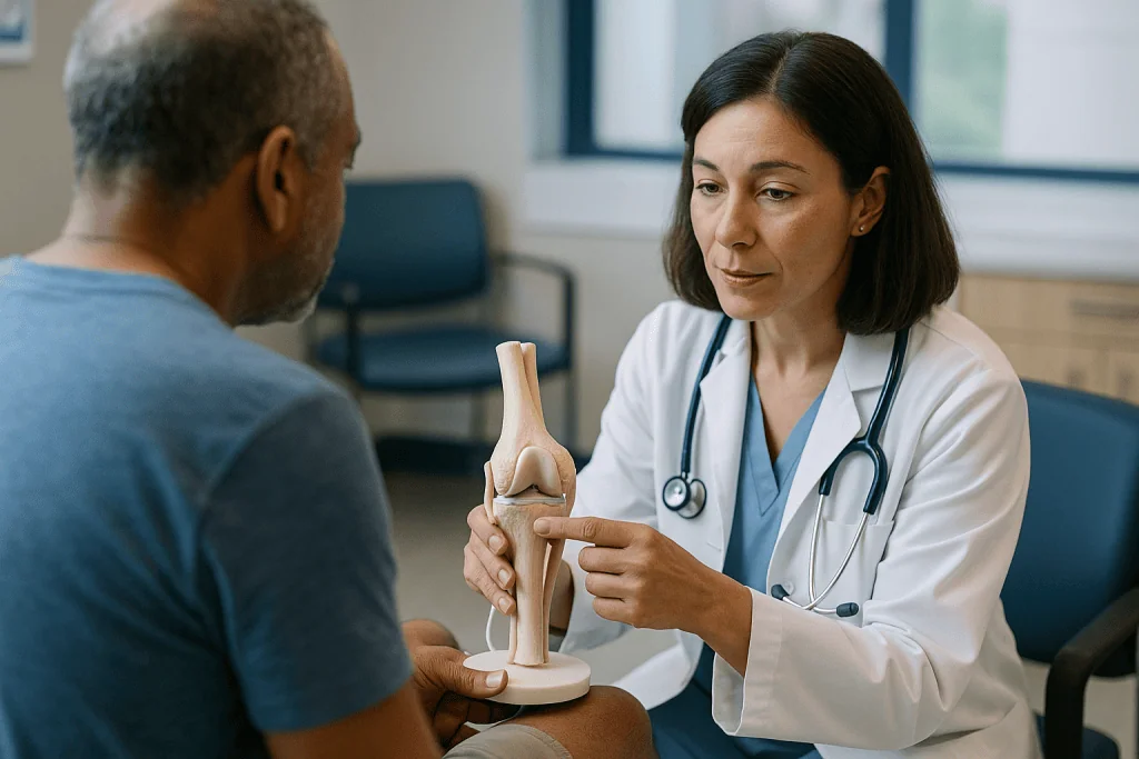
Marrow stimulation, often called microfracture, is the simplest. The surgeon cleans the edges of the defect and makes tiny holes in the underlying bone. Blood and marrow cells well up, clot, and mature into a scar-like tissue called fibrocartilage. It is a patch, not a duplicate, and it is generally softer and less durable than the original.
Osteochondral autograft transfer, sometimes called mosaicplasty or OATS, borrows small cylinders of cartilage and bone from a low-load part of the same joint and plugs them into the defect like transplanting sod. The advantage is that the plugs are true hyaline cartilage, the original type. The limit is supply: there is only so much spare surface to harvest.
Autologous chondrocyte implantation, or ACI, is a two-stage approach. A small cartilage sample is taken, the cells are grown in a laboratory, and weeks later they are implanted, often seeded on a membrane. This cell-based method suits larger defects that a few plugs cannot cover.
Osteochondral allograft uses donor cartilage and bone, matched for size and shape, to resurface larger or deeper defects where bone has also been lost. Because it replaces both layers, it is often reserved for the biggest problems.
Each option trades off tissue quality, defect size it can handle, number of surgeries, and recovery demands. Which one fits is exactly where defect size, alignment, and age come in.
Who is usually a cartilage restoration candidate, and who is usually asked to wait?
The typical candidate profile has stayed remarkably consistent across surgical guidelines and reviews. The person has a single, well-circumscribed defect, meaning clear edges with healthy cartilage all around it. The cartilage on the opposite surface of the joint, the one that rubs against the defect, is intact. Ligaments are stable, either naturally or after reconstruction. The leg is straight enough that the repaired area will not be overloaded. Symptoms match the location of the defect: pain, catching, or swelling that a clinician can reasonably attribute to that spot rather than to general wear.
People are usually asked to wait, or steered toward other options, in a mirror-image set of circumstances:
- Widespread or “kissing” lesions, where both opposing surfaces are damaged and any new tissue would be ground against a rough partner.
- Established osteoarthritis, which the NIH’s National Institute of Arthritis and Musculoskeletal and Skin Diseases describes as a whole-joint disease affecting cartilage, bone, and surrounding tissues rather than a single defect.
- Uncorrected malalignment or ligament instability, which can be addressed first or at the same time, but not ignored.
- Inflammatory arthritis, active infection, or conditions that impair healing.
- A body weight that places sustained high loads on the repair, since many programs treat weight as a modifiable factor to work on beforehand rather than a permanent exclusion.
- Inability to follow a lengthy rehabilitation plan, which is a practical concern rather than a judgment.
Waiting is not the same as being dismissed. A team may recommend physical therapy, activity modification, weight management, or a corrective osteotomy first, then revisit the cartilage question. The order matters, and it is one of the most common reasons two opinions on the same scan can differ.
Does defect size decide which cartilage procedure fits?
Size is the single variable surgeons mention most, and it works on a sliding scale rather than a hard cutoff. Small defects are usually well served by simpler techniques; large ones need methods that can supply more tissue. The exact numbers vary among surgical programs, so treat the table below as the commonly cited pattern in orthopedic literature rather than a rule your team must follow.
| Technique | Typical defect size range | Tissue produced | Common considerations |
|---|---|---|---|
| Marrow stimulation (microfracture) | Smaller, contained defects | Fibrocartilage (repair tissue) | Single surgery; durability questioned for larger or high-load areas |
| Osteochondral autograft (mosaicplasty) | Small to medium | Native hyaline cartilage with bone | Limited donor supply within the same joint |
| Autologous chondrocyte implantation | Medium to large | Hyaline-like cartilage | Two-stage procedure; longer protected rehabilitation |
| Osteochondral allograft | Large, or with bone loss | Donor hyaline cartilage and bone | Depends on graft availability and size matching |
Two nuances matter beyond the raw measurement. First, depth counts. A defect that has taken bone with it, as in osteochondritis dissecans or after a fracture, usually needs a technique that restores the bony base as well, which pushes the decision toward plugs or allograft. Second, location counts. Defects on the weight-bearing femoral condyles behave differently from those behind the kneecap, where shear forces and kneecap tracking add complexity.
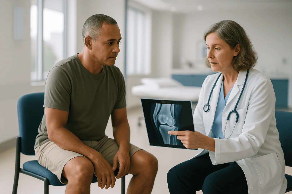
The size on your MRI report is also an estimate. Surgeons routinely find defects larger or smaller than imaging suggested once they look directly during arthroscopy, which is why many plans include a decision point during the first procedure.
Why leg alignment can matter as much as the cartilage itself
Imagine a tire wearing down on one edge. You can fit a new tire, but if the wheel is still tilted, the new one wears the same way. Knees follow the same physics. Malalignment means the leg’s mechanical axis, the line from hip to ankle along which weight travels, does not pass through the center of the knee. A bowed, varus knee shifts load to the inner compartment; a knock-kneed, valgus alignment shifts it outward.
When a cartilage defect sits in the overloaded compartment, that alignment is often part of why the defect formed and a strong predictor of whether a repair will survive. Restoring a surface under a misaligned load asks new, immature tissue to do a job the original, mature cartilage already failed at.
That is why surgical teams measure alignment on long-leg standing X-rays before recommending cartilage work. If the axis is meaningfully off, they may propose an osteotomy: a controlled cut and realignment of the tibia or femur, held with a plate, that shifts weight toward the healthier compartment. An osteotomy can be done before, alongside, or occasionally instead of cartilage restoration.
Alignment also includes the kneecap. Patellar maltracking, where the kneecap rides off-center in its groove, concentrates force on one facet. Cartilage repair behind the kneecap frequently pairs with a procedure to correct tracking, because otherwise the same shear that caused the wear returns.
Patients sometimes hear “you need your leg straightened first” as a detour. In practice, it is often the main event. The Cleveland Clinic notes that cartilage cannot repair itself effectively, so protecting whatever repair is attempted from excess load is one of the few levers a team can pull to give new tissue a fair chance.
How age and activity level shape the decision
Age matters for two reasons, and neither is a strict birthday cutoff. The first is biology: younger tissue tends to have more active cells and a healthier bony bed, and cell-based techniques rely on harvesting cells that will multiply well. The second is the state of the rest of the joint. Older knees are more likely to carry diffuse thinning, meniscal wear, and early arthritic changes that shift a person from “focal defect” toward “whole-joint disease,” where the Mayo Clinic notes management focuses on symptom control, activity, weight, and, when needed, joint replacement.
Many surgical programs describe their typical cartilage restoration patients as adolescents through middle age, but chronological age is increasingly treated as one input rather than a gate. A fit person in their fifties with a clean, isolated traumatic defect and a straight leg may be a reasonable candidate; a person in their thirties with kissing lesions and a worn meniscus may not be.
Activity level pulls in both directions. High demand makes the defect more symptomatic and raises the stakes of leaving it, but it also raises the load a repair must withstand and the patience required during months of protected rehabilitation. Athletes are often good candidates precisely because they can commit to rehab, provided they accept that return-to-sport timelines are long and not guaranteed.
Body weight is discussed frankly in these consultations because every extra unit of body weight multiplies force across the knee during walking and stairs. Teams frame it as a modifiable factor that can improve candidacy, not as a character judgment, and many will work with patients on weight and strength before committing to surgery.
What about the meniscus, ligaments and the rest of the knee?
Cartilage rarely fails alone. The meniscus, the C-shaped shock absorber between femur and tibia, spreads load across the joint surface. Lose a substantial portion of it and the pressure on the cartilage beneath rises sharply, which is one reason meniscal tears and cartilage defects so often travel together. A team evaluating a cartilage restoration candidate will look hard at the meniscus, and if too much is missing, may discuss meniscal repair or transplantation as part of a combined plan.
Ligaments matter for stability. An anterior cruciate ligament that has failed lets the tibia slide and rotate abnormally, scuffing cartilage with every pivot. Repairing cartilage in an unstable knee is a bit like repainting a wall while the roof still leaks. Surgeons generally insist on a stable joint first, either naturally or through reconstruction, sometimes performed in the same session.
The underlying bone counts too. Subchondral bone, the layer directly beneath cartilage, provides its foundation. Bone marrow edema, cysts, or collapse on MRI change the picture, because techniques that address only the cartilage layer may fail if the base is compromised.
This is why the phrase “whole-knee assessment” appears so often. Rather than asking “can the defect be filled?”, the team asks “what caused this defect, and will that cause still be present afterward?” Uncorrected alignment, instability, or meniscal loss are the usual answers, and each has its own procedure. Combined surgeries are longer and demand more rehabilitation, but addressing the cause is what separates a repair with a fighting chance from one destined to wear through.
Can knee cartilage regrow? What the evidence honestly says
This is the question underneath every other one, and it deserves a straight answer. Articular cartilage does not regrow on its own in any meaningful way in adults. The NHS and Cleveland Clinic both describe it as a tissue with very limited capacity for self-repair, because it lacks the blood vessels that bring healing cells to an injury.
What surgery can do is prompt the body to make repair tissue or physically move healthy cartilage into the gap. Microfracture produces fibrocartilage, which contains more of the tough collagen found in scar and less of the cushioning type in true articular cartilage. It fills the space and can relieve symptoms, but it is biologically different from what was lost. Cell-based techniques produce tissue described as “hyaline-like,” closer to the original, though studies examining biopsies show it is often a mixture rather than a perfect copy. Autograft and allograft plugs are the only options that place mature hyaline cartilage into the defect, and even then the border between plug and surrounding cartilage does not fully knit together the way bone does.
So has anyone ever regrown cartilage? In the strict sense of a joint spontaneously refilling a full-thickness defect with normal cartilage, no. In the practical sense of surgically restoring a functional, load-bearing surface that relieves pain for years, yes, in appropriately selected people.
Claims that supplements, injections, or unproven cell therapies regrow cartilage should be treated with caution. Mainstream reviews have not shown that any of them rebuild articular cartilage in humans, and the evidence base for symptom relief from most such products remains mixed. Ask your team specifically what the evidence shows before spending money or hope on them.
What is the cartilage regeneration success rate? Reading the numbers with care
Search this question and you will find confident percentages on many websites. Approach them skeptically. “Success” in cartilage research is measured many different ways: pain scores, function questionnaires, return to sport, MRI fill of the defect, or simply avoiding another operation. A study can report a high number on one measure and a modest one on another, and websites tend to quote the flattering figure.
Several patterns do emerge consistently across systematic reviews. Smaller, contained defects in younger, well-aligned knees tend to do better than larger, uncontained defects in older knees with other damage. Marrow stimulation often relieves symptoms well in the early years, with more concern about durability in larger or high-load defects. Cell-based and graft techniques have generally been studied for larger defects, where they appear to hold up better over longer follow-up, but they demand more surgery and longer rehabilitation. Outcomes for defects behind the kneecap have historically been less predictable than for defects on the femoral condyles.
None of this translates into a single success rate you can carry into a consultation. The honest framing is that the same technique performs very differently depending on who receives it, which is exactly why candidacy is scrutinized so closely. The selection process is the success rate.
This magazine does not print percentages unless they come from a named guideline or systematic review, and there is no single figure that fairly summarizes this field. What you can do is ask your surgeon for their own outcome data, for the specific technique proposed, in patients who resemble you. A good team will explain both the numbers and their limits. Anyone quoting a single confident percentage without those caveats is simplifying more than the evidence allows.
Osteochondritis dissecans and cartilage restoration in younger patients
Some of the most compelling cartilage restoration candidates are teenagers and young adults with osteochondritis dissecans, or OCD. The Mayo Clinic describes OCD as a condition in which a segment of bone beneath the cartilage loses blood supply, softens, and can crack or separate, most often in the knee and most commonly in children and adolescents who are active in sports.
OCD is a useful illustration of why age changes the plan. In a child whose growth plates are still open, many stable OCD lesions heal with rest and activity modification alone, so the first recommendation is often to wait and monitor with repeat imaging. Surgery is considered when a lesion is unstable, has detached, or has failed to heal after the growth plates close.
When surgery is needed, the approach tracks the same logic as adult cartilage work but with a twist. Because OCD involves bone as well as cartilage, techniques that restore the bony base, such as fixing the fragment back in place or using bone-and-cartilage plugs, are often preferred over cartilage-only repairs. Larger or fragmented lesions may need allograft.
For families, the pediatric pathway centers on preparation, comfort, and recovery rather than medicines: understanding the period of restricted weight-bearing, arranging school accommodations for crutches or a brace, planning transportation, and keeping a young athlete engaged during months away from sport. Physical therapy is central and is adjusted to growth and maturity.
Young patients usually have the biology on their side, but they also have the longest road ahead of them, and protecting a repair through decades of use is a strong argument for getting alignment, stability, and rehabilitation right the first time.
Cartilage restoration recovery time: what the following weeks and months usually look like
Recovery from cartilage surgery is slower than most people expect, and the pace is set by the biology of the repair tissue rather than by how the incision looks. Newly stimulated or implanted tissue is soft for months and must be protected from full load while it matures, which is why rehabilitation protocols are long, staged, and strict.
The early period is about protection and motion. Most protocols combine partial or non-weight-bearing on crutches with gentle range-of-motion exercise, sometimes using a continuous passive motion machine, because controlled movement helps nourish cartilage while heavy load harms it. MedlinePlus notes that even simpler knee arthroscopy involves swelling, restricted activity, and a graded return, and cartilage restoration protocols are considerably more conservative than that.
The middle phase gradually reintroduces weight, strength work, and normal walking. The NHS describes recovery from cartilage surgery as taking weeks to months, and for cell-based or graft procedures the protected phase is typically at the longer end of that range. Your team will give a schedule tailored to the technique, the defect location, and whether an osteotomy or ligament work was done at the same time.
Return to impact sport is the final phase and often the longest wait. Surgeons generally hold off on running, cutting, and jumping until strength, balance, and imaging suggest the repair can tolerate it, and some athletes are asked to accept that certain high-impact activities remain limited.
Two practical points recur in patient conversations. First, the timeline is a range tied to how your tissue heals, not a promise. Second, adherence matters: the rehabilitation program is not an afterthought to surgery but half of the treatment, and the surgeon’s judgment about whether you can commit to it is part of your candidacy.
Risks, alternatives and what "you should wait" really means
Every cartilage procedure carries the general risks of joint surgery: infection, blood clots, stiffness, persistent swelling, and nerve or vessel injury, all uncommon but real. Specific to cartilage work, the repair tissue may not fill the defect, may fail to integrate with surrounding cartilage, or may break down over time. Overgrowth of repair tissue can cause catching. Plugs harvested for autograft leave donor sites that occasionally become symptomatic. Cell-based techniques carry the added risks of a second operation, and allograft involves the small risks associated with any donor tissue.
The alternatives are not a consolation prize. Structured physical therapy strengthens the muscles that unload the joint and can meaningfully reduce symptoms from focal defects. Activity modification, weight management, and appropriate footwear or bracing change the load the defect sees every day. The NIH lists these conservative measures as the foundation of managing joint cartilage problems and osteoarthritis. Anti-inflammatory medicines and joint injections may be discussed by your prescribing clinician for symptom control; they do not rebuild cartilage, and decisions about them belong with that clinician.
For joints that have progressed to widespread wear, arthroscopic debridement, osteotomy alone, partial or total joint replacement are the mainstream surgical paths. Cartilage restoration in that setting is generally not recommended because the surrounding joint cannot support it.
When a team says “wait,” it usually means one of three things: the problem may settle without surgery, the joint needs something else fixed first, or the risk-benefit balance does not currently favor an operation. Ask which one applies to you. A clear answer turns a frustrating “not now” into a plan with a next step.
What people often get wrong about cartilage restoration candidacy
The first misconception is that a cartilage defect on MRI means surgery is needed. Many defects, including some full-thickness ones, cause no symptoms and are found incidentally. Surgical teams treat symptoms and function, not pictures, and a painless defect in a stable joint is usually watched rather than operated on.
The second is the reverse: that cartilage surgery is a way to “fix arthritis.” It is not. The NIAMS describes osteoarthritis as a disease of the whole joint, and no restoration technique resurfaces a joint with generalized wear. People with arthritis who hear about cartilage procedures are often disappointed to learn they are not candidates, but the recommendation protects them from an operation with a long recovery and little chance of durable benefit.
Third, many assume the goal is to grow back original cartilage. As discussed earlier, most techniques produce repair tissue or transplant existing cartilage; regrowth in the biological sense is not what is happening.
Fourth, people underestimate rehabilitation. It is common to hear the surgery described as “minimally invasive” and assume a quick return. The incisions may be small, but the tissue needs months of protection, and skipping that phase is one of the more common reasons repairs fail.
Fifth, an osteotomy is often heard as an unrelated extra. In reality, correcting alignment may be the most important part of the plan, and in some knees it relieves symptoms enough that cartilage work becomes unnecessary.
Finally, injections and supplements marketed as cartilage regenerators are widely believed to do what their names imply. Mainstream evidence does not support that claim, and a team that says so plainly is not being dismissive; it is being accurate.
Questions to ask your care team before deciding
A candidacy consultation goes better when you arrive with specific questions, because the answers reveal how the team is thinking about your particular joint rather than about cartilage surgery in general. Consider asking:
- How large and how deep is my defect, and did imaging show the opposite surface to be healthy?
- Is my leg alignment within a range you are comfortable with, or do you recommend an osteotomy before or alongside cartilage work?
- Are my ligaments and meniscus intact, and if not, what needs to happen first?
- Which technique are you proposing, why that one for my defect size and location, and what would change your mind during surgery?
- What does “success” mean in your experience with this technique, in people of my age and activity level, and how long has it lasted?
- What is the weight-bearing and brace plan for the first weeks, and how long before I can drive, work, and return to my sport?
- What is the realistic chance I will need another operation, and what would that look like?
- If I am not a candidate now, what specifically would change that, and what is the plan in the meantime?
- How does my body weight or fitness affect the plan, and would working on either change the recommendation?
- Which non-surgical options have I not yet fully tried?
Bring someone with you if you can. These conversations cover anatomy, staging, and timelines at once, and a second set of ears catches details you will want later. Ask for the plan in writing, including the reasoning, so you can compare it thoughtfully if you seek a second opinion. Good teams welcome that; it is part of how confident decisions get made.
When to call your doctor: red-flag signs before and after cartilage surgery
Cartilage problems are usually slow-moving, but a few situations should not wait for a scheduled follow-up. Before any surgery, contact your care team promptly if the knee locks so that you cannot straighten or bend it, if it gives way and you fall, if it swells rapidly after an injury, or if you cannot bear weight. These can signal a loose fragment, a ligament tear, or a fracture that changes both urgency and candidacy.
After surgery, the NHS and MedlinePlus both list signs that warrant a same-day call: fever or chills; increasing redness, warmth, or discharge at the incision; pain that escalates rather than settles despite prescribed measures; or the joint becoming hot and tightly swollen, which can indicate infection or bleeding into the joint.
Seek urgent care immediately for calf pain, swelling, or tenderness in one leg, or for sudden shortness of breath, chest pain, or coughing up blood. These can be signs of a blood clot in the leg or lung, a recognized risk after lower-limb surgery, especially during periods of restricted mobility.
Also call if you develop numbness or weakness in the foot, a dressing that is soaked through, a brace or cast that feels far too tight, or a sudden new catching or locking sensation during rehabilitation, which could reflect a problem with the repair.
None of these signs means something has gone wrong, but each is a reason for a clinician to look rather than for you to wait and see. Keep the after-hours number for your surgical team where you can find it, and use it. The decision about what to do next always sits with your treating team; your job is simply to make sure they know.
Frequently asked questions
What is the success rate of cartilage regeneration?
There is no single success rate, because studies define success differently: pain relief, function, MRI fill, return to sport, or avoiding another operation. Systematic reviews consistently show better results for smaller, contained defects in younger, well-aligned knees than for large defects in older joints with other damage. Ask your surgeon for outcome data on the specific technique proposed, in patients who resemble you, and how they define success.
Has anyone ever regrown cartilage?
Not in the sense of a joint spontaneously refilling a full-thickness defect with normal articular cartilage. Adult cartilage lacks a blood supply and repairs itself very poorly, as the NHS and Cleveland Clinic both note. Surgery can stimulate repair tissue, grow and implant a person’s own cartilage cells, or transplant mature cartilage plugs. These restore a functional surface in selected patients, but they are repairs and transplants, not true regrowth.
Is it possible to grow cartilage back in your knee?
Your body will not grow it back on its own, but surgical techniques can fill a defect with repair tissue or transplanted cartilage. Microfracture yields fibrocartilage, a scar-like tissue; cell-based implantation yields hyaline-like tissue; autograft and allograft place genuine hyaline cartilage into the gap. Supplements and injections marketed as cartilage regenerators have not been shown in mainstream evidence to rebuild articular cartilage.
How much does cartilage regeneration cost?
This magazine does not publish price figures, because they vary widely with technique, whether a second stage or an osteotomy is included, insurance coverage, and facility. The financial counselors or billing office at any surgical center can give you an itemized estimate for your specific plan. Ask what is included, what might change during surgery, and how rehabilitation visits are covered, since the recovery program is a large part of the total.
Who is a knee cartilage repair candidate?
Usually someone with one well-defined cartilage defect, healthy cartilage on the opposite surface, stable ligaments, a leg that is straight or can be straightened, a healthy body weight, and symptoms that fit the defect. People with widespread arthritis, kissing lesions, inflammatory arthritis, or uncorrected instability are usually asked to pursue other options first. The final judgment always rests with the treating surgical team after imaging and examination.
Can knee cartilage regrow with rest and physical therapy?
Rest and physical therapy do not regrow cartilage, but they can substantially reduce symptoms by strengthening the muscles that unload the joint and by changing how you move. In children with stable osteochondritis dissecans, the underlying bone can heal with rest, according to the Mayo Clinic, which is why watchful waiting is common in that group. For adults, conservative care is about function and comfort, not tissue regrowth.
What is the typical cartilage restoration recovery time?
Weeks to months, according to the NHS, with cell-based and graft procedures at the longer end. Early weeks involve protected weight-bearing on crutches and gentle motion; the middle phase reintroduces walking and strength; return to impact sport comes last and can take many months. Combined procedures such as osteotomy lengthen the timeline. Your team’s schedule for your specific technique is the one to follow, and it is a range rather than a promise.
Does age rule out cartilage restoration?
Not by itself. Many programs describe typical patients as adolescents through middle age, but chronological age is one input among several. Younger tissue tends to heal better and cell-based methods rely on healthy harvested cells, while older knees more often carry diffuse wear that shifts the picture toward arthritis. A fit older adult with an isolated traumatic defect and a straight, stable knee may still be considered.
Why does my surgeon want to straighten my leg before fixing the cartilage?
Because malalignment concentrates load on the compartment where the defect sits, and new repair tissue cannot withstand forces that already wore out the original cartilage. Correcting alignment with an osteotomy shifts weight toward the healthier side and gives the repair a fair chance. In some knees, realignment alone relieves symptoms enough that cartilage surgery is no longer needed, which is why it is often treated as the main event rather than an add-on.
What happens if I am told to wait?
Ask which reason applies: the problem may settle on its own, something else in the joint needs fixing first, or the risk-benefit balance currently favors non-surgical care. Each has a next step, such as physical therapy, weight management, ligament or meniscus work, or repeat imaging. Waiting is a plan, not a dismissal, and a clear explanation of what would change the recommendation is a reasonable thing to request.
References
- NHS: Cartilage damage
- Cleveland Clinic: Cartilage
- MedlinePlus: Knee arthroscopy
- NIH NIAMS: Osteoarthritis
This article is for general information only and is not a substitute for professional medical advice. Please consult a qualified doctor about your individual situation.

More from the Blog

What X-Rays and MRI Show After a Shoulder Dislocation: Fractures, Labrum and Ligament Damage
An x-ray after shoulder dislocation confirms the joint is out, shows which direction it went, and checks for associated fractures of the humeral head,…

Life After Cruciate Ligament Surgery: Stairs, Driving, Desk Work and Sport in Order
Life after cruciate surgery follows a fairly predictable order. Walking with crutches comes first, usually within days; stairs and desk work return over the…

ACL Surgery Cost: What Reconstruction Really Costs, Market by Market
ACL reconstruction typically costs USD 20,000–50,000 in the United States, GBP 7,000–14,000 for private surgery in the UK, and EUR 3,900–7,150 within our guide…

How Shoulder Impingement Is Diagnosed: The Exam Tests and When an Ultrasound or MRI Is Ordered
Shoulder impingement is diagnosed mainly by a clinician's history and hands-on examination: where the pain sits, which movements provoke it, and how the shoulder…

Tendon Surgery Recovery: Why the Splint Stays On and How Gliding Exercises Begin
Tendon surgery recovery time is usually measured in months rather than weeks. A splint typically protects a repaired hand tendon for roughly four to…

Preparing for Arthroscopy: Medication Review, Fasting and Arranging a Ride Home
Preparing for arthroscopy usually rests on three tasks: giving your surgical team a complete list of every prescription medicine, over-the-counter product and supplement you…