

Why a CT Scan Is Often Ordered for Diverticulitis and What It Shows Your Doctor

Key Takeaways
- A contrast-enhanced CT scan is the standard first test for suspected diverticulitis because it confirms inflammation and detects complications such as abscess, perforation and obstruction in one pass.
- The three classic CT findings are diverticula, thickening of the colon wall and hazy 'fat stranding' in the tissue around the inflamed segment.
- Whether the scan shows uncomplicated or complicated disease largely determines whether a person is managed at home or admitted to hospital.
- Colonoscopy is avoided during an acute attack because inflating an inflamed colon risks tearing it; it is commonly arranged around six to eight weeks after recovery to check the lining directly.
- Diverticula seen incidentally on a scan are not a diagnosis of diverticulitis; most people with diverticulosis never develop an inflamed pouch.
- Repeat CT scans are usually reserved for people who fail to improve or whose abscess is being monitored, not for confirming recovery in uncomplicated cases.
A CT scan is usually the first imaging test for suspected diverticulitis because it confirms inflammation in the colon wall and, just as importantly, shows whether complications such as an abscess, perforation or blockage are present. That single distinction shapes whether a person can typically be managed at home or needs hospital care, so doctors treat the scan as both a diagnostic and a triage tool.
The pain started low on the left side, the kind that makes you shift in your chair and then shift again. By evening there was a fever, and by midnight the emergency department clerk was asking whether you had eaten anything since lunch. Then a doctor said the words most people hear only in passing: “We’d like to get a CT scan for diverticulitis.”
Plenty of patients in that moment assume the scan is a formality, a picture to confirm what the exam already suggested. It is more than that. In diverticulitis, the story is rarely just whether the colon is inflamed; it is how inflamed, where, and whether anything has leaked, walled itself off or blocked the passage. The answers to those questions live inside a stack of cross-sectional images, and they steer nearly every decision that follows.
This explainer walks through why the scan is ordered, what a radiologist is actually looking for, what the results do and do not tell you, and how to talk with your care team about the plan.
Why doctors reach for a CT scan for diverticulitis first
Diverticulitis is inflammation or infection of small pouches, called diverticula, that bulge outward from weak spots in the colon wall. The pouches themselves are common and usually harmless; the trouble begins when one becomes inflamed. The difficulty for a clinician is that the symptoms overlap with several other conditions that call for very different treatment.
A person with left-sided abdominal pain, fever and a tender belly might have diverticulitis, but the same picture can come from appendicitis in an unusual position, inflammatory bowel disease, an ovarian problem, a kidney stone, colitis from infection, or, rarely, a colon cancer that has partially blocked the bowel. A physical exam and blood tests can raise suspicion, but they cannot reliably separate these possibilities. Mayo Clinic notes that diverticulitis is typically diagnosed during an acute attack and that a CT scan is the test most often used to confirm it.
Beyond confirming the diagnosis, the scan answers the question clinicians care about most: is this uncomplicated inflammation, or has something gone further? An inflamed pouch that has stayed contained behaves very differently from one that has burst and formed a pocket of pus. The treatment pathway, the setting of care and the urgency all hinge on that distinction, and only imaging can make it with confidence.
There is also a practical reason. CT scanners are available around the clock in most emergency departments, the scan itself takes minutes, and the images can be read quickly. For an acutely unwell person, that speed matters more than the theoretical appeal of a test that might be a little more detailed but takes hours to arrange.
What actually happens during a CT scan for diverticulitis
A CT scan, short for computed tomography, uses a rotating X-ray beam and a computer to build cross-sectional images of the body, like slices through a loaf of bread. For suspected diverticulitis the scan covers the abdomen and pelvis, from the lower ribs down to the hips.

You will usually be asked to change into a gown and remove metal such as belts or jewelry. In many cases a nurse places a small intravenous line in the arm so that iodine-based contrast dye can be given; contrast is a liquid that makes blood vessels and inflamed tissue stand out more clearly on the images. Some departments also ask you to drink a contrast liquid beforehand to outline the bowel, although many now skip this step for suspected diverticulitis to save time.
The scanner is a ring, not a tunnel. You lie on a padded table that slides through the ring while the machine hums and clicks. The technologist may ask you to hold your breath for a few seconds at a time so that breathing movement does not blur the pictures. According to MedlinePlus, the scanning itself typically takes only a few minutes, though the whole visit, including preparation and waiting for the contrast to circulate, is longer.
When intravenous contrast goes in, most people feel a brief warm flush spreading through the body and sometimes a metallic taste. Both fade within a minute or two and are not allergic reactions. Afterward you can normally get up and move around straight away. The images go to a radiologist, a physician who specializes in interpreting scans, and a report goes back to the treating team, often within the hour in an emergency setting.
CT scan diverticulitis findings: what the radiologist is looking for
A radiologist reading a scan for suspected diverticulitis looks for a small cluster of specific signs, then checks systematically for anything that would change the picture.
The first is the presence of diverticula themselves, seen as small air- or stool-filled outpouchings along the colon, most often in the sigmoid colon on the lower left. The second is thickening of the colon wall in the affected segment. A healthy colon wall is thin; an inflamed one swells noticeably, and the radiologist measures it.
The third, and often the most telling, is what radiologists call fat stranding. The colon sits in a cushion of fat, and on a normal scan that fat looks dark and uniform. When the wall beneath it is inflamed, fluid seeps into the fat and creates hazy, streaky lines. Stranding around a thickened segment containing diverticula is the classic signature of the condition.
With those three in view, the search shifts to complications. Is there a collection of fluid with a rim that lights up with contrast, suggesting an abscess, a walled-off pocket of pus? Are there tiny bubbles of air outside the bowel, which point to a perforation, meaning a hole in the wall? Is there free fluid in the abdomen? Is the bowel upstream dilated, hinting at obstruction? Is there an abnormal channel connecting the colon to the bladder or another organ, known as a fistula?
Finally, the report usually comments on everything else in view: the appendix, the ovaries, the kidneys, the liver. Cleveland Clinic points out that one advantage of CT is this ability to rule other conditions in or out at the same time.
Uncomplicated or complicated? How the scan grades severity
Clinicians commonly sort diverticulitis into two broad groups based on what the CT shows, and the sorting drives nearly every decision that follows.
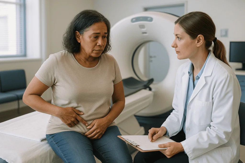
Uncomplicated diverticulitis means the inflammation is confined to the colon wall and the surrounding fat. There is no abscess, no free air, no obstruction and no fistula. Complicated diverticulitis means at least one of those features is present. Surgeons often go a step further and use a staging system that separates a small abscess next to the bowel from a larger one deeper in the pelvis, and both of those from a perforation that has spilled pus or bowel contents into the abdominal cavity.
| What the CT shows | What it usually means | How it commonly shapes care |
|---|---|---|
| Wall thickening and fat stranding only | Uncomplicated diverticulitis | Often manageable outside hospital in otherwise healthy adults; the team decides on antibiotics case by case |
| Small abscess close to the colon | Complicated, contained | Frequently treated with antibiotics and close monitoring; drainage sometimes considered |
| Larger or distant abscess | Complicated, contained | Image-guided drainage is often discussed alongside antibiotics |
| Free air or free fluid beyond the colon | Perforation | Urgent surgical assessment |
| Dilated upstream bowel | Obstruction | Hospital care; surgical review depending on severity |
The table describes typical patterns described by Mayo Clinic and Johns Hopkins Medicine, not fixed rules. A frail person with uncomplicated findings may still be admitted, and a robust person with a tiny abscess may be watched closely at home. The scan supplies the anatomy; the treating team weighs it against age, other illnesses, how the person looks, and how they respond over the first day or two.
What is the most accurate test for diverticulitis?
For an acute attack, contrast-enhanced CT of the abdomen and pelvis is the test most guidelines treat as the reference standard, and it is the one that both Mayo Clinic and Cleveland Clinic describe as the usual route to a confirmed diagnosis. Its strength is not just that it detects inflammation but that it does so across the whole abdomen in a single pass, catching complications and alternative diagnoses at the same time.
Other tests have roles. Ultrasound uses sound waves rather than X-rays, involves no radiation and can be done at the bedside. In experienced hands it can show a thickened colon wall and inflamed fat, and it is sometimes chosen first for pregnant patients or young adults. Its limitations are that it depends heavily on the operator, that bowel gas and body habitus can obscure the view, and that it is less reliable for finding deep abscesses or small amounts of free air.
MRI, which uses magnetic fields, offers excellent soft-tissue detail without radiation. It is not the everyday choice in emergency settings because it takes longer, is less available at night and is harder for someone in pain to lie still for, but it is a reasonable alternative when radiation avoidance is a priority.
Blood tests support the picture rather than making the diagnosis. A raised white cell count and an elevated C-reactive protein, a marker of inflammation the liver produces, make an infection or inflammatory process more likely but cannot say where it is. A plain X-ray of the abdomen is largely a historical tool here; it can show free air from a perforation but misses most of what matters.
Put simply, accuracy in this setting means answering three questions at once: is it diverticulitis, is it complicated, and is it something else entirely? CT answers all three more consistently than any other single test.
Colonoscopy vs CT scan for diverticulitis: is one better?
People often ask whether a colonoscopy, a camera examination of the inside of the colon, would give a clearer answer than a scan. During an acute attack, the answer from major sources is no, and the reasons are about safety as much as accuracy.
Colonoscopy looks at the lining from the inside. Diverticulitis is primarily a disease of the outer wall and the tissue around it. An inflamed pouch can appear as little more than a reddened opening from within, while the abscess sitting just outside it is invisible to the camera. A CT scan, by contrast, sees the outside of the bowel, the surrounding fat and the whole pelvis.
The safety concern is more pressing. Colonoscopy requires the colon to be inflated with gas so that the camera can see, and the bowel is also cleaned out beforehand with a strong laxative preparation. An acutely inflamed, weakened segment of colon is at greater risk of tearing under that pressure. For this reason, Cleveland Clinic and Mayo Clinic both describe colonoscopy as a test for after recovery, not during the attack.
Where colonoscopy earns its place is in the weeks that follow. The inflamed segment on a CT can occasionally hide, or mimic, a colon cancer, because both can thicken the wall. Once the inflammation has settled, a colonoscopy examines the lining directly and can take biopsies, small tissue samples, if anything looks unusual. Cleveland Clinic notes that this follow-up examination is commonly arranged around six to eight weeks after symptoms resolve, particularly after a first episode or a complicated one, and for anyone not already up to date with colon cancer screening.
The two tests, then, are not competitors. One answers the acute question of what is happening now; the other answers the later question of whether anything else is going on.
Who is usually scanned right away, and who is asked to wait
Not everyone with lower abdominal pain needs a CT, and not everyone who has had diverticulitis before needs one each time. The decision follows a loose but sensible logic.
Scanning promptly is the norm when the diagnosis is uncertain, when it is a first episode, when the person has a fever or a raised heart rate, when the abdomen is very tender or rigid, or when the exam and blood tests suggest something more than simple inflammation. Older adults and people whose immune systems are weakened, whether by medication or illness, also tend to be scanned early because their symptoms can be muted even when a complication is brewing. Anyone who is not improving after a day or two of treatment is another clear candidate.
Some clinicians will hold off in a narrow group: a person who has had several confirmed episodes before, whose current symptoms feel identical and mild, who has no fever, and who is otherwise well. In that setting a doctor may decide to treat and monitor closely, with a low threshold to scan if anything changes. The NHS notes that a GP may sometimes diagnose based on symptoms and examination, referring for tests when there is doubt.
Pregnancy calls for a different conversation. Ultrasound or MRI is usually preferred to avoid radiation to the fetus, and the team weighs the small radiation risk of CT against the danger of missing a complication.
A known severe allergy to contrast dye or significantly reduced kidney function does not rule out CT; the scan can be done without intravenous contrast, at the cost of some detail, or the team may choose another modality. In every case the decision belongs with the treating clinicians, who can see the whole picture in front of them.
Contrast dye, radiation and the real risks of the scan
Two worries come up almost every time: the dye and the radiation. Both deserve a straight answer.
Iodine-based contrast is given through a vein to sharpen the images. The warm flush and metallic taste are expected and harmless. True allergic-type reactions occur in a small minority of people and are usually mild, such as hives or itching; severe reactions are uncommon and emergency departments are equipped to treat them. Anyone with a previous reaction to contrast, or with asthma or multiple allergies, should say so before the scan, because the team may adjust the plan or take precautions.
The kidneys clear contrast from the body, so people whose kidney function is already reduced face a small risk of temporary worsening. Departments commonly check a recent blood test for kidney function before giving contrast when time allows, and they encourage drinking fluids afterward. People taking certain diabetes medicines may be given specific instructions about pausing them briefly; follow the team’s guidance rather than deciding alone.
Radiation is the longer-term concern. A CT of the abdomen and pelvis delivers considerably more radiation than a chest X-ray. MedlinePlus describes the added lifetime cancer risk from a single scan as very small, and most radiologists frame the trade-off this way: the danger of missing a perforation or abscess today is far larger and far more immediate than the theoretical risk from one scan. Modern scanners also adjust the dose to body size. The risk becomes more relevant for people who accumulate many scans over years, which is one reason clinicians try not to re-scan mild recurrent episodes reflexively.
The other practical risks are minor: bruising at the needle site, and the discomfort of lying still while in pain. Claustrophobia is rarely an issue because the ring is open at both ends.
What a CT scan can miss or mistake
For all its usefulness, a CT is a picture, not a verdict, and honest interpretation means knowing where it falls short.
The most discussed pitfall is the overlap between inflamed diverticulitis and colon cancer. Both can thicken the colon wall over a short segment. Radiologists use clues to tell them apart, such as the length of the thickened segment, whether the surrounding fat is inflamed out of proportion to the wall change, and whether enlarged lymph nodes are present, but the distinction is not always certain. This is precisely why a follow-up colonoscopy is recommended after recovery in many cases: to look directly at the lining once the swelling has settled.
Very early or very mild inflammation can produce subtle changes that a scan may not clearly show. A small perforation that has already sealed may leave only a few bubbles of air that are easy to overlook. Right-sided diverticulitis, which is less common in Western populations but more frequent in people of East Asian descent, can be mistaken for appendicitis when the appendix itself is hard to see.
Scans without intravenous contrast, sometimes necessary because of kidney function or allergy, lose some ability to define abscess walls and to assess blood flow to the bowel. And a person’s body shape, previous surgery or a large amount of bowel gas can degrade image quality.
The more important limitation is conceptual. A CT shows anatomy at one moment. It cannot tell you how a person will feel tomorrow, and a reassuring scan does not remove the need for clinical follow-up. Teams treat the report as one input alongside the physical exam, blood tests and, above all, how the person changes over time.
After the scan: what the following days and weeks usually look like
The report reaches the treating team quickly, and the plan tends to fall into one of a few patterns.
When the scan shows uncomplicated diverticulitis in someone who is otherwise well, many people are managed at home. Mayo Clinic describes this typically involving rest, a period of clear liquids or a low-fiber diet while the bowel calms, and a decision about antibiotics that is increasingly individualized rather than automatic. Symptoms commonly begin to ease within a few days; the NHS notes that most people with uncomplicated diverticulitis start to feel better within two to three days of starting treatment. A check-in, by phone or in person, is usually arranged within that window.
When the scan shows a complication, or when the person is older, frail, immunocompromised or simply too unwell to eat and drink, hospital admission is the norm. Intravenous fluids and intravenous antibiotics are standard in this setting. An abscess above a certain size may be drained by a radiologist using CT or ultrasound guidance to place a thin tube through the skin, avoiding an operation. Free perforation with widespread contamination usually means emergency surgery.
Over the following weeks, diet is gradually widened back to normal, with fiber reintroduced as comfort allows. A follow-up colonoscopy is commonly scheduled around six to eight weeks after recovery, as Cleveland Clinic outlines, especially after a first or complicated episode.
Beyond that, the conversation shifts toward prevention and long-term planning. Recurrence is possible, and for people who have had multiple or complicated episodes, a surgeon may discuss elective removal of the affected segment. That is a considered, unhurried decision made with the treating team, not one the scan makes for you.
What are the newest treatments for diverticulitis, and how does CT guide them?
The biggest change in diverticulitis care over the past decade is not a new drug or device. It is a shift toward matching the intensity of treatment to what the scan shows, and toward doing less when the imaging is reassuring.
For years, antibiotics were prescribed for nearly every episode. Randomized trials in otherwise healthy adults with CT-confirmed uncomplicated diverticulitis have found that withholding antibiotics does not clearly lead to worse outcomes, and Mayo Clinic now describes antibiotics as something that may not be needed in mild, uncomplicated cases. That approach depends entirely on a CT that has excluded complications; nobody proposes skipping antibiotics on the basis of symptoms alone. Antibiotics remain standard for complicated disease, for people who are immunocompromised, and for those with significant other illnesses.
Outpatient management has expanded for the same reason. A scan showing uncomplicated inflammation lets the team send a well person home with a clear plan and a low threshold to return, rather than admitting everyone.
For abscesses, image-guided percutaneous drainage, meaning drainage through the skin using a needle and tube directed by CT or ultrasound, has replaced open surgery for many contained collections. It is less invasive and often allows a later, planned decision about whether surgery is needed at all.
Surgical techniques have also moved toward minimally invasive approaches when an operation is required, and toward reserving elective surgery for people whose episodes are frequent, complicated or affecting quality of life, rather than operating after a set number of attacks. Dietary advice has softened too: older instructions to avoid nuts, seeds and popcorn are no longer supported by evidence, as Harvard Health notes.
None of these is a promise of a particular result. Each is a way of using better information, much of it from the CT, to avoid overtreatment in some people and to act decisively in others.
How is diverticulitis diagnosed without a CT scan?
A CT is the usual route, but it is not the only one, and understanding the alternatives helps explain why a doctor might choose differently.
Clinical diagnosis rests on the classic triad of left lower abdominal pain, fever and a raised white cell count. In a person with a prior confirmed episode and an identical mild presentation, a doctor may reasonably diagnose on this basis and monitor closely. The trade-off is that clinical diagnosis alone is wrong often enough that most guidelines recommend imaging for first episodes and whenever there is any doubt.
Ultrasound, particularly a technique called graded compression in which the sonographer presses the probe gently over the tender area, can identify a thickened bowel wall and inflamed fat. In parts of Europe it is used as a first-line test, with CT reserved for cases where ultrasound is unclear or a complication is suspected. Its appeal is the absence of radiation and contrast; its weakness is dependence on operator skill and its lower reliability for deep complications.
MRI is the other radiation-free option. It gives excellent detail of the bowel wall and surrounding tissues and is a preferred alternative in pregnancy when ultrasound is inconclusive. Availability and scan time limit its everyday use in emergencies.
Blood and urine tests narrow the field rather than settle it. A urine test helps exclude infection or kidney stones; a pregnancy test is routine in people who could be pregnant; inflammatory markers gauge severity.
Diverticulosis, the mere presence of pouches without inflammation, is frequently discovered incidentally on a colonoscopy or a CT done for another reason. Johns Hopkins Medicine notes that most people with diverticulosis never develop symptoms at all. Finding pouches on a scan is not a diagnosis of diverticulitis; the inflammation is what defines the disease.
What people often get wrong about CT scans and diverticulitis
A handful of misunderstandings show up repeatedly in clinic conversations, and clearing them up changes how people read their own results.
The first is that diverticula on a scan mean disease. They do not. Harvard Health notes that diverticulosis becomes increasingly common with age and that most people who have it never develop an inflamed pouch. A report that mentions “scattered colonic diverticula” in someone scanned for a kidney stone is describing normal aging anatomy, not a problem.
The second is that a normal CT rules out all gut trouble. It rules out diverticulitis and its complications with high reliability, but it is not a screening test for polyps or early cancer, which is why colonoscopy still has a role.
The third is the reverse: that an abnormal CT means surgery. Most episodes, even some complicated ones, are managed without an operation, and surgeons increasingly reserve surgery for perforation, failed drainage or repeated severe attacks.
The fourth is that the scan must be repeated to confirm recovery. In uncomplicated disease that has settled clinically, repeat imaging usually adds radiation without adding information. Follow-up scans are reserved for people who are not improving, whose abscess is being monitored, or whose diagnosis is in doubt.
The fifth concerns diet. People often believe the scan will tell them which foods caused the attack. It cannot, and the old advice to avoid seeds and nuts has not held up in research.
The last is that a colonoscopy would have been the better first test. As explained above, during an attack it is both less informative about the outside of the bowel and riskier because of the gas and preparation involved. Timing is what makes it valuable.
Questions to ask your care team
Reports are written for clinicians, and the words in them can sound alarming out of context. A few direct questions turn a dense paragraph into a plan you understand.
- Did the scan confirm diverticulitis, and which part of the colon is affected?
- Is it uncomplicated or complicated? If complicated, what exactly did the scan show: an abscess, free air, a blockage or a fistula?
- Did the scan show anything else that needs follow-up, even if unrelated to today’s problem?
- Was intravenous contrast used, and is there anything I should do afterward, such as drinking extra fluids or pausing a medicine?
- Will I be treated at home or in hospital, and what specifically would make you change that decision?
- Are antibiotics being prescribed, and if not, why is that reasonable in my case?
- What symptoms should prompt me to come back before my scheduled review?
- Do I need a follow-up colonoscopy, and roughly when?
- Is a repeat scan planned, or only if I fail to improve?
- Given my history, is elective surgery something we should discuss down the line?
It helps to ask for a copy of the radiology report and to write down the answers. Bring the report to your follow-up appointment and to any colonoscopy, because the clinician performing it will want to know which segment was inflamed. If English is not your first language, or if you find medical conversations hard to follow, asking for a relative to sit in or for an interpreter is entirely ordinary and encouraged.
Above all, remember that the scan describes anatomy at one moment. The people who examined you, reviewed the images and watched how you responded are the ones who can put it in context, and every decision about your care sits with them.
When to call your doctor
Most people with uncomplicated diverticulitis begin to improve within a few days, as the NHS describes. The point of knowing the warning signs is to catch the minority whose course changes, and to act early rather than wait for the next appointment.
Contact your care team the same day if pain is getting worse rather than better after two or three days of treatment, if fever returns or climbs, if you cannot keep fluids down, if you notice new pain in a different part of the abdomen, or if you develop a burning sensation when passing urine or pass air or stool in the urine, which can signal a fistula to the bladder.
Seek emergency care immediately for any of the following:
- Sudden, severe abdominal pain, especially if the whole belly becomes hard, rigid or exquisitely tender to touch.
- Persistent vomiting, or a swollen abdomen with no bowel movement or gas passing, which may indicate obstruction.
- Heavy rectal bleeding or passing large amounts of dark or bright red blood.
- Fever with shaking chills, confusion, a racing heart, fainting or feeling unusually cold and clammy, all of which can point to a spreading infection affecting the whole body.
- Difficulty breathing or chest pain.
People taking medicines that suppress the immune system, including long-term steroids, and adults over 65 should have a particularly low threshold for calling, because in these groups pain and fever can be muted even when a complication is developing. The same applies to anyone whose scan showed a complication and who was sent home with a plan for close review.
If you are unsure whether a symptom counts, call. A brief conversation is far better than a delayed diagnosis of a perforation, and clinicians would always rather hear from you early.
Frequently asked questions
What is the most accurate test for diverticulitis?
Contrast-enhanced CT of the abdomen and pelvis is the test most guidelines treat as the reference standard during an acute episode. It confirms inflammation, identifies complications such as abscess or perforation, and checks for other conditions that mimic diverticulitis. Ultrasound and MRI are alternatives when radiation avoidance matters, but neither is used as routinely in emergency settings.
Is a colonoscopy better than a CT scan for diverticulitis?
No, not during an attack. Colonoscopy looks only at the inner lining and requires inflating the colon with gas, which risks tearing an inflamed wall. CT sees the outside of the bowel and the surrounding tissue, where the disease actually sits. Colonoscopy becomes useful after recovery, when it can examine the lining for anything the inflammation may have hidden.
What are the CT scan diverticulitis findings a radiologist looks for?
The core findings are diverticula along the colon, thickening of the bowel wall in that segment, and fat stranding, which is hazy streaking in the fat around the colon caused by inflammation. The radiologist then searches for complications: a rim-enhancing fluid collection suggesting abscess, bubbles of air outside the bowel suggesting perforation, free fluid, a dilated upstream bowel, or a fistula.
What are the warning signs of diverticulosis?
Diverticulosis, the presence of pouches without inflammation, usually has no warning signs at all and is most often found incidentally on a scan or colonoscopy. Some people report mild cramping, bloating or a change in bowel habit, but these are non-specific. Symptoms such as persistent lower abdominal pain, fever or rectal bleeding suggest diverticulitis or bleeding from a pouch and warrant medical assessment.
How is diverticulitis diagnosed without a CT scan?
A doctor may diagnose clinically in someone with prior confirmed episodes and an identical mild presentation, using the pattern of left-sided pain, fever and raised inflammatory markers. Ultrasound performed by an experienced operator can show wall thickening and inflamed fat, and MRI is a radiation-free alternative often preferred in pregnancy. Imaging is still recommended for first episodes and whenever complications are suspected.
Do I need contrast dye for a diverticulitis CT scan?
Intravenous contrast is usually preferred because it sharpens the outline of abscess walls and inflamed tissue. If you have significantly reduced kidney function or a previous severe reaction, the scan can be done without contrast at the cost of some detail, or the team may choose ultrasound or MRI instead. Tell the team about any prior reactions before the scan.
How long does a CT scan for diverticulitis take?
The scanning itself typically takes only a few minutes, according to MedlinePlus. The full visit is longer because of changing, placing an intravenous line and allowing contrast to circulate. In an emergency department the radiologist’s report often comes back within an hour, allowing the treating team to decide quickly whether home management or admission is appropriate.
Can a CT scan tell the difference between diverticulitis and colon cancer?
Often, but not always. Both can thicken a segment of colon wall, and radiologists use clues such as segment length, degree of surrounding inflammation and lymph node appearance to distinguish them. Because the overlap is real, a follow-up colonoscopy is commonly recommended after the inflammation settles, particularly after a first or complicated episode, to examine the lining directly.
What are the newest treatments for diverticulitis?
The main changes are about using CT results to tailor care rather than new drugs. In otherwise healthy adults with CT-confirmed uncomplicated disease, antibiotics are no longer automatic. Contained abscesses are often drained through the skin under image guidance rather than by open surgery, and elective surgery is reserved for people with frequent, complicated or life-limiting episodes rather than after a set number of attacks.
Will I need another CT scan after recovering from diverticulitis?
Usually not for uncomplicated disease that settles as expected; repeat imaging adds radiation without new information. A follow-up scan is more likely if symptoms fail to improve, if an abscess is being monitored after drainage, or if the original diagnosis was uncertain. Colonoscopy, rather than CT, is the typical follow-up test to check the colon lining once inflammation has resolved.
References
- NHS – Diverticular disease and diverticulitis
- Cleveland Clinic – Diverticulitis
- MedlinePlus – CT Scans
This article is for general information only and is not a substitute for professional medical advice. Please consult a qualified doctor about your individual situation.

More from the Blog

Gastroenteritis in Older Adults and People With Diabetes: Why Fluids Are Watched Closely
Gastroenteritis in older adults and people with diabetes is watched closely because the fluid and salt losses from vomiting and diarrhea are less forgiving…

When Cirrhosis Leads to Transplant Evaluation: The Signs That Prompt a Referral
Cirrhosis usually prompts a transplant evaluation at the first sign of decompensation: fluid in the abdomen (ascites), bleeding from swollen veins in the esophagus,…

Life After Hepatitis C Treatment: Liver Health Habits, Vaccines and Ongoing Checks
Life after hepatitis C treatment starts with an HCV RNA test at least 12 weeks after the course ends to confirm the virus is…

Laparoscopic Gallbladder Removal for Cholecystitis: Step by Step in the Operating Room
Laparoscopic cholecystectomy for cholecystitis is keyhole surgery that removes an inflamed gallbladder through three or four small abdominal incisions. Under general anesthesia the surgeon…

Eating With Crohn’s Disease: How Diet Works Alongside Treatment, Not Instead of It
Diet can ease Crohn's disease symptoms and correct nutritional shortfalls, but it does not replace medical treatment. Current evidence supports using food alongside prescribed…

Flying Home After Gallbladder Surgery: How Long Cholecystitis Patients Stay in Istanbul
There is no single safe date for flying after gallbladder surgery; the wait depends on the type of operation and how recovery is going.…