

Why Myasthenia Gravis Evaluation Includes Thymus Imaging and Breathing Tests
Key Takeaways
- The thymus is imaged in nearly everyone newly diagnosed because a thymoma, found in roughly 10 to 15 percent of people with myasthenia gravis, does not cause symptoms of its own and changes the treatment pathway.
- Chest CT with contrast is the usual first scan; MRI is added when the picture is unclear, when contrast is a concern, or to separate fatty normal gland from cellular hyperplasia.
- No scan shows myasthenia gravis itself; diagnosis rests on fatigable weakness, antibody blood tests, and electrical studies, with single-fiber EMG the most sensitive test for a transmission problem.
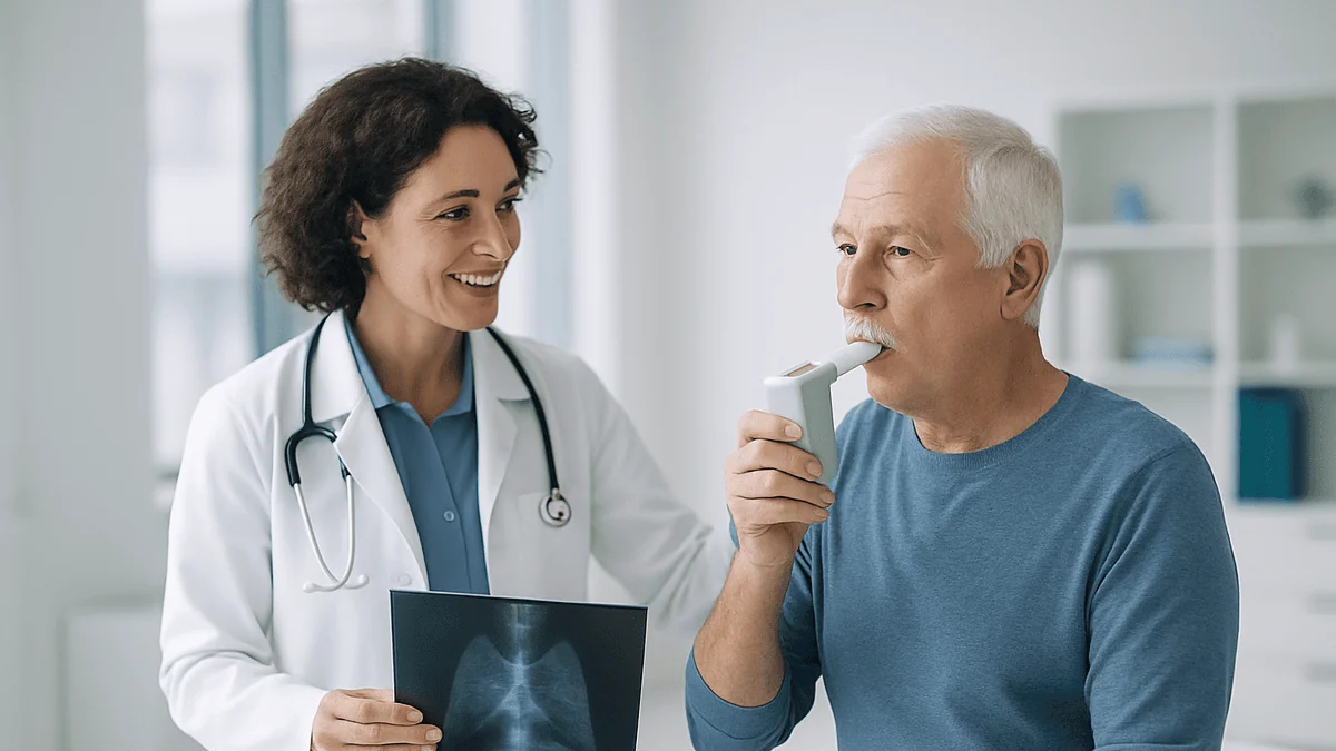
- Forced vital capacity and inspiratory pressure are measured at diagnosis to create a personal baseline, because respiratory muscles can weaken without noticeable breathlessness.
- In a randomized trial of 126 antibody-positive adults aged 18 to 65 without thymoma, thymectomy plus a corticosteroid reduced disease severity and steroid needs over three years compared with the corticosteroid alone.
- Facial weakness can leak air around a spirometry mouthpiece and falsely lower results, so mention it before testing and ask about a mask or cheek support.
Myasthenia gravis evaluation includes thymus imaging because the thymus gland, which sits behind the breastbone, is abnormal in many people with the condition and a chest CT or MRI can reveal a thymoma or an enlarged gland that changes treatment planning. Breathing tests are added because the same weakness can quietly affect the muscles that move air, and measuring lung volumes early gives the team a safety baseline.
The appointment was supposed to be about droopy eyelids. Instead, the neurologist mentioned a chest scan, then a nurse handed over a plastic mouthpiece and asked for the biggest breath possible. For someone whose main complaint is seeing double by dinnertime, that sequence can feel like the clinic changed the subject. It did not.
Myasthenia gravis thymus imaging and breathing tests are two of the least intuitive parts of the workup, and two of the most useful. One looks for a gland most adults have never thought about since biology class. The other measures a set of muscles that, in this condition, can weaken without anyone noticing until the numbers slip. Understanding why each test is ordered turns a confusing week of appointments into a plan with a logic behind it, and it helps you ask better questions when the results arrive.
How does the thymus affect myasthenia gravis?
The thymus is a small, soft gland tucked behind the breastbone, in the space between the lungs called the mediastinum. In childhood it is busy: it trains immune cells called T cells to tell the body’s own tissue from outside threats. After puberty it shrinks and much of it is gradually replaced by fat, which is why an adult thymus is often hard to pick out on a scan at all (Cleveland Clinic).
Myasthenia gravis is an autoimmune condition, meaning the immune system mistakenly attacks the body’s own tissue. In most people with the disease, antibodies (immune proteins) block or destroy acetylcholine receptors, the docking sites on muscle where a nerve signal is received. Fewer working receptors means weaker, more tiring contractions. Roughly 85 percent of people with generalized myasthenia gravis carry these acetylcholine receptor antibodies (Cleveland Clinic).
Where does the thymus fit? Researchers believe the gland is one place where the faulty immune instruction is written. In many people with the condition the thymus stays large and active into adulthood, a state called thymic hyperplasia, and it appears to contain the very cells that teach the immune system to target the receptor (NIH, NINDS). A smaller group has a thymoma, a tumor of the thymus that is usually slow growing but can occasionally be invasive. Published estimates put thymoma at about 10 to 15 percent of people with myasthenia gravis (Cleveland Clinic; NIH, NINDS).
That link works in the other direction too. Some people are found to have a thymoma first, on a scan done for another reason, and are only then assessed for the muscle weakness they had put down to stress or age. The gland and the disease are entangled, which is exactly why the gland gets its own picture.
What actually happens during myasthenia gravis thymus imaging
For most people the test is a CT scan of the chest, usually with an intravenous contrast dye that makes blood vessels and soft tissue easier to tell apart. You lie on a narrow table that slides through a ring-shaped scanner while you hold your breath for a few seconds at a time. The scanning itself takes minutes; the check-in, cannula placement and waiting take longer than the pictures.
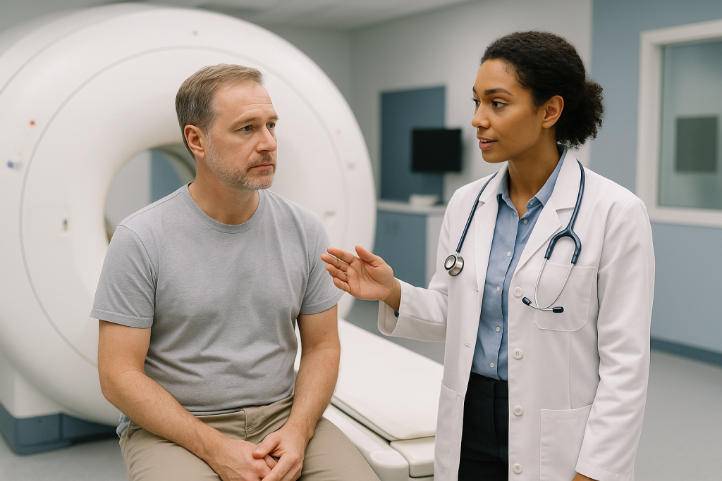
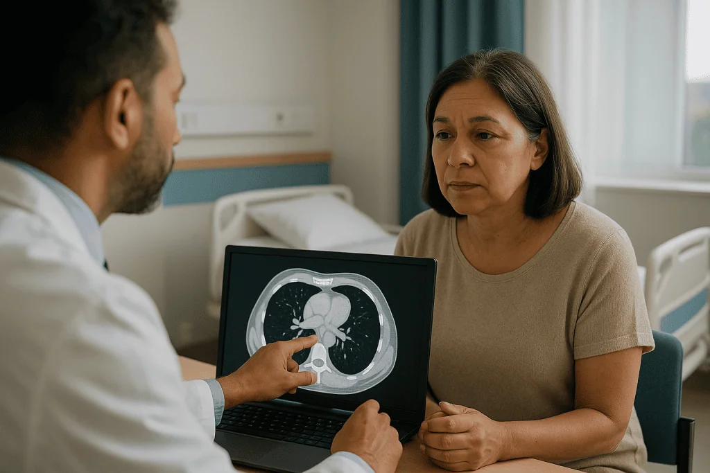
The radiologist is looking at a specific address. The thymus sits in the front part of the mediastinum, just behind the sternum and above the heart. On the images they assess whether there is any soft tissue where fat is expected, how big it is, whether its edges are smooth or irregular, and whether it stays within its own borders or seems to lean on neighboring structures such as the large veins or the lining of the lung. Those features, read together, help distinguish a normal involuted gland, a diffusely enlarged one, and a discrete mass (Mayo Clinic).
An MRI may be used instead of, or after, a CT. MRI uses magnets and radio waves rather than X-rays, involves a longer time in a tube-shaped scanner, and is noisier. It is often chosen when the CT finding is ambiguous, when contrast dye is a concern because of kidney function or allergy, or in younger people where avoiding radiation matters more (Mayo Clinic).
Preparation is light. You may be asked not to eat for a few hours before a contrast CT, to mention any kidney problems or past reactions to dye, and to remove metal for MRI. Tell the team if you have a pacemaker, implanted device or claustrophobia. Nothing about the scan itself treats the disease; it is a map-making exercise, and the map decides the next conversation.
Can myasthenia gravis be seen on imaging?
Not directly. Myasthenia gravis lives at the junction between nerve and muscle, at a scale far below what any hospital scanner resolves. A chest CT cannot show blocked receptors, and a brain MRI in this condition is typically normal. If a scan of the head is ordered, it is usually to rule out other explanations for double vision or facial weakness, such as a stroke or a nerve compression, not to confirm myasthenia (NHS).
What imaging can see is the thymus, and the thymus is where the condition often leaves a visible footprint. Two patterns matter:
- Thymic hyperplasia: the gland is larger or denser than expected for a person’s age, but keeps its normal two-lobed, arrowhead shape. This is the more common finding and is not a tumor.
- Thymoma: a distinct rounded or lobulated mass arising from the gland. Most are contained within a capsule; a minority spread into nearby tissue, which is why the radiologist comments on borders, not just size.
There is a real gray zone. A young adult’s normal thymus can look bulky, and a small thymoma can hide inside a gland that has partly turned to fat. That overlap is one reason the radiology literature on thymus imaging in this condition is so extensive: distinguishing the two on CT alone is not always possible, and MRI techniques that separate fat from cellular tissue are sometimes added to sharpen the call.
The honest summary is that imaging answers a narrower question than most people expect. It does not say whether you have myasthenia gravis; the antibody blood tests and nerve studies do that. It says what your thymus looks like, and that answer can move a person from one treatment pathway to another (NIH, NINDS).
CT scan for myasthenia gravis, or MRI? Which scan and why
People often ask why they had a CT while someone in an online group had an MRI. Both look at the same gland; they trade off different strengths. The table below summarizes the typical roles, though your team may reasonably choose differently based on your history.
| Feature | Chest CT with contrast | Chest MRI |
|---|---|---|
| What it uses | X-rays and, usually, iodine-based contrast dye | Magnets and radio waves; sometimes gadolinium contrast |
| Time in scanner | Minutes | Often longer; several breath-hold sequences |
| Typical first choice? | Yes, in most adults | Usually a second step or a substitute |
| Strength | Fast, widely available, shows the whole mediastinum and lungs clearly | Better at separating fat from cellular tissue, helpful for hyperplasia versus thymoma; no radiation |
| Common reasons to prefer it | Initial survey; assessing a mass’s relationship to vessels | Indeterminate CT; kidney concerns or dye allergy; younger patients; follow-up of a known gland |
| Limits | Radiation dose; overlap between a plump normal gland and hyperplasia | Longer, noisier, not suitable with some implants; motion can blur images |
Two practical points follow. First, a normal CT does not close the file forever; if symptoms change or antibody results shift the picture, a repeat or an MRI can be requested. Second, the report wording matters more than the modality. Phrases such as “prominent thymic tissue, likely hyperplasia” and “anterior mediastinal mass, thymoma not excluded” lead to very different next conversations, and it is fair to ask your neurologist to translate the exact sentence.
Occasionally a PET scan, which highlights metabolically active tissue, is added when a mass is suspected to be more aggressive. That is the exception rather than the routine, and it is a decision for the treating team based on the CT or MRI appearance (Mayo Clinic).
Why breathing tests belong in a nerve and muscle workup
The diaphragm and the muscles between the ribs are skeletal muscles, wired to nerves through the same acetylcholine receptors that the immune system is attacking. In a condition defined by muscles tiring with use, the muscles that never get to rest are a natural point of vulnerability. Yet breathing weakness is easy to miss. Unlike a drooping eyelid, it does not show up in a mirror, and people adapt: they talk in shorter sentences, sleep propped up, and describe themselves as “out of shape” rather than short of breath.
Clinicians care about this because of myasthenic crisis, the term for weakness severe enough that a person cannot breathe or clear secretions adequately without mechanical help. It is a medical emergency, and it can be triggered by infection, certain medicines, surgery, or simply an unpredictable flare (NIH, NINDS; Mayo Clinic). Crisis is uncommon relative to the whole population with the disease, but it is the outcome every myasthenia plan is built to prevent.
A breathing test at diagnosis therefore does three jobs at once:
- It establishes a personal baseline, so that a future number can be judged against your own normal rather than a population average.
- It can reveal early respiratory involvement in someone who reports no breathlessness at all.
- It provides an objective trigger point. Teams commonly agree in advance what fall in lung volume would prompt closer monitoring or hospital care, which takes guesswork out of a stressful moment.
The measurements are also repeated whenever the picture changes: before any planned operation, including thymus surgery, during a flare, or when a new medicine that can affect neuromuscular transmission is being considered. Think of the first test less as a diagnosis and more as calibrating an instrument you will use for years.
What a myasthenia gravis breathing test actually involves
Most people picture a treadmill. The reality is a chair, a nose clip and a technician who is unusually encouraging. The core test is spirometry, which measures how much air you can move. The number that matters most in this condition is forced vital capacity, or FVC: the total volume you can blow out after the deepest breath you can take. Because myasthenia weakens muscles rather than narrowing airways, FVC tends to fall while the airways themselves stay open, a pattern that distinguishes it from asthma or emphysema.
Two further measurements are often added. Maximal inspiratory pressure (sometimes called negative inspiratory force) records how hard you can suck in against a closed mouthpiece, which reflects diaphragm strength directly. Maximal expiratory pressure does the same for blowing out, which relates to how well you can cough. Each takes a few repeated efforts, and the best attempt is recorded.
Weak facial muscles complicate things. If your lips cannot seal around the mouthpiece, air leaks and the numbers read falsely low; technicians may use a cushioned mask or ask you to support your cheeks with your hands. Mention facial weakness before you start so the result is interpreted fairly.
At the bedside, when equipment is not to hand, clinicians sometimes use a single-breath count: take a full breath and count aloud steadily, and note how high you reach before needing another breath. It is a rough screen, not a substitute for spirometry, and its value lies in comparing your own counts over time rather than in any universal cutoff.
Results are reported against predicted values for your age, sex and height, and, more usefully, against your previous tests. A single reading below prediction is a prompt for attention; a downward trend across days is what changes management. Timing also matters: because weakness fluctuates, an afternoon test can read lower than a morning one, so teams try to repeat measurements at similar times (Mayo Clinic).
What is the gold standard for diagnosing myasthenia gravis?
There is no single one, and anyone who says otherwise is simplifying. Diagnosis rests on three legs that support each other: a clinical pattern, an antibody blood test, and an electrical study of the nerve-muscle junction (Mayo Clinic; NHS).
The clinical pattern is fatigable weakness, meaning strength that is reasonable after rest and drains with repeated use, then partly recovers. The eye muscles are involved first in many people, producing drooping lids and double vision that worsen by evening; in others the pattern extends to chewing, swallowing, speech, the neck, the limbs and breathing. In the clinic a neurologist may test this directly, for example by asking you to hold an upward gaze or repeatedly rise from a chair, and may perform an ice pack test, placing a cold pack on a drooping eyelid for a couple of minutes to see whether cooling temporarily restores lid height, which is characteristic of this condition (Mayo Clinic).
The antibody test is the most specific piece. A positive acetylcholine receptor antibody result in someone with a fitting story is close to conclusive. When that test is negative, laboratories look for antibodies to muscle-specific kinase (MuSK), a different protein at the same junction; a smaller group remains seronegative, meaning no measurable antibody is found even though the disease is present (Cleveland Clinic).
The electrical studies fill that gap. Repetitive nerve stimulation delivers a series of small shocks and watches whether the muscle response fades. Single-fiber electromyography, which records from individual muscle fibers with a very fine needle, is the most sensitive test available for a transmission problem, though it is less specific and requires a specialist to perform and interpret (Mayo Clinic; Cleveland Clinic).
Notice what is not on the list: thymus imaging and breathing tests. They do not diagnose the condition. They characterize it once the diagnosis is likely, which is precisely why they are ordered after, not instead of, the tests above.
Who is usually asked to have thymus imaging, and who may be asked to wait
Guideline-level practice is straightforward on the first point: almost everyone with newly diagnosed myasthenia gravis is offered chest imaging of the thymus, regardless of how mild the weakness seems (NHS; Mayo Clinic). The reason is that thymoma does not announce itself through symptoms; a person with eyelid-only disease can harbor one, and a person with severe generalized weakness can have an entirely fatty gland. Because the presence of a thymoma changes the treatment pathway, the scan is done up front.
Within that broad rule, timing and modality vary:
- Acetylcholine receptor antibody positive: imaging is a routine early step, since both hyperplasia and thymoma cluster in this group.
- MuSK antibody positive: thymic abnormalities are much less common, so imaging is still typically performed to exclude a mass, but thymus surgery is not usually pursued on the basis of the gland alone.
- Seronegative: imaging is generally done, partly because an unexpected thymoma would strengthen an uncertain diagnosis.
- Late-onset disease: the gland has often fully involuted, but thymoma remains possible at any age, so the scan is not skipped.
Who is asked to wait? Two situations come up. If someone is acutely unwell, with swallowing or breathing difficulty, stabilizing them takes precedence; lying flat for a contrast CT is uncomfortable and occasionally unsafe when the diaphragm is weak, so the scan is scheduled once they can tolerate it. And when a first CT shows a plump but normally shaped gland in a young adult, the team may choose to repeat imaging after an interval rather than act immediately, because a normal adolescent-pattern thymus and hyperplasia can look alike (Mayo Clinic).
Pregnancy is a special case: CT is usually deferred or replaced with MRI without contrast, and the decision is shared between the neurology and obstetric teams. In every scenario the order of tests belongs to the treating clinicians, who weigh the value of the information against the timing that keeps you safe.
Does an abnormal thymus scan mean surgery? Thymoma and myasthenia gravis
Not automatically, and the two findings lead to different reasoning.
When imaging shows a thymoma, removal of the gland, an operation called thymectomy, is generally recommended for the tumor itself, whether or not it improves the muscle disease. Thymomas can enlarge and, in a minority of cases, invade surrounding structures, and complete removal allows the tissue to be examined under a microscope and staged. The surgical approach, timing, and any additional treatment are decisions for a thoracic surgeon working alongside the neurologist, and they depend on the mass’s size and relationship to nearby vessels (Mayo Clinic; Cleveland Clinic).
When imaging shows hyperplasia or an unremarkable gland, thymectomy becomes a choice rather than a necessity, and the evidence is worth stating plainly. A randomized trial that followed 126 adults aged 18 to 65 with acetylcholine receptor antibody positive, non-thymomatous generalized myasthenia gravis found that, over three years, those who had thymectomy in addition to a corticosteroid had lower average disease severity scores and needed less of the steroid than those treated with the steroid alone (NIH, PubMed). That is a benefit, but a gradual one, measured in months and years rather than weeks, and it was studied in a defined group: people outside that age range, those with ocular-only disease, and those with MuSK antibodies were not part of the trial, so the evidence for them is weaker (NIH, NINDS).
Alternatives exist. Many people are managed with medicines alone, particularly when weakness is mild, when antibodies are of the MuSK type, or when age or other health problems make surgery less attractive. Risks of thymectomy are those of chest surgery in general, including bleeding, infection, and a temporary worsening of weakness around the operation, which is one of the reasons breathing tests are repeated beforehand.
The scan opens the door to this conversation. It does not walk through it for you; that is done with your care team, ideally with time to think.
How imaging and breathing results shape the treatment conversation
Once the thymus picture and the lung numbers are on the table, the treatment discussion usually organizes itself around three questions: how much weakness is there, how quickly does it need to be controlled, and is there a structural reason to operate. Medicines are chosen by mechanism, and it helps to know the categories without needing to know any doses, which remain entirely the prescriber’s domain.
Acetylcholinesterase inhibitors slow the breakdown of acetylcholine at the junction, so more of the chemical messenger is available to the receptors that remain. They tend to act within the hour and wear off within hours, which makes them a symptom-relief tool rather than a treatment of the immune process (NHS; Mayo Clinic).
Immunosuppressive therapies act on the underlying attack. Corticosteroids dampen immune activity relatively quickly, typically over weeks; steroid-sparing agents such as certain antimetabolite drugs work more slowly, often taking several months to show their full effect, which is why they are frequently started alongside a faster agent (Mayo Clinic). Newer targeted therapies, including antibodies that block complement, a cascade of immune proteins that damages the receptor region, or that lower circulating antibody levels, are options in specific situations defined by antibody type and disease severity.
For rapid control, two approaches work within days: intravenous immunoglobulin, a pooled antibody preparation that appears to blunt the harmful antibodies’ effects, and plasma exchange, which filters antibodies out of the blood. Both are temporary and are typically used for a crisis, a severe flare, or to strengthen someone before surgery (NIH, NINDS).
Where do the tests fit? A thymoma pushes surgery up the list. Hyperplasia in a younger, antibody-positive adult makes thymectomy a reasonable discussion. Low or falling breathing numbers argue for faster-acting treatment and closer monitoring, and against elective procedures until strength improves. None of these are rules a patient applies alone; they are the inputs your neurologist weighs when suggesting a plan, and they are the numbers worth understanding when you agree to it.
What the following days and weeks usually look like
The rhythm after these tests is more waiting than doing, which surprises people who expected a chest scan to bring an instant verdict.
Imaging reports are typically available within a few days, though complex findings may be reviewed at a multidisciplinary meeting where radiologists, neurologists and thoracic surgeons look at the pictures together, which can add a week or two. If the report is indeterminate, a follow-up MRI or a repeat scan after an interval may be scheduled rather than an immediate decision. Ask when and how you will hear the result, and who will explain it.
Breathing test results are usually discussed the same day. If your numbers are in a comfortable range, you may simply be given a baseline and told when they will be rechecked. If they are lower than expected, expect a more active plan: earlier follow-up, instructions about what changes should prompt a call, and sometimes a hospital stay for monitoring while treatment takes hold.
Medicines follow their own timelines. Symptom-relief drugs act quickly, so many people notice a difference in daily function within days of starting them; immune-directed treatments are judged over weeks to months, and clinicians generally warn against reading too much into the first fortnight (Mayo Clinic; NHS). If a steroid is started, some people experience a temporary dip in strength in the first weeks before improvement, which is one reason initiation is sometimes done under observation.
If thymectomy is planned for a non-urgent reason such as hyperplasia, it is usually timed for when weakness is well controlled and breathing tests are reassuring, which can mean weeks or months of optimization first. After surgery, any effect on the myasthenia itself is expected to unfold slowly, over months and into the following years, rather than at the first outpatient review (NIH, PubMed).
The practical advice for this stretch is unglamorous: keep a simple diary of good and bad times of day, note infections or new prescriptions from any clinician, and bring it to appointments. Patterns in that notebook often steer decisions as much as the scan does.
What people often get wrong about the thymus and myasthenia gravis
“If the scan is normal, the thymus is not involved.” Hyperplasia can be microscopic. Glands that look ordinary on CT have been found to contain the immune structures associated with the disease when examined after removal. A normal-looking scan rules out a visible mass; it does not rule the gland innocent.
“A thymoma means cancer in the usual sense.” Thymomas are tumors and are treated seriously, but most are slow growing and contained within a capsule. The distinction between a non-invasive thymoma and the rarer, more aggressive thymic carcinoma is made by pathologists after removal, not by the word “thymoma” on a radiology report (Cleveland Clinic).
“Removing the thymus makes the disease go away.” The best trial evidence shows a gradual reduction in severity and in steroid requirement over three years in a defined group of patients, not a return to a pre-disease state for everyone (NIH, PubMed). Some people improve a great deal, some modestly, and the effect takes time.
“If I can climb stairs, my breathing must be fine.” Limb and respiratory muscles can weaken out of step with each other. The reason spirometry is done in people who feel fine is precisely that feeling fine is not a reliable guide in this condition.
“The disease will show on a brain MRI.” It will not. Head imaging, when done, is to exclude other causes of similar symptoms (NHS).
“Adults do not have a thymus.” They do; it has mostly turned to fat, but the tissue persists and can still be the site of a tumor at any age, which is why late-onset patients are scanned too.
“Once diagnosed, I will get weaker every year.” The course is variable. Many people reach a stable, well-controlled state with treatment, and symptoms in some fluctuate rather than steadily progress (NIH, NINDS). The tests described here exist to steer that course, not to predict a decline.
Questions to ask your care team
A good consultation leaves you knowing not just what was ordered but why, and what each result will change. These prompts are meant to be adapted, not read out as a script.
- What did the thymus report actually say, and is the team confident it distinguishes hyperplasia from thymoma? If not, what is the next step to tell them apart?
- Which antibody was found, if any, and how does that change what the thymus findings mean for me?
- Based on my scan and my age, is thymectomy something you would raise now, later, or not at all? What evidence applies to someone in my situation?
- What were my forced vital capacity and inspiratory pressure numbers, and what values would prompt you to want to see me urgently?
- How often will breathing tests be repeated, and should I learn a home check such as the single-breath count?
- Which of my current medicines, including over-the-counter and antibiotic prescriptions from other doctors, could worsen neuromuscular weakness, and how should I flag this to other clinicians?
- If I develop an infection or need an operation for an unrelated reason, what should happen differently because of this diagnosis?
- Who is my point of contact between appointments, and what is the out-of-hours route if swallowing or breathing worsens?
- Is there anything in my scan unrelated to the thymus that needs follow-up?
- What would you consider a good response to treatment at three months, and at a year?
Bring someone with you if you can. Fatigue, including in the muscles used for speech, makes long consultations harder, and a second set of ears catches details that matter later. Writing down the exact report wording and your baseline numbers gives you a reference point that no amount of searching online can replace, because it is about you.
When to call your doctor
Most of this condition is managed in clinics, on a timescale of weeks. A small number of situations are not, and recognizing them quickly is the most important safety skill a person with myasthenia gravis and their household can have.
Seek emergency care immediately, by calling emergency services rather than driving to a clinic, if any of the following develop (NIH, NINDS; Mayo Clinic):
- Difficulty breathing, breathlessness at rest, or needing to sit upright to breathe.
- Trouble swallowing saliva, choking on liquids, or a new inability to clear secretions with a cough.
- Speech that becomes rapidly slurred, nasal, or too weak to be heard.
- Weakness that is worsening hour by hour rather than day by day.
- A single-breath count or home spirometry reading that has dropped sharply from your usual, especially alongside any of the above.
Contact your neurology team the same day, or the on-call service outside clinic hours, if weakness is clearly worse than your recent baseline, if you have developed a fever or infection, if a new medicine has been started by another prescriber, or if double vision or drooping has spread to new muscles. Infections and certain antibiotics, cardiac and anesthetic drugs are recognized triggers of flares, and early adjustment by the treating team is far preferable to waiting for a crisis (NHS).
After a thymus scan, call if you develop a rash, swelling or breathing difficulty within hours of contrast dye, which can indicate an allergic reaction. After thymectomy, report increasing chest pain, fever, wound redness or discharge, or any return of breathing or swallowing weakness.
A practical rule some teams suggest: if you are debating whether a change is serious enough to call about, that debate is itself a reason to call. Reassurance costs little; a missed crisis costs a great deal. Every one of these decisions, from adjusting treatment to admitting you for monitoring, rests with the clinicians who know your history, and the tests described in this article are what give them the numbers to decide well.
Frequently asked questions
How does the thymus affect myasthenia gravis?
The thymus is thought to be one place where the immune system learns to attack acetylcholine receptors on muscle. In many people with the condition the gland remains enlarged and active into adulthood, a state called hyperplasia, and it contains cells involved in producing the harmful antibodies. A smaller group has a thymoma, a tumor of the gland. Both findings help explain why the gland is scanned and sometimes removed.
What is the gold standard for diagnosing myasthenia gravis?
There is no single gold standard. Diagnosis combines a characteristic pattern of weakness that worsens with use, a blood test for acetylcholine receptor or MuSK antibodies, and electrical studies such as repetitive nerve stimulation or single-fiber EMG. A positive antibody result with a fitting history is highly specific; electrical studies are most useful when antibodies are negative. Thymus imaging and breathing tests characterize the disease rather than confirm it.
What is the hallmark symptom of myasthenia gravis?
Fatigable weakness is the hallmark: strength that is reasonable after rest and drains with repeated use, then partly recovers. It often affects the eye muscles first, causing drooping eyelids or double vision that worsens by evening, and may involve chewing, swallowing, speech, limb or breathing muscles. Because many conditions cause tiredness, this pattern needs formal testing by a clinician rather than self-assessment.
Can myasthenia gravis be seen on imaging?
Not directly. The disease occurs at the microscopic junction between nerve and muscle, far below the resolution of CT or MRI, and brain scans are usually normal. What imaging can show is the thymus gland, which may be enlarged or contain a thymoma in people with the condition. A chest scan therefore answers a different question from the antibody and nerve tests that establish the diagnosis.
What does a CT scan for myasthenia gravis look for?
It examines the front of the mediastinum, the space behind the breastbone, for thymic tissue. The radiologist notes whether soft tissue is present where fat is expected, its size and shape, whether edges are smooth or irregular, and whether it touches nearby vessels or the lung lining. These features help separate a normal involuted gland, diffuse hyperplasia, and a discrete thymoma, though some cases remain ambiguous and need MRI or follow-up.
Is thymic hyperplasia in myasthenia gravis dangerous?
Hyperplasia is not a tumor and does not spread. It describes a gland that is larger or more active than expected for age, and it is the more common thymic finding in younger antibody-positive people. Its significance lies in treatment planning: trial evidence suggests thymectomy can gradually reduce disease severity in selected adults with non-thymomatous disease. Whether surgery is worthwhile for an individual is a decision for the neurology and surgical teams.
What is a myasthenia gravis breathing test?
It is usually spirometry, measuring forced vital capacity, the total air you can blow out after a maximal breath, together with maximal inspiratory and expiratory pressures that reflect diaphragm and cough strength. Results are compared with predicted values and, more importantly, with your own previous tests. A falling trend is more meaningful than any single number, and repeat testing is common before surgery or during a flare.
Why do I need breathing tests if I am not short of breath?
Respiratory muscles can weaken out of step with limb or eye muscles, and people often adapt without noticing. Testing at diagnosis provides a personal baseline so that later changes can be judged accurately, and it can reveal early involvement that would otherwise go undetected until a crisis. It also gives your team an agreed objective threshold for closer monitoring, which removes guesswork from a stressful situation.
What is the connection between thymoma and myasthenia gravis?
About 10 to 15 percent of people with myasthenia gravis have a thymoma, and a proportion of people found to have a thymoma for other reasons turn out to have the muscle condition. When a thymoma is present, surgical removal is generally recommended for the tumor itself, regardless of its effect on weakness, and the tissue is examined to determine whether it is contained or invasive.
Does thymus removal treat myasthenia gravis?
It can help in selected people, but slowly and not universally. In a randomized trial of adults aged 18 to 65 with acetylcholine receptor antibodies and no thymoma, thymectomy plus a corticosteroid produced lower disease severity and lower steroid requirements over three years than the steroid alone. Benefits emerge over months to years, and the evidence is weaker for ocular-only disease, MuSK antibodies, and older adults.
References
- Myasthenia Gravis, National Institute of Neurological Disorders and Stroke (NIH)
- Myasthenia Gravis (MG), Cleveland Clinic
- Myasthenia gravis, NHS
- Myasthenia Gravis, MedlinePlus
This article is for general information only and is not a substitute for professional medical advice. Please consult a qualified doctor about your individual situation.

More from the Blog

How Long Before Preventive Migraine Treatment Shows Results? What Follow-Up Visits Assess
Preventive migraine treatment usually needs a fair trial before anyone can judge it. Most oral preventives are assessed after roughly two to three months…

Trigeminal Neuralgia Surgery Recovery: The First Days, Incision Care and Returning to Work
Recovery after microvascular decompression usually means a hospital stay of a few days, then several weeks at home while a small incision behind the…

What to Gather Before a Motor Neuron Disease Consultation: EMG Results, Scans and Medications
Before a motor neuron disease consultation, gather the actual EMG and nerve conduction reports (not just the summary letter), any MRI of the brain…

Flying Home After a Craniotomy: How Long International Patients Usually Stay and Why
Most surgical teams ask patients to avoid flying after craniotomy until a follow-up scan shows any trapped air has cleared, the wound has healed,…

Cerebrovascular Disease Follow-Up: What Is Normal and When to Call Your Neurology Team
After a stroke or TIA, lingering fatigue, mild headaches that ease with rest and slow, uneven improvement in strength or speech are usually part…
Multiple Sclerosis and Pregnancy Planning: How Neurologists Adjust Treatment Around It
Multiple sclerosis and pregnancy planning usually starts months before conception. Neurologists review which disease-modifying therapy someone takes, because some are continued in pregnancy while…