

Why New Moles Appear Suddenly: Normal Causes, Warning Signs and When to Get Checked

Key Takeaways
- A typical adult has between 10 and 40 moles, and most of them appear by around age 40, new spots in young adulthood are usually your genes running on schedule.
- Ultraviolet exposure is the biggest controllable driver of new moles, and sun protection in childhood pays the largest dividends decades later.
- Hormonal surges during puberty and pregnancy commonly trigger new moles and darken existing ones, though a single asymmetric or fast-changing spot still needs checking.
- Many 'new moles' after age 50 are actually seborrheic keratoses, sun spots, or cherry angiomas, benign growths that multiply with age and get mislabeled in everyday speech.
- Early melanoma almost never causes symptoms you can feel; visible change on the skin: the 'E for evolving' in ABCDE, is the signal that matters most.
- Home mole-removal pastes and 'one-day' hacks cause scarring and can destroy the very tissue a pathologist needs to rule out cancer, removal belongs in a clinic.
New moles usually appear because of a mix of inherited genes, cumulative sun exposure, and hormonal shifts such as puberty or pregnancy. Most people keep developing moles into their 30s and 40s, and the vast majority are harmless. A truly new or fast-changing spot after age 40, or one that looks unlike your others, deserves a professional skin exam.
It happens in ordinary moments: you’re rolling up a sleeve in September and there it is: a small brown dot on your forearm that you’d swear wasn’t there in June. Your first instinct is to search your memory. Your second, often around midnight, is to search the internet, where the answers range from “totally normal” to genuinely alarming.
Here’s the honest middle ground. Skin is a living organ that keeps remodeling itself for decades, and pigment cells are part of that story. Most new moles are the skin doing something unremarkable, on a schedule set largely by your DNA and your lifetime tally of sunny afternoons.
But “most” is not “all,” and the difference matters. What follows is what the evidence actually says about why fresh spots show up, which ones can be safely watched, and which ones have earned a spot on a doctor’s calendar.
Is it normal for moles to show up out of nowhere?
Through childhood, the teenage years, and well into adulthood, yes, it’s normal. The Mayo Clinic notes that most moles develop by around age 40, and a typical adult carries somewhere between 10 and 40 of them scattered across the body. Your skin doesn’t hand you that collection at birth; it builds it gradually, one spot at a time, over roughly four decades.
A useful distinction hides inside the word “suddenly.” A true mole takes weeks to months to become visible as pigment cells slowly multiply and cluster. What feels sudden is usually sudden noticingthe spot sat quietly on the back of a shoulder or behind a knee until a mirror, a partner, or a haircut brought it to your attention. Genuinely overnight arrivals are more often something else entirely: a tiny bruise or blood blister, an irritated hair follicle, a splinter of dried blood under the skin, or a seborrheic keratosis you’ve only just registered.
Context changes the math, though. New pigmented spots are common and expected at 19, less common at 45, and worth a closer look at 65, not because age makes every spot sinister, but because the ordinary mole-making phase of life has largely wound down by then. The same brown dot carries different meaning at different ages, which is why the rest of this article keeps circling back to that timeline.
What a mole actually is, and how one forms
Every square inch of your skin contains melanocytes, the cells that manufacture melanin, the pigment that gives skin its color. Normally these cells sit spread out like fence posts along the base of the epidermis, quietly passing pigment to their neighbors. A mole, a melanocytic nevus, in clinical language, forms when a group of melanocytes stops spacing out and instead grows in a tight cluster, or “nest,” as Cleveland Clinic describes it. Concentrated pigment in a concentrated spot: that’s the brown dot you see.
Moles also have a life cycle, which surprises many people. A young mole often starts flat and evenly brown, with the cell nests sitting at the junction of the epidermis and dermis. Over years, those nests can migrate deeper into the dermis, and the mole may become raised, softer, and sometimes paler, even flesh-colored. Some eventually shrink and disappear altogether in later life. That slow, decades-long arc is normal biology, not a warning sign.
What flips a spread-out melanocyte into a nest-builder? Three forces do most of the work: the genetic instructions you inherited, ultraviolet light acting on skin cells over time, and hormonal signals that can nudge melanocytes into an active phase. Each deserves its own explanation, because each answers a different version of the question “why now?”
Sunlight is the biggest driver you can actually control
Ultraviolet radiation does two things to melanocytes. In the short term it tells them to produce more pigment: that’s a tan, which is skin damage wearing a flattering disguise. Over the longer term, UV exposure encourages melanocytes to proliferate and can damage their DNA, and both effects favor mole formation. MedlinePlus and Mayo Clinic list sun exposure among the established contributors to new moles, and dermatology research has consistently found that children who get more sun, especially intense, intermittent doses that end in sunburn, grow up with higher mole counts.
You can often read the pattern on your own body. Moles tend to concentrate on sun-exposed real estate: forearms, upper back, shoulders, calves, the face. A new spot appearing on your shoulder the autumn after a beach-heavy summer is not a coincidence; it’s frequently a delayed receipt for UV already spent. Tanning beds run the same biological tab, only faster and more intensely: the CDC classifies indoor tanning as a source of skin-damaging UV with no safe threshold.
One honest caveat: sun exposure explains a lot, but not everything. Moles also turn up on skin that rarely sees daylight: the scalp under thick hair, the buttocks, between toes. Genetics can seed melanocyte nests anywhere. So while covering up genuinely slows new mole formation, nobody should feel they failed at sunscreen because a spot appeared under a waistband.
Hormones can wake melanocytes up
Melanocytes carry receptors that respond to hormonal signals, which is why the body’s big hormonal chapters often show up on the skin. Puberty is the classic example: teenagers routinely sprout new moles and watch existing ones darken as hormone levels climb. The NHS and Cleveland Clinic both note that moles commonly appear or change during these years, and it’s considered part of normal development rather than a red flag.
Pregnancy is the other headline event. Rising hormone levels stimulate pigment production broadly, many pregnant people notice darker nipples, a dark line down the abdomen, and moles that deepen in color. Some moles also appear for the first time during pregnancy. Mild, uniform darkening across many moles is generally expected. What the evidence does not support is the old reassurance that “pregnancy changes everything, so ignore it”: a single mole that changes shape, grows asymmetrically, or develops irregular borders during pregnancy still deserves the same evaluation it would get at any other time.
Smaller hormonal shifts, starting or stopping hormone-based medications, thyroid changes, menopause, are sometimes blamed for new moles, and the mechanism is plausible, but the evidence here is thinner and less consistent. If you notice a cluster of new spots after a medication change, mention it to your prescribing clinician rather than assuming a connection either way. That’s a case where saying “the evidence is uncertain” is simply the truthful answer.
Genetics deal the hand before the sun ever plays it
Look at your parents’ arms and you’ll get a rough preview of your own. How many moles a person develops is strongly heritable, twin studies have shown that mole count tracks closely with shared genes, and families visibly cluster into “many moles” and “few moles” camps. If a new spot appears on you at 28, and your mother collected moles steadily through her 30s, you’re most likely watching an inherited program run on schedule.
Some of that programming is set before birth. Congenital moles, present at delivery or emerging in the first months of life, occur in roughly 1 in 100 newborns, according to Cleveland Clinic. The rest of the genetic influence unfolds slowly, determining not just how many moles you’ll grow but where they cluster, how dark they run, and how your melanocytes respond when UV comes calling. Genes load the dice; sunlight rolls them.
A small subset of families carries a pattern doctors watch more carefully: large numbers of moles, many of them atypical in appearance, alongside relatives who have had melanoma. People from these families benefit from regular professional skin exams and often from baseline photography, because their starting risk is genuinely higher. If melanoma runs in your close family and you have many unusual-looking moles, that combination is worth raising with a doctor even if nothing is currently changing.
Why do you get more moles as you get older, until you don't?
The trajectory is more interesting than a straight line. Mole counts climb through childhood and adolescence, keep rising through the 20s and 30s, and typically peak around the fourth decade: Mayo Clinic puts the arrival of most moles before age 40. After that, something counterintuitive happens: the number often starts to fall. Moles gradually lose pigment, flatten or soften, and some vanish entirely, so a person in their 70s may carry noticeably fewer moles than they did at 35. The NHS notes this slow fading in later adulthood as a normal pattern.
Why the decline? Melanocyte nests aren’t permanent structures. Over decades, the cells within a mole can descend deeper into the dermis, mature, and eventually stop producing pigment. Think of moles less like tattoos and more like long-running performances that eventually close.
Here’s where language trips people up. Many adults say they’re “getting more moles” with age when what’s actually multiplying is a different cast of characters: seborrheic keratoses (waxy, stuck-on-looking growths), solar lentigines (flat sun spots, often miscalled liver spots), cherry angiomas (small red dots), and skin tags. All of these become more common with age, all are benign, and all get lumped under “moles” in everyday speech. So the honest answer to “why more moles as I get older?” is often: those newcomers aren’t moles at all, which is reassuring, but only after someone qualified has confirmed what they are.
A new mole after 40 deserves more scrutiny, here's why
Nothing magical happens on a 40th birthday, but the statistics quietly shift. Because the skin’s natural mole-making phase largely concludes by that age, a genuinely new pigmented spot in midlife or beyond is swimming against the biological current. Most such spots still turn out to be benign, often a seborrheic keratosis or a lentigo rather than a true mole, but the probability that a new pigmented lesion is something that needs attention rises with each decade.
There’s a second reason doctors lean in. Melanoma, the most serious skin cancer, does not always grow out of an existing mole; a substantial share arises on previously unmarked skin as a brand-new spot. That’s precisely why the “E” in the ABCDE checklist, evolving, includes newness itself. A fresh dark spot at 55 isn’t automatically worrisome, but it has skipped the reassuring backstory that a 20-year-old mole carries.
The practical takeaway is proportionate, not fearful. Under 40, a new mole that looks like your other moles and behaves calmly can usually be watched. Over 40, the threshold for showing a new pigmented spot to a clinician should be lower, especially if you have fair skin, a history of sunburns, many existing moles, or a family history of melanoma. One short appointment, often with a magnified look through a dermatoscope, settles the question far better than months of mirror-checking and worry.
That new 'mole' might not be a mole at all
Skin produces a whole gallery of small growths, and to an untrained eye most of them read as “mole.” Learning the look-alikes does two useful things: it explains why spots seem to multiply with age, and it sharpens your eye for the changes that genuinely matter.
- Seborrheic keratosisthe great impersonator of later adulthood. Waxy, slightly warty, tan to nearly black, with a distinctive “pasted-on” appearance, as if you could flick it off with a fingernail. Harmless, extremely common after 50, and frequently mistaken for a fast-growing mole.
- Solar lentigoa flat, evenly brown patch on sun-exposed skin: backs of hands, face, shoulders. It’s a sun-damage souvenir, not a melanocyte nest, though a changing one still merits a look.
- Skin taga soft, flesh-colored flap on a tiny stalk, favoring the neck, armpits, and skin folds. Annoying where clothing rubs; medically insignificant.
- Cherry angiomaa small, bright-red dome made of dilated blood vessels. These accumulate steadily from middle age onward and are benign.
- Dermatofibromaa firm, brownish bump, often on the legs, that dimples inward when pinched. Frequently the ghost of an old insect bite or minor injury.
The catch: melanoma can occasionally mimic the mimics, particularly a dark seborrheic keratosis. Pattern recognition is genuinely difficult, even for clinicians without magnification. Use this list to understand your skin, not to self-clear a spot that’s new, dark, or changing.
The ABCDE check: how to read a new spot like a clinician
Doctors screen pigmented spots with a five-letter checklist, and it works just as well at home. No single letter is a verdict; the checklist flags spots that deserve professional eyes, and “E” carries extra weight for anything new.
| Letter | What to look for | Why it matters |
|---|---|---|
| A: Asymmetry | One half doesn’t match the other in shape | Benign moles usually grow evenly from the center outward |
| B: Border | Ragged, notched, or blurred edges | Irregular borders suggest uneven, disorganized cell growth |
| C: Color | Multiple shades, brown, black, red, white, or blue, in one spot | Healthy moles tend to be a single, uniform color |
| D: Diameter | Larger than about 6 mm: a pencil eraser | Size alone isn’t decisive, but bigger spots earn closer review |
| E: Evolving | Any change in size, shape, color, height, or a spot that is simply new | Change over weeks to months is the single most telling sign |
Add one more tool the checklist doesn’t capture: the “ugly duckling” sign. Most of a person’s moles resemble one another, like siblings. A spot that looks like it wandered in from another family, darker, larger, or just different from your personal pattern, warrants attention even if it passes every ABCDE letter. Mayo Clinic and the CDC both emphasize evolution and outlier appearance as the features most worth acting on.
Atypical moles: unusual doesn't mean dangerous
Somewhere between a textbook mole and a worrying lesion sits the atypical mole, also called a dysplastic nevus. These run larger than average, often bigger than that 6 mm eraser benchmark, with borders that fade into surrounding skin rather than stopping cleanly, and color that shifts from tan at the edges to darker brown at the center. Some people have one; some have dozens, a pattern that tends to run in families.
Two facts about atypical moles are worth holding at the same time, because clinics spend a lot of energy correcting the popular misreading of each. First: the overwhelming majority of atypical moles never become anything. They are not “pre-melanomas” ticking toward transformation, and routinely removing all of them doesn’t eliminate risk, melanoma can still arise on unmarked skin. Second: having many atypical moles is a genuine marker of elevated melanoma risk for the whole person, not just for those particular spots. Mayo Clinic lists numerous or unusual moles among the established risk factors.
The practical consequence is surveillance rather than surgery. People with multiple atypical moles benefit from periodic professional skin exams, and many clinicians use whole-body photography so that next year’s exam compares against this year’s map instead of memory. If you suspect your moles fit this description, large, fuzzy-edged, multi-toned, numerous, that’s a reasonable trigger for a baseline appointment, even with nothing actively changing.
How do you stop getting moles? What actually works
Start with the honest part: you cannot stop new moles entirely. The genetic share of mole formation is not negotiable, and no cream, supplement, or diet has evidence behind it for preventing melanocyte nests. Anyone selling mole prevention in a bottle is selling past the science.
The UV share, however, is very much negotiable, and it’s substantial. The CDC’s sun-safety guidance maps directly onto slowing new mole formation, because the same UV exposure drives both moles and skin cancer risk:
- Seek shade during peak UV hours, roughly 10 a.m. to 4 p.m., when the sun’s rays do the most damage.
- Use broad-spectrum sunscreen with SPF 30 or higher on exposed skin, reapplying after swimming, sweating, or about every two hours outdoors.
- Let clothing do the heavy lifting: long sleeves, a wide-brimmed hat, and UV-blocking sunglasses outperform sunscreen you forgot to reapply.
- Skip tanning beds entirely, indoor UV carries the same risks as outdoor UV, concentrated.
One finding deserves special emphasis for parents: childhood is when sun protection buys the most. Mole counts are heavily shaped by UV exposure in the first two decades of life, and blistering sunburns in childhood are a well-documented risk factor for melanoma decades later. Protecting a seven-year-old’s shoulders this July is, quite literally, editing the skin they’ll examine at fifty. For adults, the payoff is smaller but real, fewer new spots, less photoaging, and lower cancer risk, all from the same habits.
When to see a doctor: red flags that shouldn't wait
Most new moles can be watched calmly. A short list of findings should skip the watching phase and go straight to an appointment. See a doctor promptly if a mole or new spot is changing noticeably over weeks to a few monthsgrowing, darkening, developing new colors, or changing shape; if it bleeds, crusts, itches persistently, becomes painful, or won’t heal; if it’s a clear ugly duckling that looks unlike everything else on your skin; if it’s a brand-new pigmented spot after age 40; or if you notice a new dark streak under a fingernail or toenail, or a dark spot on a palm or solelocations where melanoma is rarer but easily missed, particularly in people with darker skin tones. The NHS advises getting any mole checked if it changes, and that advice is deliberately low-threshold: clinicians would far rather examine ten benign spots than miss one that mattered.
A word about the search phrase “signs your body is fighting melanoma”: early melanoma almost never announces itself through how you feel. There’s no characteristic fatigue, fever, or ache in the early stages: the skin itself is the signal, which is exactly why visual checks carry so much weight. Occasionally the immune system does leave visible fingerprints, such as a pale halo forming around a mole or a mole fading unevenly; a halo around an otherwise normal mole is usually benign, but irregular fading within a changing spot should be examined. When in doubt, the rule is simple and boring: changing spot, timely appointment.
What actually happens at a skin check
Knowing the process removes most of the dread, because the process is brief and painless. A clinician will ask when you noticed the spot, whether it has changed, and about your sun history and family history. Then comes the dermatoscope: a handheld magnifier with polarized light that reveals pigment patterns invisible to the naked eye. In experienced hands, dermoscopy sorts the clearly benign from the genuinely uncertain within minutes, and most new-mole appointments end right there, with reassurance.
If a spot lands in the uncertain zone, the next step is usually one of two things. Some lesions are photographed and rechecked after a set interval, so any evolution is documented rather than remembered. Others go to biopsy: after numbing the area with a local anesthetic, the clinician removes all or part of the spot and sends it to a pathology lab, where the tissue is examined under a microscope. That microscopic reading, not the visual impression, and certainly not a phone photo, is what actually determines whether a lesion is benign. Result timelines vary by lab and case, so ask your clinician what to expect and how you’ll be notified.
Two useful things to bring: a list or phone photos of any spots you’re worried about (they hide efficiently once you’re in a gown), and honesty about tanning-bed use and past sunburns. Neither earns a lecture; both sharpen the risk assessment.
Skip the one-day mole removal hacks
Search results for new moles sit uncomfortably close to pages promising natural removal “in one day”, pastes, acids, threads, and creams sold for burning or strangling moles off at home. The evidence-based verdict is blunt: don’t. These methods have no place in reputable medical guidance, and the problems they cause are well documented in the dermatology literature.
The first problem is cosmetic. Caustic “mole removal” products don’t distinguish mole from surrounding skin; they burn both, and the frequent result is a scar larger and more noticeable than the spot it replaced. The second problem is medical, and it’s the serious one: destroying a pigmented lesion at home destroys the tissue a pathologist would have examined. If that spot happened to be an early melanoma, home removal can delay diagnosis while abnormal cells remain beneath the scar: the worst possible trade, swapping a five-minute biopsy for months of false reassurance.
Legitimate mole removal exists, and it’s straightforward: a clinician numbs the area, removes the mole by shave or excision, and sends the tissue for microscopic review. The NHS notes that harmless moles generally don’t need removal at all, though people choose it for irritation, a mole that catches on a collar or razor, or for cosmetic reasons. Whether removal makes sense, and by which technique, is a decision for the examining clinician. What’s not negotiable is the pathology step; every removed mole deserves a look under the microscope.
Frequently asked questions
Is it normal for moles to show up out of nowhere?
Yes, especially before age 40, most people keep developing moles from childhood through their 30s, and a typical adult ends up with 10 to 40 of them. What feels like ‘out of nowhere’ is usually a spot that grew slowly over weeks and was only just noticed. A genuinely new pigmented spot in midlife or later is less expected and worth showing to a doctor, even though most still turn out benign.
Why do you get more moles as you get older?
Mole counts naturally climb through the 20s and 30s and peak around age 40, driven by genetics, cumulative sun exposure, and hormonal phases. After that, true moles often fade or disappear. Many spots that appear later in life aren’t moles at all, seborrheic keratoses, sun spots, skin tags, and cherry angiomas all multiply with age, are benign, and get called ‘moles’ in everyday conversation.
How do you stop getting moles?
You can’t prevent them entirely, because a large share of mole formation is genetic. You can meaningfully reduce new ones by limiting UV exposure: shade during the 10 a.m. to 4 p.m. peak, broad-spectrum SPF 30 or higher sunscreen, protective clothing and hats, and no tanning beds. The effect is strongest in childhood, when sun exposure shapes lifetime mole counts most, so protecting kids’ skin matters most of all.
What are the signs your body is fighting melanoma?
Early melanoma rarely causes any symptoms you can feel, no telltale fatigue or fever. The skin itself is the warning system: a spot that changes size, shape, or color over weeks to months, develops irregular borders or multiple colors, bleeds, itches, or simply looks unlike your other moles. Occasionally the immune system visibly attacks a lesion, causing patchy fading; irregular fading within a changing spot should be examined promptly.
Can stress cause new moles?
There’s no solid evidence that stress directly creates moles. Melanocytes respond to genetics, UV light, and hormones, and while stress alters hormone levels, studies haven’t shown a reliable link between stressful periods and new mole formation. People often notice their skin more during anxious stretches, which can make existing spots feel new. If a spot is genuinely new and changing, its cause matters less than getting it looked at.
Do new moles during pregnancy mean something is wrong?
Usually not. Pregnancy hormones stimulate pigment production broadly, so mild, uniform darkening of many moles and the appearance of a few new ones are common and expected. What pregnancy does not do is excuse concerning changes: a single mole that grows asymmetrically, develops irregular borders or mixed colors, or bleeds deserves the same prompt evaluation it would get at any other time. Mention any changing spot at a prenatal visit.
Is a new mole always cancerous?
No: the vast majority of new moles are benign, particularly in people under 40, where new moles are part of normal skin development. That said, melanoma often arises as a brand-new spot rather than from an existing mole, which is why ‘new’ counts as a change under the ABCDE checklist’s ‘evolving’ criterion. Age, appearance, and behavior determine concern: a new spot that’s symmetric, evenly colored, and stable is reassuring; one that’s changing is not.
How fast does a normal mole grow?
Slowly: a benign mole typically takes weeks to months to become visible and then stays remarkably stable for years, evolving only gradually over decades as it flattens, softens, or fades. Growth you can track month to month is not typical mole behavior. A pigmented spot that visibly enlarges, darkens, or changes shape over a few weeks to a few months should be examined by a clinician rather than watched at home.
Can I remove a new mole at home?
No, home removal with creams, pastes, or cutting is unsafe on two counts. Caustic products burn surrounding skin and commonly leave scars worse than the mole. More seriously, destroying the tissue eliminates the pathology exam that rules out melanoma, so a dangerous lesion can be partially removed and missed. Clinical removal is quick, uses local numbing, and every removed mole is checked under a microscope, which is the entire point.
What's the difference between a mole and a freckle?
A freckle is extra pigment; a mole is extra pigment cells. Freckles are flat, appear on sun-exposed skin, often darken in summer and fade in winter, and are most common in fair-skinned people. Moles are clusters of melanocytes, can be flat or raised, hold their color year-round, and follow a decades-long life cycle. Freckles don’t become moles, and neither requires action unless a spot starts changing or stands out from its neighbors.
References
This article is for general information only and is not a substitute for professional medical advice. Please consult a qualified doctor about your individual situation.

More from the Blog
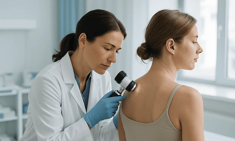
Mole Removal: How Dermatologists Do It and What to Expect
The only safe way to get rid of a skin mole is professional removal. A dermatologist typically uses shave excision or surgical excision under…
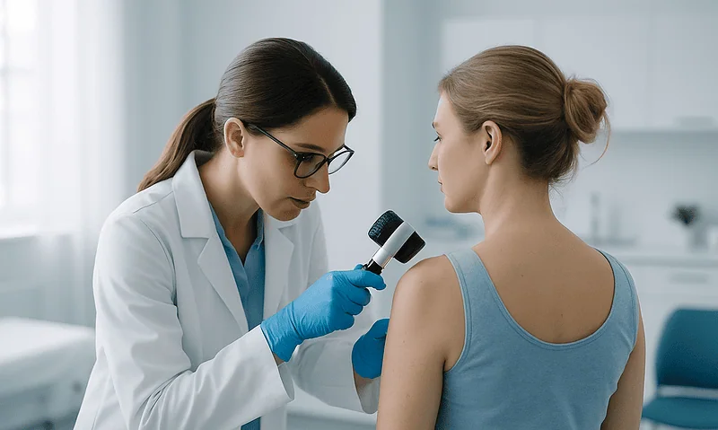
How Long Does Contact Dermatitis Take to Clear Once the Trigger Is Removed?
Once the trigger is fully removed, contact dermatitis usually clears within two to four weeks, according to Mayo Clinic, and many milder cases settle…

What Vitiligo Repigmentation Really Looks Like: Why Color Returns Slowly and Unevenly
Vitiligo repigmentation usually appears as small brown dots around hair follicles and a slow creep of color inward from patch edges. With treatment, first…

What Causes Hives, and Can Stress Cause Them?
Hives (urticaria) appear when immune cells in the skin called mast cells release histamine, making small blood vessels leak fluid into the skin and…
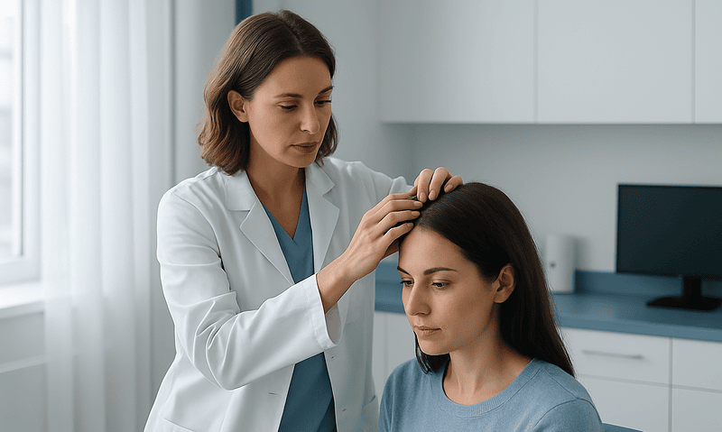
How Fast Does Hair Grow? The Timeline and What Influences It
Scalp hair grows about half an inch (1.25 centimeters) per month, or roughly six inches per year, though genetics, age, hormones, and overall health…
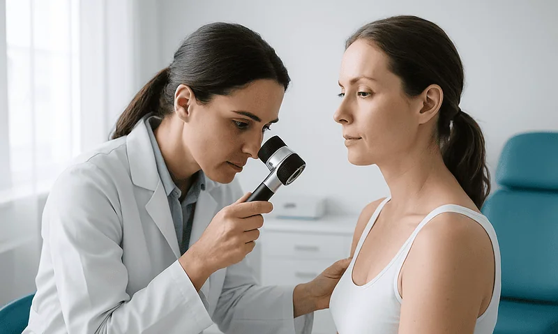
Is It Rosacea or Acne? Why Dermatologists Look at Flushing, Age and Blackheads Before Treating
Rosacea and acne both cause red bumps on the face, but they are different conditions. Rosacea usually begins after age 30, brings flushing and…