Arachnoid Cysts
Arachnoid cysts are fluid-filled sacs near the brain or spine, often found by chance. Learn about symptoms, causes, diagnosis and treatment options.

Quick answer
Arachnoid cysts are noncancerous, fluid-filled sacs that form between the brain or spinal cord and the arachnoid membrane, one of the protective layers covering them. Most are present from birth, cause no symptoms, and are found by chance on a scan. Larger cysts may cause headaches, seizures, or balance problems and are sometimes treated surgically.
What are arachnoid cysts?
Arachnoid cysts are sacs filled with clear fluid that form between the brain or spinal cord and one of the thin protective layers that cover them. That layer is called the arachnoid membrane, one of the three coverings known together as the meninges. The fluid inside an arachnoid cyst is usually cerebrospinal fluid, the same watery liquid that normally surrounds and cushions the brain and spinal cord.
Arachnoid cysts are not tumors. They are not cancerous, they do not spread, and the cells that line them are not abnormal in the way cancer cells are. They are best thought of as a pocket of fluid trapped within a fold or split of the arachnoid membrane. Most arachnoid cysts occur inside the skull, and the most common location is a region behind the eye socket and beneath the temple called the middle cranial fossa. Cysts can also develop along the spinal canal, although this is less common.
Arachnoid cysts are the most common type of fluid-filled cyst found inside the skull. Many are present from birth and stay the same size for a lifetime without ever causing a problem. A large proportion are discovered by chance when a person has a brain scan for an unrelated reason, such as a headache, a head injury, or dizziness. Arachnoid cysts are reported more often in males than in females and are frequently identified in childhood, although they can be found at any age. Because so many cause no symptoms, the true number of people who have one is not known with certainty.
Arachnoid cysts symptoms
Most arachnoid cysts cause no symptoms at all. When symptoms do occur, they usually happen because the cyst is large enough to press on nearby brain or spinal tissue, or because it interferes with the normal flow of cerebrospinal fluid. The exact symptoms depend on where the cyst is located and how much pressure it is causing. Common arachnoid cysts symptoms reported in people with brain cysts include:
- Headaches, which may be persistent or worse at certain times of day
- Nausea or vomiting, sometimes linked with headache
- Dizziness or problems with balance
- Seizures
- Hearing or vision changes, depending on the location of the cyst
- Weakness or numbness on one side of the body
- Difficulty with coordination or walking
- Problems with memory, attention, or behavior
- In infants, an unusually rapid increase in head size or a bulging soft spot
In children, especially very young ones, symptoms may be harder to notice. Developmental delay, irritability, or a head that grows faster than expected may be the first signs that something is putting pressure on the developing brain. In many cases, however, children with arachnoid cysts develop entirely normally.
Arachnoid cysts of the spine tend to produce different symptoms because they press on the spinal cord or the nerves leaving it. These may include back pain, pain that travels down an arm or leg, weakness or tingling in the limbs, stiffness, or changes in bladder or bowel control. Spinal cysts are less common than cysts inside the skull.
A small number of arachnoid cysts bleed into themselves or rupture, sometimes after a minor head injury. When this happens, symptoms can appear suddenly and may include a severe headache, vomiting, confusion, or drowsiness. This is uncommon but needs urgent medical assessment.
Causes and risk factors
Doctors divide arachnoid cysts into two groups based on how they are thought to develop, and this is the clearest way to understand arachnoid cysts causes.
Primary arachnoid cysts are congenital, meaning they are present at birth. They are believed to form during early fetal development, when the arachnoid membrane splits or folds abnormally and a small pocket of cerebrospinal fluid becomes trapped inside. The pocket may stay the same size for life, or it may slowly enlarge if fluid continues to enter but cannot easily leave. Primary cysts are by far the most common type. The precise reason why the membrane develops this way in some babies and not others is not fully understood.
Secondary arachnoid cysts develop later in life as a result of something that damages or scars the meninges. Possible triggers include:
- Head or spinal injury
- Meningitis, an infection of the membranes covering the brain and spinal cord
- Bleeding around the brain, such as a subarachnoid hemorrhage
- Previous brain or spinal surgery
- Tumors or other conditions that disturb the normal flow of cerebrospinal fluid
Secondary cysts are less common than primary cysts, and in some people the two types are difficult to tell apart on a scan.
Known risk factors are limited. Being male appears to be associated with a somewhat higher chance of having an arachnoid cyst, and a history of head trauma, meningitis, or brain surgery can increase the risk of a secondary cyst. Arachnoid cysts do not appear to run strongly in families in most cases, although rare familial cases have been described. They are not caused by diet, lifestyle, or anything a parent did during pregnancy, and there is no known way to prevent a primary cyst from forming.
Arachnoid cysts diagnosis
Arachnoid cysts diagnosis relies almost entirely on imaging, because a cyst cannot be detected by a physical examination alone. In many cases the cyst is first noticed on a scan ordered for another reason. When a cyst is suspected or has been found, doctors typically use one or more of the following:
- Magnetic resonance imaging (MRI) uses a strong magnetic field and radio waves to produce detailed pictures of the brain or spine. It is the preferred test for arachnoid cysts because it shows the cyst wall, its relationship to surrounding structures, and whether the fluid inside matches normal cerebrospinal fluid. Special MRI sequences can help distinguish an arachnoid cyst from other fluid-containing lesions.
- Computed tomography (CT) uses X-rays to create cross-sectional images. It is quick, widely available, and often the first scan performed in an emergency. CT can show the size and location of a cyst and any thinning or remodeling of the nearby skull bone, but it gives less detail than MRI.
- Prenatal ultrasound can sometimes detect a cyst before birth. If so, follow-up imaging after birth is usually recommended.
- Cerebrospinal fluid flow studies are specialized MRI techniques that show how fluid moves around the cyst. They may help doctors decide whether the cyst is connected to the normal fluid spaces.
An MRI or CT scan usually gives a confident diagnosis without the need for a tissue sample. Doctors may also arrange additional tests to assess how the cyst is affecting you, rather than to confirm the cyst itself. These can include a neurological examination to check strength, sensation, reflexes, and coordination; an electroencephalogram (EEG), which records electrical activity in the brain, if seizures are suspected; and hearing or vision testing if the cyst lies near the relevant nerves. In children, a doctor may track head circumference over time and assess developmental milestones.
Part of the diagnostic process is ruling out other conditions that can look similar on a scan, such as other types of cysts, certain slow-growing tumors, or enlarged normal fluid spaces. The pattern on MRI is often distinctive enough to settle the question, but a follow-up scan after several months may be suggested to confirm that the cyst is stable.
Arachnoid cysts treatment options
There is no single correct approach to arachnoid cysts treatment. The decision depends on whether the cyst is causing symptoms, its size and location, whether it is growing, and the age and general health of the person. In many hospitals these decisions are made jointly by neurologists, who specialize in disorders of the nervous system, and neurosurgeons, who perform operations on the brain and spine. At Acibadem, arachnoid cysts are typically assessed and managed through the Neurosurgery department in collaboration with neurology and radiology teams.
Observation. For a cyst that causes no symptoms, the most common recommendation is to do nothing beyond monitoring. Your doctor may suggest a repeat MRI or CT scan after a period of time to confirm that the cyst is not enlarging. If it remains stable and you remain well, further scans may be spaced out or stopped. Many people never need any other treatment.
Medication. There is no medicine that shrinks or removes an arachnoid cyst. However, medication may be used to manage symptoms. Anti-seizure drugs can help control seizures linked to a cyst, and pain relievers may be used for headaches. These treat the effects of the cyst rather than the cyst itself.
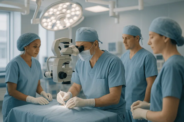
Surgery. Surgery is generally considered only when a cyst is clearly causing symptoms, is growing, is associated with a buildup of fluid in the brain (a condition called hydrocephalus), or has bled or ruptured. The goal of surgery is to relieve pressure by draining the cyst or creating a permanent pathway for the fluid to escape. The main surgical approaches are:
- Fenestration, in which the surgeon makes openings in the cyst wall so the fluid can drain into the normal cerebrospinal fluid spaces. This can be done through an open operation, called a craniotomy, in which a section of skull is temporarily removed, or through a smaller opening using an endoscope, a thin tube with a camera. The endoscopic approach is less invasive but is not suitable for every cyst.
- Shunt placement, in which a thin tube is placed into the cyst and tunneled under the skin to drain fluid into the abdominal cavity, where the body absorbs it. A shunt provides continuous drainage but is a permanent device that can block or become infected and may need revision over time.
- Needle aspiration, in which fluid is drawn out through a needle. This is used less often because cysts commonly refill.
For spinal arachnoid cysts that press on the spinal cord or nerves, surgery to remove or open the cyst and relieve pressure is the usual approach when symptoms warrant it.
All surgery carries risks, including bleeding, infection, leakage of cerebrospinal fluid, seizures, and the possibility that the cyst returns. Your surgeon should explain the specific risks and expected benefits for your situation. Many people experience relief of pressure-related symptoms after successful surgery, but recovery of function that was already lost is not guaranteed.
Rehabilitation. If a cyst has caused weakness, balance problems, speech difficulty, or developmental delay, physical therapy, occupational therapy, or speech therapy may be recommended, either instead of or after surgery, to help regain skills and adapt to any lasting effects.
Living with arachnoid cysts and outlook
For most people, an arachnoid cyst is an incidental finding that never affects daily life. Cysts that do not cause symptoms and do not grow generally have an excellent outlook, and the main task is simply to keep any follow-up appointments your doctor suggests and to be aware of warning signs. Learning that you have something in your brain can be unsettling, and it is reasonable to ask your doctor to explain the scan and to discuss how likely the cyst is to ever cause trouble.
There is usually no need to change your diet, exercise routine, or work because of a stable cyst. Some doctors advise people with large cysts, particularly children and adolescents, to be cautious with high-impact contact sports because of the rare risk of bleeding or rupture after a head blow. This advice varies, so it is worth discussing your individual situation rather than assuming restrictions are necessary.
When a cyst does cause symptoms and is treated surgically, many people see their pressure-related symptoms improve. Outcomes are generally better when the problem is identified before permanent damage to brain or spinal tissue has occurred. Some symptoms, such as seizures, may persist even after the cyst has been drained, and long-term medication may still be needed. Cysts can also recur or refill after treatment, so follow-up imaging is commonly recommended for several years.
Children who have had a symptomatic cyst treated early often go on to develop normally, although some may benefit from ongoing developmental support. Adults living with an untreated cyst that occasionally causes headaches may find it helpful to keep a symptom diary so that any change over time can be reviewed with their doctor.
Frequently asked questions
Are arachnoid cysts dangerous?
In most cases, no. The great majority of arachnoid cysts are small, stable, and never cause symptoms. A cyst can become a concern if it grows large enough to press on the brain or spinal cord, blocks the flow of cerebrospinal fluid, or bleeds. These situations are uncommon, and when they do occur they can usually be recognized on imaging and treated. Your doctor can give you a sense of how much your particular cyst is likely to matter based on its size and location.
What are the most common arachnoid cysts symptoms?
When arachnoid cysts do cause symptoms, headache is the one most often reported, followed by nausea, dizziness, seizures, and balance problems. Symptoms depend heavily on where the cyst sits. It is important to remember that many people with a cyst also have headaches for entirely unrelated reasons, and a scan that shows a cyst does not necessarily mean the cyst is the cause. Doctors weigh the size and position of the cyst against the pattern of symptoms before drawing conclusions.
What causes arachnoid cysts to form?
Most arachnoid cysts are congenital, meaning they form before birth when the arachnoid membrane develops abnormally and traps a pocket of fluid. The underlying reason this happens is not fully understood. A smaller number are secondary cysts that develop after head injury, meningitis, bleeding around the brain, or surgery. Arachnoid cysts causes do not include diet, stress, or lifestyle, and nothing a parent does during pregnancy is known to cause them.
How is arachnoid cysts diagnosis confirmed?
Diagnosis is confirmed with imaging, most reliably MRI. The scan shows a well-defined, fluid-filled sac whose contents match normal cerebrospinal fluid. CT can also show the cyst and is often the first test done in an emergency. A biopsy or tissue sample is almost never needed because the appearance on MRI is usually characteristic. Your doctor may arrange a follow-up scan to make sure the cyst is stable.
Do arachnoid cysts always need treatment?
No. Arachnoid cysts treatment is only recommended when the cyst is causing symptoms, is enlarging, or has led to complications such as hydrocephalus or bleeding. A cyst that is not causing problems is usually left alone and monitored with periodic scans. Surgery is reserved for cases where the expected benefit clearly outweighs the risks of operating on the brain or spine.
Can an arachnoid cyst go away on its own?
Occasionally, yes. There are reports of arachnoid cysts shrinking or disappearing without treatment, sometimes after a rupture allows the fluid to drain into the surrounding spaces. This is not common, however, and most cysts stay roughly the same size over time. Cysts do not respond to medication, and there is no reliable way to make one disappear other than surgery.
Can arachnoid cysts grow or come back after surgery?
Some cysts do grow slowly over time, which is one reason doctors may suggest repeat imaging. After surgery, a cyst can refill, particularly if the openings created during fenestration close over or a shunt stops working. For this reason, follow-up scans are usually recommended for a period after treatment, and new or returning symptoms should be reported promptly.
When to see a doctor
If you have been told you have an arachnoid cyst and it is being monitored, you should keep the follow-up appointments your doctor has suggested and let them know about any new or worsening symptoms. You should also arrange a medical review if you develop persistent headaches, recurring dizziness, hearing or vision changes, weakness or numbness, or, in a child, unexpected developmental delay or rapid head growth.
Seek emergency medical care immediately if you or someone with a known arachnoid cyst experiences any of the following red-flag signs, especially after a head injury:
- A sudden, severe headache unlike any previous headache
- Repeated vomiting, especially with headache
- A seizure, particularly a first-ever seizure
- Sudden confusion, unusual drowsiness, or difficulty staying awake
- Sudden weakness, numbness, or paralysis of the face, arm, or leg
- Sudden loss of vision, double vision, or slurred speech
- Loss of balance or inability to walk
- Loss of bladder or bowel control with back pain or leg weakness
- In an infant, a bulging soft spot, high-pitched crying, or extreme sleepiness
These symptoms can indicate bleeding into or around the cyst, a rise in pressure inside the skull, or compression of the spinal cord. They are not common, but they require urgent assessment.
Medically reviewed by the Acıbadem International Medical Board September 13, 2026
See our medical review board →
Update history
- PublishedSeptember 13, 2026
- Medical review approvedSeptember 13, 2026
- Last content updateSeptember 13, 2026