

Proliferative Vitreoretinopathy
Proliferative vitreoretinopathy is scar tissue that forms after retinal detachment or its repair. Learn about symptoms, causes, diagnosis and treatment.
Quick answer
Proliferative vitreoretinopathy (PVR) is a complication in which scar-like membranes grow on, under or around the retina after a retinal detachment or its surgical repair. The membranes contract and pull on the retina, causing wrinkling or re-detachment. It is the most common reason detachment surgery fails and is usually treated with further retinal surgery.
What is proliferative vitreoretinopathy?
Proliferative vitreoretinopathy, often shortened to PVR, is a complication that can develop after a retinal detachment or after surgery to repair one. The retina is the thin layer of light-sensing tissue that lines the back of the eye. The vitreous is the clear, jelly-like substance that fills the middle of the eye and sits against the retina. A retinal detachment happens when the retina separates from the wall of the eye, most often because a tear in the retina allows fluid to pass underneath it.
In proliferative vitreoretinopathy, the eye responds to the detachment or to the surgery with an exaggerated healing reaction. Cells that normally stay in place move onto the surface of the retina, underneath it, or into the vitreous, and form sheets of scar-like tissue called membranes. Over weeks, these membranes shrink and pull on the retina. This pulling, known as traction, can wrinkle the retina, hold retinal tears open, cause a repaired retina to detach again, or prevent a detached retina from settling back into position.
PVR is widely recognized as the most common reason that retinal detachment surgery does not succeed on the first attempt. It mainly affects people who have had a rhegmatogenous retinal detachment (a detachment caused by a retinal tear), particularly after repair surgery. It can also follow severe eye injuries and, less often, other operations inside the eye. It is not the same as proliferative diabetic retinopathy, which is a separate condition caused by diabetes, even though the names sound similar.
The condition is managed by retina specialists, who are ophthalmologists (eye doctors) with additional training in surgery on the retina and vitreous. Within Acibadem, care for this condition is provided through the Ophthalmology department.
Proliferative vitreoretinopathy symptoms
Proliferative vitreoretinopathy symptoms are often similar to the symptoms of a retinal detachment itself, because the scar tissue is causing the retina to lift or fold. Many people notice them in the weeks or first few months after retinal detachment surgery. Symptoms may include:
- Vision that fails to improve, or begins to worsen again, after retinal detachment repair
- A shadow, curtain or dark area returning at the edge of vision
- Blurred or dim central vision
- Straight lines looking wavy, bent or distorted
- New or increasing floaters (small specks or threads drifting across vision)
- Flashes of light
- A general sense that the vision in the affected eye is deteriorating
Symptoms can vary with the stage and location of the scar tissue. Early, mild PVR may cause few or no symptoms if the membranes sit in the far periphery of the retina, away from the macula (the central part of the retina responsible for detailed vision). As the membranes contract, the retina may wrinkle, and distortion or blurring may develop. In more advanced stages, fixed folds form, and the retina may detach again, producing the curtain-like shadow that many people remember from their original detachment.
Because eyes are often blurry and uncomfortable in the normal recovery period after surgery, patients may find it hard to judge whether a change is expected or worrying. In general, vision after retinal surgery is expected to slowly improve or stabilize. Vision that clearly gets worse, or a shadow that comes back, should be reported promptly to the surgical team.
Proliferative vitreoretinopathy causes and risk factors
Proliferative vitreoretinopathy causes come down to an overactive wound-healing response inside the eye. When the retina tears or detaches, cells from the retinal pigment epithelium (the pigmented layer beneath the retina) and supporting cells called glial cells can escape through the tear. Inflammation and breakdown of the normal barrier between the blood and the retina release signaling substances that encourage these cells to multiply, change their character, and produce collagen. The result is contractile membranes that behave like scar tissue and pull on the retina.
Not everyone with a retinal detachment develops PVR. Certain features of the detachment, the eye and the treatment are known to raise the risk:
- Large retinal tears, giant retinal tears or multiple tears
- A retinal detachment that has been present for a long time before repair
- Bleeding into the vitreous (vitreous hemorrhage)
- Detachment of the choroid, the blood-vessel layer beneath the retina
- Inflammation inside the eye (uveitis) before or after surgery
- Penetrating or blunt eye injury (trauma)
- Previous failed retinal detachment surgery or a history of PVR in the same eye
- Multiple operations on the same eye
- Extensive use of freezing treatment (cryotherapy) during repair
- Absence of the natural lens (aphakia) or complications involving the lens
Some studies have also examined general factors such as smoking and genetic differences in inflammatory pathways, but these are less well established than the eye-specific factors above. Your surgeon may consider your individual risk when planning the type of surgery and follow-up.
Proliferative vitreoretinopathy diagnosis
Proliferative vitreoretinopathy diagnosis is made by an ophthalmologist through a detailed eye examination, usually supported by imaging. There is no blood test for PVR. The key steps typically include:
- Dilated fundus examination: drops are used to widen the pupil so the doctor can view the retina with an indirect ophthalmoscope, a bright head-mounted light with a handheld lens. This allows inspection of the whole retina, including the far periphery.
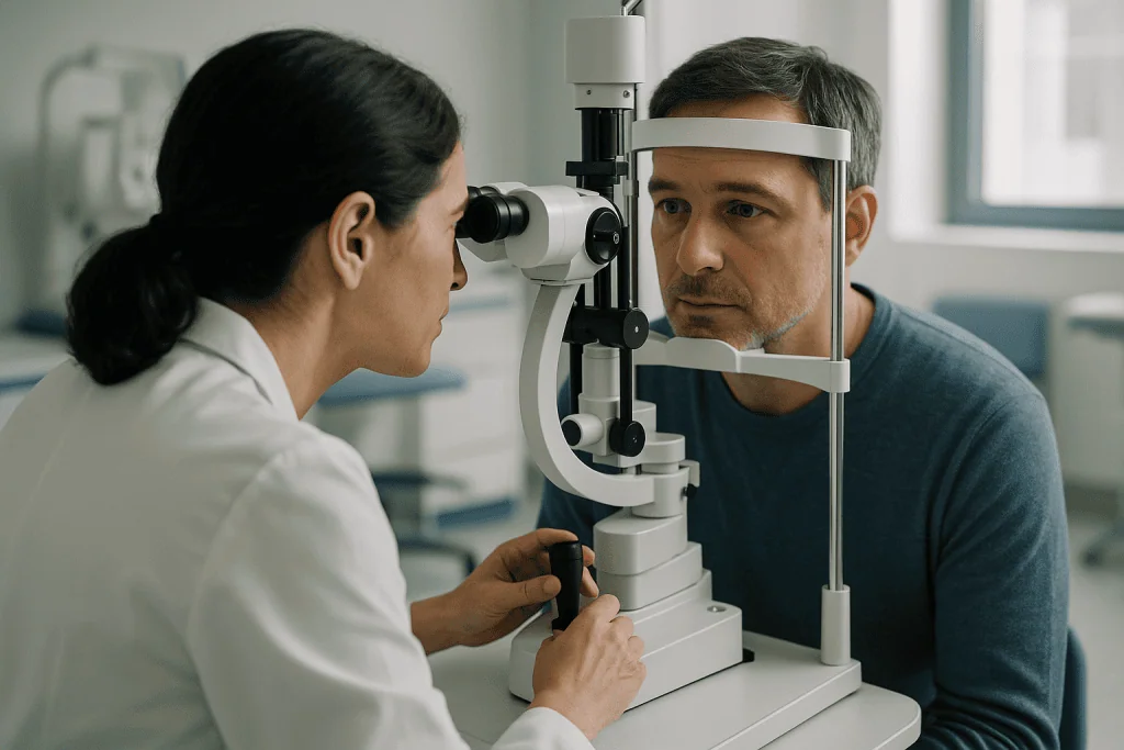
- Slit-lamp examination: a microscope with a narrow beam of light, often combined with a special contact or handheld lens, gives a magnified view of the vitreous and retinal surface.
- Optical coherence tomography (OCT): a painless scan that uses light waves to produce cross-sectional pictures of the retina. It can show membranes on the retinal surface, wrinkling, and fluid under the retina, especially near the macula.
- B-scan ultrasound: a sound-wave scan used when the view into the eye is blocked by blood, cataract or cloudy media. It can show whether the retina is detached and whether it is being pulled into folds.
- Wide-field retinal photography: digital images that document the extent of the membranes and allow comparison over time.
Doctors describe the severity of PVR using a grading system. In simplified terms, grade A refers to haze and pigment clumps in the vitreous; grade B refers to wrinkling of the inner retinal surface, rolled edges of retinal tears and stiffness of the retina; and grade C refers to full-thickness fixed folds of the retina, which may be described as anterior (toward the front of the eye) or posterior (toward the back). The grade, the location of the folds and how many sections of the retina are involved all help guide treatment decisions.
In many cases the diagnosis becomes apparent during routine follow-up visits after retinal detachment surgery, which is one reason these visits are important even if the eye feels fine.
Treatment options for proliferative vitreoretinopathy
Proliferative vitreoretinopathy treatment depends on the grade of scarring, whether the retina is detached, whether the macula is threatened, and how much useful vision the eye is expected to have. Options range from careful observation to complex surgery.
Observation
Mild membranes in the peripheral retina that are not pulling on the macula and are not causing a detachment may simply be monitored. Your doctor may arrange closer follow-up so that any progression is caught early. In some situations, particularly when an eye has very limited visual potential or the person has serious health problems, observation may also be chosen instead of further surgery.
Medication
At present there is no medication that is widely accepted as a proven cure for established PVR. Anti-inflammatory eye drops and, in some cases, steroid injections are commonly used around the time of surgery to reduce inflammation, which is one of the drivers of scar formation. Several drugs that aim to interrupt the scarring process have been studied and continue to be investigated, but they are not standard therapy everywhere. Your surgeon can explain whether any adjunctive medicine is appropriate in your case.
Surgery
Surgery is the main treatment when PVR is causing or threatening a retinal detachment. It is usually more complex than the original detachment repair. The procedure most often used is a pars plana vitrectomy, in which the surgeon removes the vitreous gel through tiny openings in the wall of the eye. This is combined with one or more of the following:
- Membrane peeling: the scar membranes are carefully lifted off the surface of the retina, and sometimes from underneath it, using fine instruments.
- Relaxing retinotomy or retinectomy: if the retina has become so shortened and stiff that it cannot be flattened, the surgeon may cut or remove a portion of scarred peripheral retina so the rest can settle back into place.
- Scleral buckle: a silicone band placed around the outside of the eye to support the retina and relieve traction, sometimes added to vitrectomy.
- Perfluorocarbon liquid: a heavy liquid used temporarily during the operation to flatten the retina from the inside; it is removed before the surgery ends.
- Laser treatment (retinopexy): laser burns create small scars that seal the retina to the eye wall around tears and cut edges.
- Tamponade: at the end of surgery the eye is filled with a long-acting gas bubble or silicone oil to hold the retina in position while it heals. Gas absorbs on its own over weeks; silicone oil usually stays in the eye for several months and is often removed in a second, smaller operation, although in some eyes it is left in place long term.
After surgery, you may be asked to keep your head in a specific position for a period of time so that the gas or oil presses against the right part of the retina. Air travel and travel to high altitude are usually restricted while a gas bubble is present, because the bubble can expand and raise the eye pressure dangerously. Your surgical team will give you individual instructions.
Rehabilitation and vision support
Even when the retina is successfully reattached, vision in the affected eye may remain reduced. Low-vision services can help with magnifiers, lighting, and strategies for reading and daily tasks. Glasses prescriptions often change after surgery, especially while silicone oil is in the eye, and a cataract (clouding of the natural lens) frequently develops after vitrectomy and may need its own treatment later.
Living with proliferative vitreoretinopathy and outlook
PVR is a serious condition, and it is fair to say that the outlook is guarded rather than reassuring. With modern surgical techniques, the retina can be reattached in many eyes, sometimes after more than one operation. However, an attached retina does not always mean good vision. The macula may have been damaged by the original detachment or by the scarring, and vision often remains limited compared with the other eye. In general, eyes with less extensive scarring, a macula that was never detached, and a shorter history of detachment tend to do better, while eyes with advanced scarring, trauma or repeated surgery tend to do less well.
Recovery is usually slow and involves multiple follow-up appointments over many months. Recurrence of scar tissue is possible, and your doctor will watch for it. Long-term issues can include raised eye pressure or glaucoma, low eye pressure, cataract, changes to the cornea, and problems related to silicone oil. Many people also find the emotional side of a long and uncertain recovery difficult, particularly when one eye is doing most of the work. Protecting the better-seeing eye with appropriate eyewear, attending all scheduled reviews, and telling the care team about any new symptoms are practical steps that help.
It is important to remember that outcomes vary widely between individuals. Your surgeon is the best person to discuss what is realistic for your eye, based on the findings at examination and surgery.
Frequently asked questions
What are the first proliferative vitreoretinopathy symptoms after retinal detachment surgery?
The earliest sign is often that vision stops improving as expected, or that a shadow or curtain returns at the edge of vision in the weeks or months after surgery. Some people notice new distortion or blurring instead. Because early membranes in the far periphery may cause no symptoms at all, follow-up examinations are the main way early PVR is detected.
What causes proliferative vitreoretinopathy to develop in some eyes and not others?
The basic cause is an excessive healing response in which cells escape through a retinal tear, multiply and form contracting scar tissue. Why this happens in some eyes and not others is not fully understood. Known contributors include large or multiple tears, long-standing detachment, bleeding, inflammation, trauma and previous surgery, but PVR can also occur in eyes without obvious risk factors.
How is proliferative vitreoretinopathy diagnosis confirmed?
An ophthalmologist confirms the diagnosis by examining the retina through a dilated pupil, looking for characteristic membranes, wrinkling and fixed folds. OCT scans can show membranes and fluid under the retina, and ultrasound is used when the view is blocked. The findings are then graded to describe how severe and widespread the scarring is.
Is proliferative vitreoretinopathy treatment always surgery?
Not always. Mild scarring that is not threatening the retina or the macula may be observed closely. When PVR is causing or threatening a detachment, surgery is the standard treatment, usually a vitrectomy with membrane peeling and a gas or silicone oil tamponade. Medications are generally used to support surgery rather than replace it, and no drug is yet widely accepted as a stand-alone cure.
Can proliferative vitreoretinopathy come back after treatment?
Yes, recurrence is possible. The same healing response that caused the first episode can be triggered again by further surgery, which is why repeat operations carry a higher risk. Surgeons try to reduce this risk by removing membranes as completely as possible, relieving traction and controlling inflammation, but no method eliminates the risk entirely.
Will my vision return to normal after proliferative vitreoretinopathy?
In many cases, vision does not return fully to what it was before the retinal detachment, even if the retina is successfully reattached. The degree of recovery depends on how much the macula was affected, how long the retina was detached, and whether complications develop. Some people regain useful vision, while others are left with significant impairment in that eye.
Can proliferative vitreoretinopathy be prevented?
There is no reliable way to prevent PVR. Prompt treatment of retinal tears and detachments, careful surgical technique, and control of inflammation around surgery may lower the risk, and research into preventive medicines is ongoing. Attending follow-up visits allows early scarring to be found and addressed before it causes more damage.
When to see a doctor
Anyone who has had a retinal detachment or retinal surgery should keep all scheduled follow-up appointments, even if the eye seems to be recovering well, because early PVR may cause no symptoms. In addition, seek same-day advice from your eye care team or an emergency department if you notice any of the following, as they may signal a new or recurrent retinal detachment or another serious complication:
- A shadow, curtain or dark area appearing or spreading in any part of your vision
- A sudden drop in vision, or vision that was improving and is now clearly getting worse
- A sudden increase in floaters or new flashes of light
- New distortion, where straight lines look wavy or bent
- Severe eye pain, especially with nausea or vomiting, which can indicate very high eye pressure
- Increasing redness, discharge or a feeling that the eye is much more sensitive to light than before
- Any of these symptoms after a blow to the eye or after a fall
Sudden changes in vision after retinal surgery should never be assumed to be a normal part of healing. A prompt examination allows the doctor to decide whether the change is expected or whether urgent treatment is needed to protect your sight.
Medically reviewed by the Acıbadem International Medical Board September 13, 2026
See our medical review board →
Update history
- PublishedSeptember 13, 2026
- Medical review approvedSeptember 13, 2026
- Last content updateSeptember 13, 2026

