Breast Calcification Biopsy: Procedure, Recovery and Results
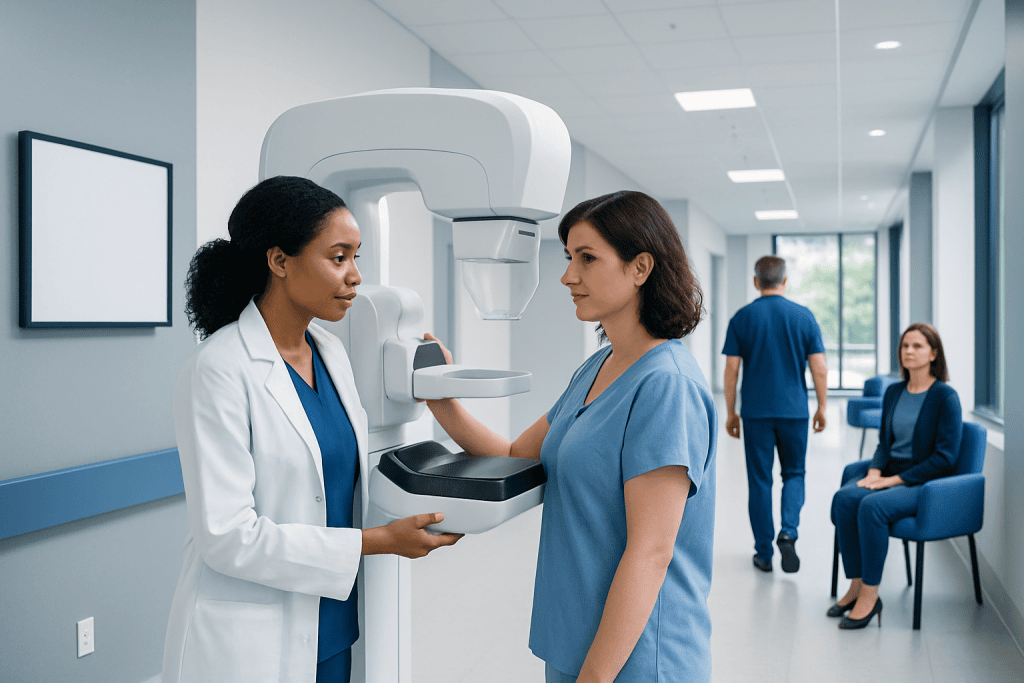
Most breast calcifications are noncancerous and are found during routine mammography. A stereotactic or tomosynthesis-guided needle biopsy is commonly used when calcifications cannot be felt or seen on ultrasound.
Key Takeaways
- Most breast calcifications are noncancerous and are found during routine mammography.
- A stereotactic or tomosynthesis-guided needle biopsy is commonly used when calcifications cannot be felt or seen on ultrasound.
- The procedure is usually performed with local anaesthetic, takes about an hour, and does not usually require an overnight stay.
- Mild bruising, soreness and swelling are common for several days; most people return to usual light activities within 24 hours.
- Results may show benign changes, high-risk cell changes or cancer, and are interpreted alongside the mammogram findings.
A breast calcification biopsy is an image-guided procedure that removes small tissue samples from an area of calcifications seen on a mammogram. Most calcifications are benign, but biopsy can clarify the cause and guide any needed follow-up or treatment.
Overview: what a breast calcification biopsy involves
A breast calcification biopsy is a procedure used to examine tiny calcium deposits seen on a mammogram. It is usually recommended when the pattern, shape or distribution of calcifications needs closer assessment. The biopsy removes small samples of tissue so a pathologist can identify the cause under a microscope.
Breast calcifications are common, particularly with increasing age, and most are benign. They may develop because of normal breast changes, past inflammation or injury, benign cysts, previous surgery, or changes within breast ducts. Some patterns can be associated with ductal carcinoma in situ (DCIS) or, less commonly, invasive breast cancer, which is why a tissue diagnosis may be advised.
Calcifications are often too small to feel during a breast examination. For this reason, the procedure is generally guided by mammography or breast tomosynthesis, rather than by touch. The most commonly used approach is a vacuum-assisted stereotactic breast biopsy, which allows several samples to be collected through one small skin opening.
How it works and who may need a biopsy
A radiologist first reviews the mammogram and assigns a level of concern based on the appearance of the calcifications. Features such as their size, shape, number and arrangement help determine whether short-term imaging follow-up is appropriate or whether biopsy is the safer next step. Imaging can estimate the likelihood of a concerning finding, but it cannot always provide a definite diagnosis.
A breast calcification biopsy may be recommended when calcifications are new, increasing, grouped closely together, irregular in shape or arranged in a pattern that follows a breast duct. It may also be advised if a previous mammogram did not show the area, or if imaging findings do not clearly fit a benign explanation.
Before the procedure, the care team should review medicines, allergies, bleeding disorders, pregnancy status and any implanted devices. Blood-thinning medicines and supplements may need individual instructions from the prescribing clinician; they should not be stopped without medical advice. A person who is pregnant or might be pregnant should tell the radiology team, as mammography guidance uses a small amount of radiation.
- Calcifications that look typically benign often need no biopsy.
- Probably benign findings may be monitored with planned mammograms.
- Suspicious or unclear calcifications are commonly assessed with image-guided biopsy.
Step-by-step: how a breast calcification biopsy is performed
On the day of the procedure, the person is usually awake and can normally eat and drink unless the team gives different instructions. They may be asked to avoid deodorant, talcum powder or lotion on the breast and underarm because these products can create imaging artefacts. Comfortable clothing and a supportive bra can be helpful after the biopsy.
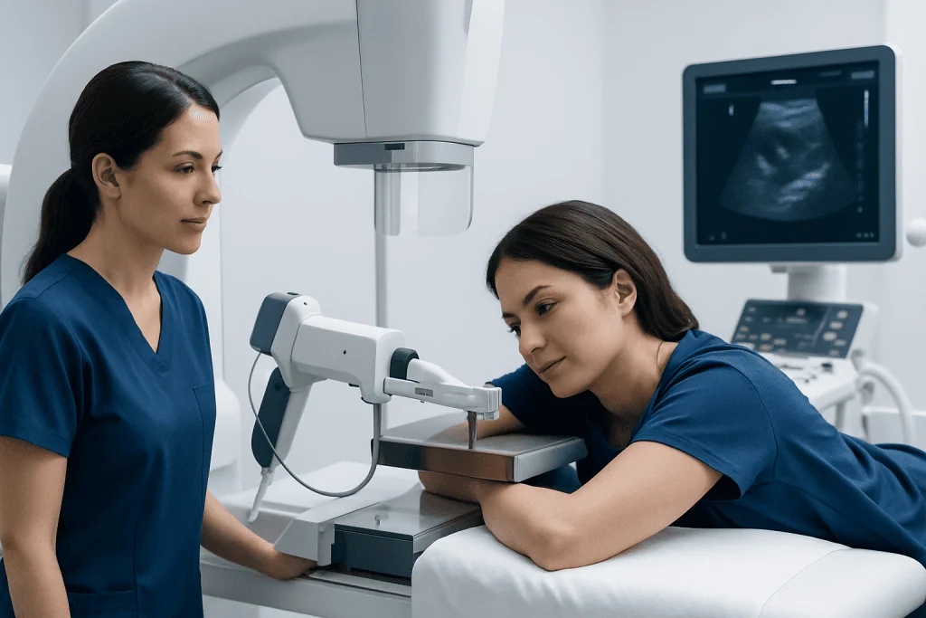
For a stereotactic biopsy, the breast is positioned and gently compressed between mammography plates to keep the area still. Several images are taken to precisely locate the calcifications in three dimensions. The skin is cleaned, and local anaesthetic is injected; there may be brief stinging or pressure, but sharp pain should be reported promptly.
The radiologist makes a very small incision and advances a biopsy needle to the targeted area using imaging guidance. Multiple tissue samples are collected, often using a vacuum-assisted device. An X-ray of the samples may confirm that calcifications were obtained. A tiny marker clip is usually left at the biopsy site so it can be identified on future imaging; it does not usually cause problems and is designed to remain safely in place.
After the needle is removed, firm pressure is applied to reduce bleeding. Stitches are usually unnecessary. The site is covered with small adhesive strips and a dressing, followed by a post-procedure mammogram to document the marker position. Image-guided breast biopsy is part of breast cancer treatment and diagnosis planning when results show cancer or a high-risk change.
Recovery timeline, benefits and possible risks
Most people go home shortly after the procedure. The breast may feel tender or bruised once the local anaesthetic wears off, and a small amount of swelling is common. Applying a cold pack over the dressing at intervals during the first day and wearing a supportive bra can improve comfort. The clinical team can advise which pain-relief medicines are suitable for the individual.
Light daily activities can usually resume the next day, but strenuous exercise, heavy lifting and vigorous upper-body activity may need to be avoided for 24 to 48 hours, or longer if advised. Dressings and showering instructions vary by centre, so it is important to follow the instructions provided. Adhesive strips are generally left in place until they loosen naturally.
The key benefit of biopsy is clarity: it can avoid unnecessary surgery for benign findings and ensure that significant abnormalities receive timely specialist assessment. Possible complications are uncommon but include bleeding, a collection of blood under the skin called a haematoma, infection, prolonged pain or an inadequate sample. Seek advice if there is increasing redness, warmth, swelling, fever, persistent bleeding or worsening pain.
Results are usually available within several working days, although timing varies depending on the laboratory and whether additional testing is needed. The radiologist and pathology team also check that the biopsy result matches the imaging appearance. If results and imaging do not agree, further sampling or surgical assessment may be recommended.
How many days should I rest after a breast biopsy?
Most people need only one day of relative rest after a needle biopsy for breast calcifications. Routine activities, desk work and gentle walking are often possible the following day, provided the person feels well and the treating team has not given different instructions.
For the first 24 to 48 hours, it is generally sensible to avoid heavy lifting, running, swimming, strenuous exercise and activities that cause breast movement or pressure. Bruising and tenderness may last several days and occasionally up to one or two weeks, especially if a larger area was sampled or a haematoma developed.
Recovery needs differ from person to person. Anyone whose work is physically demanding, who takes medicines affecting clotting, or who has ongoing swelling or pain should ask their clinician when it is safe to return to full activity. A supportive bra and the aftercare plan provided by the biopsy unit can make recovery more comfortable.
What biopsy results tell you about breast calcifications
Pathology results identify the type of cells and tissue changes present in the sampled area. A benign result may show ordinary calcium deposits, fibrocystic changes, cyst-related changes, fibroadenoma, inflammation, fat necrosis or other noncancerous causes. When the result is benign and matches the mammogram appearance, the person may return to routine screening or have a planned follow-up mammogram.
Some results identify changes that are not cancer but may require additional discussion, surveillance or surgery because they can be associated with a higher future breast cancer risk or may be close to an unsampled area of cancer. Examples can include atypical ductal hyperplasia, atypical lobular hyperplasia, lobular carcinoma in situ or certain papillary lesions. Management depends on the exact finding, sample adequacy and imaging-pathology agreement.
A result may also show DCIS, an early non-invasive breast cancer that is often detected as calcifications, or invasive breast cancer. These diagnoses require referral to a breast multidisciplinary team to discuss treatment options. Further information about breast cancer can help patients understand how diagnosis, staging and treatment planning are approached.
How serious is calcification in the breast, and can a radiologist tell if it is cancerous?
Calcification in the breast is not automatically serious. In fact, most calcifications are benign and do not cause symptoms or require treatment. Their significance depends less on the presence of calcium itself and more on the mammographic pattern and whether it has changed over time.
A radiologist can often recognise calcifications that have a clearly benign appearance. However, even experienced radiologists cannot confirm cancer from mammography alone when calcifications have a suspicious or indeterminate pattern. Biopsy is used to establish the diagnosis and prevent both unnecessary alarm and missed disease.
It is helpful to remember that a recommendation for biopsy does not mean cancer has been diagnosed. It means the imaging findings deserve a more precise answer. The final interpretation combines the pathology report with the imaging findings, medical history and, when needed, specialist input from breast radiology, pathology, surgery and oncology.
When to seek medical care
A person should contact the biopsy team promptly after the procedure if bleeding does not stop with firm pressure, the breast becomes increasingly swollen, there is spreading redness or warmth, pus-like drainage, fever, or pain that is worsening rather than improving. These signs do not always indicate a serious problem, but they should be assessed.
Medical review is also important for a new breast lump, nipple discharge that is bloody or spontaneous, skin dimpling, persistent focal breast pain, nipple changes or new underarm swelling. These symptoms often have benign explanations, but they should not be self-diagnosed and may need clinical examination and imaging.
People should attend all follow-up appointments, even after a benign biopsy result, because the care team may recommend a specific imaging schedule. Acibadem International’s multidisciplinary specialists and JCI-accredited hospitals support international patients with breast imaging, biopsy assessment and coordinated treatment planning when needed.
Frequently asked questions
Is a breast calcification biopsy painful?
Local anaesthetic is used to numb the skin and deeper breast tissue, so most people feel pressure, pulling or vibration rather than sharp pain during the sampling. Mild soreness and bruising are common after the anaesthetic wears off. The radiology team should be told immediately if pain is significant during the procedure.
How long does a breast calcification biopsy take?
The imaging-guided part of the procedure commonly takes about 30 to 60 minutes, although preparation and post-biopsy imaging can make the appointment longer. The exact time depends on the location of the calcifications and the biopsy method used. Most people return home on the same day.
What do biopsy results tell me about breast calcifications?
Results show whether the sampled calcifications are due to benign breast changes, a high-risk abnormality, DCIS or invasive cancer. The pathology result is compared with the mammogram to ensure the samples explain the imaging finding. If they do not match, further assessment may be needed.
How serious is calcification in the breast?
Most breast calcifications are benign and not dangerous. Some patterns need additional imaging or biopsy because they can occasionally be linked with DCIS or breast cancer. Their seriousness can only be judged from the full imaging assessment and, when required, tissue results.
Can a radiologist tell if breast calcifications are cancerous?
A radiologist can identify patterns that appear benign and patterns that are more suspicious, but mammography alone cannot always confirm whether calcifications are cancerous. A biopsy provides the definitive tissue diagnosis when imaging is uncertain or concerning. This is why a biopsy recommendation is a diagnostic step, not a cancer diagnosis.
Will the marker clip set off airport security or need to be removed?
The small marker clip used after a needle biopsy is usually made of materials that are safe for the body and generally does not set off airport security systems. It is normally left in place to identify the biopsy site on future breast imaging. It does not usually need to be removed unless there is a separate clinical reason for surgery.
References
- American Cancer Society
- American College of Radiology
- National Cancer Institute
- Breast Cancer Now
- Radiological Society of North America
This article is for general information only and is not a substitute for professional medical advice. Please consult a qualified doctor about your individual situation.
Oncology care in Turkey — second opinion and treatment plan
JCI-accredited · board-certified surgeons · reply within 24h
Add us as a Preferred Source to see more of our trusted health content across Google Search, AI Overviews and Discover.

More from the Health Library


Related Specialists

Prof. Dr. İsmail Çepni
Gynecology & Obstetrics
Assoc. Prof. Dr. Emine Şatır
Gastroenterology
Dr. Ayşe Yemişçi
Dermatology
Prof. Dr. Solmaz Balcı Akar
Ophthalmology