Breast Papilloma Treatment: How It Works, Results and What to Expect

A breast papilloma is usually a benign growth inside a milk duct, often near the nipple. Treatment decisions depend on symptoms, imaging, biopsy results, lesion size and whether atypical cells are present.
Key Takeaways
- A breast papilloma is usually a benign growth inside a milk duct, often near the nipple.
- Treatment decisions depend on symptoms, imaging, biopsy results, lesion size and whether atypical cells are present.
- Surgical excision removes the papilloma and allows the tissue to be examined fully in a laboratory.
- Recovery after breast papilloma surgery is commonly measured in days to a few weeks, although healing differs between individuals.
- A papilloma itself is not usually cancer, but atypia or imaging-biopsy disagreement may require additional evaluation or removal.
Breast papilloma treatment is individualized: some papillomas can be monitored after a reassuring biopsy, while others are removed to relieve symptoms, confirm the diagnosis or address atypical cells. Most breast papillomas are benign, and a breast specialist can help patients understand whether observation or surgery is the safest next step.
Breast Papilloma Treatment: How It Works
Breast papilloma treatment may involve careful follow-up or removal of the affected milk duct area. The right approach depends on whether the papilloma is causing nipple discharge or a lump, what imaging shows, and whether a core needle biopsy found only benign papilloma tissue or cells with atypia. Treatment is designed both to manage symptoms and to make sure the diagnosis is clear.
A breast papilloma, also called an intraductal papilloma, is a small wart-like growth that develops within a breast duct. It is made of fibrous tissue and blood vessels and is usually benign. A solitary papilloma commonly occurs close to the nipple and may cause clear or bloody discharge; multiple papillomas can occur farther from the nipple and may be found during breast imaging.
When removal is recommended, the surgeon usually performs an excisional biopsy or duct excision. The goal is to remove the lesion, sometimes with a small amount of surrounding duct tissue, while preserving as much normal breast tissue as possible. The removed tissue is examined by a pathologist, which provides more complete information than a small biopsy sample alone.
How Doctors Decide Whether Treatment Is Needed
Not every biopsy-proven benign papilloma requires surgery. In selected people, imaging surveillance may be appropriate when the imaging findings and biopsy results agree, the lesion appears fully sampled, there are no concerning symptoms and no atypical cells are identified. Follow-up commonly includes clinical review and scheduled breast imaging recommended by the treating team.
Removal is more likely to be advised if there is spontaneous bloody or persistent nipple discharge, a palpable mass, a larger lesion, multiple lesions, abnormal imaging that does not match the needle-biopsy result, or atypical cells on pathology. Atypia means cells look unusual under the microscope; it is not the same as cancer, but it can change the level of concern and the recommended next steps.
Decisions are ideally made by a multidisciplinary breast team that may include a breast surgeon, radiologist and pathologist. This approach helps ensure that mammogram, ultrasound or MRI findings are considered alongside the pathology report and a person’s individual risk factors, including family history of breast cancer.
- Observation: appropriate for selected low-risk benign papillomas with reliable follow-up.
- Vacuum-assisted removal: may be considered for certain imaging-detected lesions in experienced centers.
- Surgical excision: used when symptoms, pathology or imaging findings make complete removal or further diagnosis advisable.
What Happens During Breast Papilloma Surgery
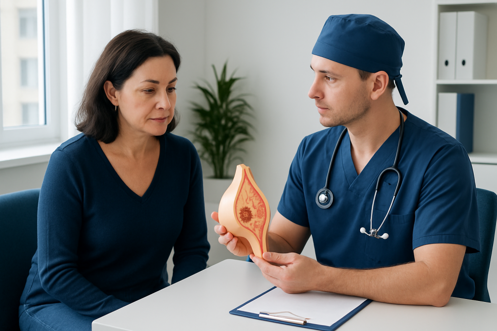
Before surgery, the team reviews imaging and biopsy findings and explains the planned procedure. If the papilloma cannot be felt, a radiologist may place a small localization marker or wire, or use another guidance technique, to help the surgeon identify the exact area. Preoperative assessment also includes discussing medicines, allergies, anesthesia and any medical conditions that may affect surgery.
Breast papilloma excision is commonly performed as an outpatient procedure. Depending on the location and extent of surgery, it may be done with local anesthesia and sedation or with general anesthesia. The surgeon makes a small incision, often placed near the edge of the areola when appropriate, removes the targeted tissue or involved duct, and sends it for laboratory analysis.
The incision is then closed with sutures, skin adhesive or small wound-closure strips, and a dressing is applied. In most uncomplicated excisions, a drain is not needed. The procedure itself is often relatively short, but patients should allow time for preparation, recovery from anesthesia and discharge instructions.
The final pathology report is important because it confirms whether the removed tissue is benign papilloma, papilloma with atypia, or another breast condition. If additional findings are identified, the breast team will explain what they mean and whether further assessment, imaging or treatment is needed.
Benefits, Risks and Expected Results
The main benefit of removing a breast papilloma is diagnostic certainty from examining the whole lesion. Surgery may also stop troublesome nipple discharge, remove a lump and reduce worry about a persistent abnormal finding. For people whose lesion was benign on final pathology, no cancer treatment is needed for that papilloma.
As with any procedure, excision has potential risks. These include temporary pain, bruising, swelling, bleeding, infection, delayed wound healing, scar formation and changes in breast or nipple sensation. Less commonly, surgery can alter the appearance of the breast or nipple, particularly if tissue close to the nipple or several ducts must be removed.
Removing a duct near the nipple may affect the ability to breastfeed from that duct in the future. The overall impact varies with the procedure and the amount of tissue removed, so this is worth discussing before surgery for people who may wish to breastfeed. A breast surgeon can also explain how the incision location may affect sensation and cosmetic outcome.
Most people have a good outcome after treatment. However, a new breast symptom, a new papilloma or an unrelated breast condition can still occur later. Continuing routine breast awareness and attending recommended screening appointments remain important.
How Long Does It Take to Recover From Breast Papilloma Surgery?
Recovery from breast papilloma surgery is often straightforward. Many people go home the same day and can manage light daily activities within several days. Soreness, mild swelling and bruising are common during the first week, while complete comfort and wound healing may take a few weeks.
The care team will give individualized instructions for showering, dressing care, driving, exercise and return to work. In general, avoiding heavy lifting, strenuous exercise and activities that pull on the incision until the surgeon approves can support healing. A supportive bra may improve comfort for some patients.
Follow-up is usually arranged to review the incision and discuss the final pathology report. Patients should contact their clinical team promptly if they develop increasing redness, warmth, swelling, fever, worsening pain, pus-like drainage, significant bleeding or a wound that opens. These symptoms do not always mean a serious problem, but they need timely assessment.
Are Breast Papillomas Serious?
Breast papillomas are usually not serious in the sense that most are benign rather than cancerous. Even so, they should be properly evaluated because they can cause nipple discharge or a lump and because a small needle biopsy may not always show every part of the lesion. Imaging and pathology findings together guide whether follow-up or removal is appropriate.
A papilloma that contains atypia, is associated with suspicious imaging or does not match the biopsy findings needs more careful assessment. This does not mean cancer is present, but it may mean excision is recommended to obtain a complete diagnosis. A breast specialist can explain the individual level of concern clearly and calmly.
Any new spontaneous nipple discharge, especially if it is bloody or occurs from one breast, should be assessed by a healthcare professional. Most causes are not cancer, but evaluation helps identify the cause and determine the appropriate care.
How Fast Do Breast Papillomas Grow?
Breast papillomas often grow slowly, and many are detected because of symptoms or imaging rather than because a person notices rapid growth. There is no single growth rate for all papillomas. Their appearance can also be influenced by how they are measured and whether they are located within a dilated milk duct.
A change in the size of a breast lump, new discharge or a new imaging finding should not be assumed to be a papilloma. Repeat examination and imaging may be needed to clarify what is happening. For people managed with surveillance, attending planned follow-up appointments is the best way to monitor a known lesion safely.
Rapidly developing breast changes deserve clinical assessment, particularly when accompanied by skin changes, nipple inversion, persistent pain or enlarged lymph nodes. These findings have many possible explanations, and timely review helps ensure appropriate testing.
Can Breast Papilloma Become Cancer?
A typical solitary breast papilloma without atypia is benign and does not usually become cancer. However, papillomas can occasionally be found alongside atypical changes or, less commonly, an area of cancer. This is why doctors consider the pathology report, imaging appearance and whether the tissue sample adequately explains the imaging finding.
The term “upgrade” may be used when surgical excision identifies atypia or cancer that was not seen in the original core needle biopsy. The likelihood varies according to the characteristics of the lesion and the biopsy, so it should be interpreted by the treating breast team rather than applied as a general prediction for every patient.
People with multiple papillomas or papillomas associated with atypia may be advised to have a personalized follow-up plan. This may include more attentive imaging surveillance and discussion of overall breast cancer risk factors. It is important to remember that increased risk is not the same as a diagnosis of cancer.
When to Seek Medical Care
Medical assessment is recommended for a new breast lump, spontaneous nipple discharge, a change in nipple shape, skin dimpling, persistent focal breast pain or a change seen on screening imaging. Bloody or clear discharge from one duct, especially when it happens without squeezing, should be evaluated by a clinician with breast-care experience.
After a biopsy or operation, patients should seek advice urgently for heavy bleeding, severe swelling, fever, worsening redness, foul-smelling drainage, breathing difficulty or symptoms of an allergic reaction. For less urgent concerns, such as ongoing discomfort or questions about activity, contacting the surgical team is appropriate.
Acibadem International’s multidisciplinary breast specialists and JCI-accredited hospitals support international patients with diagnosis, imaging, pathology review and treatment planning for breast conditions. The most suitable plan should always be based on an in-person clinical assessment and the individual pathology and imaging results.
Frequently asked questions
What is the usual treatment for a breast papilloma?
Treatment may be observation with follow-up imaging or removal of the lesion. The choice depends on symptoms, imaging findings, biopsy results, the presence of atypia and whether the imaging and pathology results match. A breast specialist can recommend the safest option for the individual situation.
Is breast papilloma surgery painful?
The procedure is performed with anesthesia, so pain should be controlled during surgery. Mild soreness, swelling or bruising is common afterward and is usually managed with the pain-relief plan provided by the clinical team. Severe or worsening pain should be reported.
Will nipple discharge stop after papilloma removal?
When discharge is caused by the removed papilloma or affected duct, surgery often improves or stops it. However, nipple discharge can have more than one cause, so the result depends on the underlying condition and the extent of duct involvement. The surgeon can discuss what to expect in an individual case.
Can a breast papilloma come back after surgery?
The same papilloma is unlikely to return if it has been completely removed. However, another papilloma or a different breast condition can develop later. Continued routine screening and evaluation of new symptoms are still important.
Do all breast papillomas need to be removed?
No. Some benign papillomas without atypia can be safely monitored when imaging and biopsy findings agree and there are no concerning symptoms. Others are best removed because of symptoms, atypical cells, suspicious imaging or uncertainty about the diagnosis.
Can breast papilloma affect breastfeeding?
A papilloma itself may cause nipple discharge, but it does not always affect breastfeeding. Surgery involving ducts near the nipple can affect milk flow from those ducts, with the impact varying by procedure. Patients planning future breastfeeding should discuss this with their breast surgeon before treatment.
References
- American Cancer Society
- National Cancer Institute
- American Society of Breast Surgeons
- Radiology Society of North America
- Mayo Clinic
This article is for general information only and is not a substitute for professional medical advice. Please consult a qualified doctor about your individual situation.
Oncology care in Turkey — second opinion and treatment plan
JCI-accredited · board-certified surgeons · reply within 24h
Add us as a Preferred Source to see more of our trusted health content across Google Search, AI Overviews and Discover.