CT Scan Stroke: Preparation, Procedure and Results
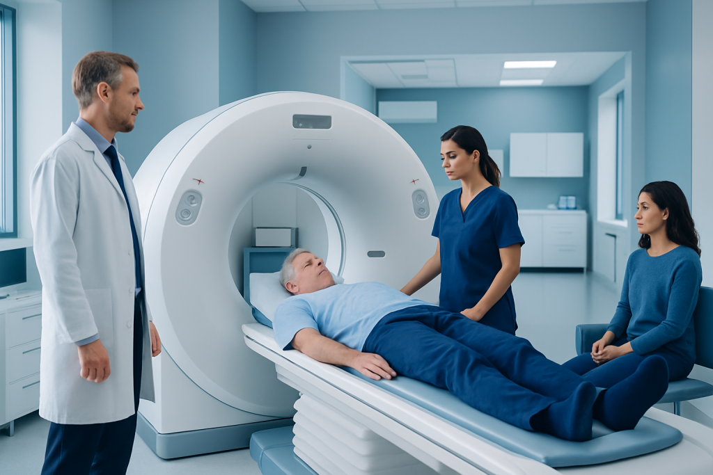
A non-contrast head CT is commonly the first imaging test when stroke is suspected because it is fast and widely available. CT can quickly detect brain bleeding and may show early signs of ischemic stroke or established brain injury.
Key Takeaways
- A non-contrast head CT is commonly the first imaging test when stroke is suspected because it is fast and widely available.
- CT can quickly detect brain bleeding and may show early signs of ischemic stroke or established brain injury.
- CT angiography and CT perfusion may be added to evaluate blocked arteries and potentially salvageable brain tissue.
- A normal early CT scan does not always rule out an ischemic stroke; doctors interpret it alongside symptoms, examination findings and other tests.
- Sudden facial drooping, arm weakness, speech difficulty, vision changes or severe imbalance require emergency assessment immediately.
A CT scan for stroke is usually performed urgently to look for bleeding in the brain and signs of blocked blood flow. It helps the stroke team choose the safest next steps, although early ischemic stroke may not always be visible on an initial non-contrast scan.
CT Scan Stroke: An Overview
A ct scan stroke assessment is an urgent imaging evaluation used when a person has symptoms that could be caused by a stroke. It most often begins with a non-contrast CT scan of the head, which uses X-rays and computer processing to create detailed cross-sectional images of the brain. The main early purpose is to identify or exclude bleeding in the brain, because treatment differs substantially between bleeding-related and blockage-related strokes.
Stroke symptoms can begin suddenly and may include weakness or numbness on one side of the body, facial drooping, trouble speaking or understanding language, loss of balance, new vision problems, or a severe sudden headache. Imaging is only one part of emergency stroke care. The healthcare team also considers the time symptoms began, medical history, medicines, blood tests, blood pressure and neurological examination.
Most strokes are ischemic, meaning a blood vessel supplying the brain has become blocked. A smaller proportion are hemorrhagic, meaning a blood vessel has ruptured and caused bleeding. CT imaging helps distinguish these possibilities promptly and may support decisions about time-sensitive treatments, including stroke treatment.
How a CT Scan Works and Who May Need One
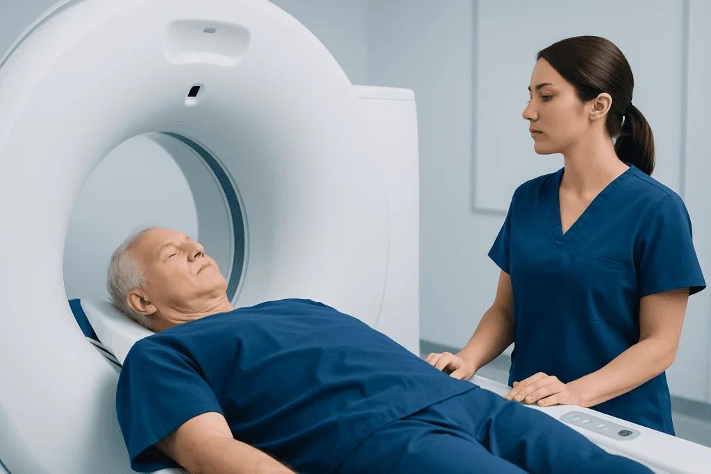
CT, or computed tomography, combines multiple X-ray images taken from different angles. A computer reconstructs these images into slices of the brain, allowing clinicians to look for blood, swelling, large areas of injury, masses or other conditions that can resemble stroke. The scan itself is painless and does not involve entering a narrow tunnel like a conventional MRI examination.
Emergency clinicians may request a head CT for anyone with new symptoms that suggest a possible stroke or transient ischemic attack. It may also be used after head injury, new seizures, sudden confusion, an unexplained severe headache, or a rapid decline in consciousness when brain bleeding must be considered.
Some people undergo additional CT studies with an iodinated contrast agent injected through a vein. CT angiography shows the arteries of the head and neck and can identify certain large-vessel blockages. CT perfusion examines how blood is moving through brain tissue and may help selected patients when the time of symptom onset is uncertain or prolonged. The exact imaging plan is individualized and should not delay essential emergency care.
Pregnancy, kidney disease, prior reactions to contrast material and use of certain medicines should be shared with the care team. These factors do not automatically prevent scanning, but they help clinicians select the most appropriate and safe approach.
Preparing for a CT Scan for Suspected Stroke
In a suspected stroke, preparation is intentionally limited because speed matters. The patient should not drive themselves to an imaging center or wait for a routine appointment. Emergency services can begin assessment on the way to hospital and alert the stroke team before arrival.
Whenever possible, a family member or witness should tell the team when the person was last known to be well, what symptoms were noticed, and whether symptoms have changed. This timing can affect treatment eligibility. Bringing a medication list is helpful, particularly information about anticoagulants, antiplatelet medicines, diabetes treatments and any known contrast allergy.
Patients should not eat or drink if they have swallowing difficulty, reduced alertness or have been advised not to do so. They may be asked to remove jewelry, hairpins, hearing aids, dentures or other removable items around the head and neck. Hospital staff will give clear instructions and will prioritize stabilization of breathing, circulation and blood glucose when needed.
If contrast-enhanced CT is planned, the team may check kidney function or ask about prior contrast exposure where this can be done without delaying urgent care. In a life-threatening situation, clinicians carefully balance potential contrast-related risks against the benefit of identifying a treatable artery blockage.
Step-by-Step: What Happens During the Procedure
On arrival at the scanning area, the patient is usually positioned lying on their back on a padded table. Staff may place a support beneath the head and use gentle positioning aids to reduce movement. The table then moves through the short, doughnut-shaped CT scanner while the images are acquired.
For a basic non-contrast head CT, no injection is required. The patient is asked to remain as still as possible, but the image acquisition itself is generally very brief. The scanner may make soft whirring sounds, and a radiographer can communicate with the patient throughout the examination.
If CT angiography or CT perfusion is needed, staff insert or use an existing intravenous line to inject contrast material. A brief warm sensation, metallic taste or feeling of needing to urinate can occur and usually passes quickly. The scanner then takes images timed to the flow of contrast through the blood vessels and brain.
After the images are completed, a radiologist and stroke clinicians review them urgently. If a large blocked artery is found, the patient may be evaluated for mechanical thrombectomy, a catheter-based procedure that can remove an eligible clot in selected patients. Imaging findings are always considered together with the patient’s symptoms and clinical condition.
What Can CT Scan Results Tell Me About a Stroke?
CT scan results can show whether there is bleeding inside or around the brain, which is essential before treatments that could increase bleeding risk are considered. CT may also reveal swelling, pressure effects, a large established area of infarction, or another explanation for symptoms, such as a brain tumor or certain types of infection.
In ischemic stroke, the earliest non-contrast CT can appear normal, especially soon after symptoms start. This does not mean that a stroke has been excluded. Subtle early changes may be visible to experienced readers, while clearer areas of low density can develop over time as injured tissue becomes more apparent.
CT angiography may show narrowing or blockage in important arteries supplying the brain. CT perfusion can provide information about reduced blood flow and may help identify tissue that is severely affected versus tissue that could still be saved in carefully selected circumstances. These tools may support decisions about urgent reperfusion therapy.
Results should be explained by the stroke team in context. Imaging cannot always predict an individual person’s recovery on its own. Location and extent of brain injury, treatment timing, age, overall health and early response to treatment all contribute to outlook. For more information about the condition itself, see stroke.
How Long Does a CT Scan Take for a Stroke Patient?
The actual image acquisition for a non-contrast CT head scan commonly takes only a few minutes. Positioning the patient, transferring safely to the table, placing an intravenous line if contrast is required and performing additional imaging can make the overall imaging visit longer, but stroke teams work to complete this process as efficiently as possible.
CT angiography and CT perfusion add time, although they are still designed for rapid emergency assessment. The total time varies according to the patient’s ability to lie still, whether contrast is needed, local emergency protocols and whether other immediate medical needs must be addressed first.
Preliminary results may be communicated quickly to the emergency and stroke teams because decisions can be time-sensitive. A formal radiology report may include additional detail after the initial urgent interpretation. The patient or family should ask the treating clinician what the scan shows and how it affects the next treatment step.
What Is the CT Protocol for Stroke Diagnosis?
A CT protocol for stroke diagnosis is a planned sequence of imaging studies selected according to the person’s symptoms, timing and clinical stability. In many hospitals, the first examination is a non-contrast CT of the head. Its key role is to detect acute hemorrhage and evaluate for major early signs of established brain injury.
When clinicians suspect a blockage in a major artery, they may add CT angiography of the head and neck. This evaluates the blood vessels and can identify a possible large-vessel occlusion, narrowing, arterial dissection or other vascular abnormality. In selected patients, CT perfusion may also be used to assess regional blood flow.
Protocols differ between healthcare systems and are updated as evidence and guidelines evolve. They are not a single test that applies identically to every person. The goal is to obtain enough reliable information to make urgent treatment decisions without unnecessary delay or exposure to contrast or radiation.
Other tests may follow, including ECG, blood tests, ultrasound of the neck arteries, echocardiography or MRI. These investigations help clarify the stroke mechanism and plan prevention after the immediate emergency has been managed.
Which Is Better for Stroke, MRI or CT Scan?
Neither test is universally better for every stage and type of stroke. CT is often used first in an emergency because it is fast, accessible and highly effective for identifying acute bleeding. It can also be paired with angiography and perfusion imaging to assess blood vessels and blood flow rapidly.
MRI is more sensitive than non-contrast CT for detecting many small or early ischemic strokes, particularly in some areas such as the brainstem and cerebellum. Diffusion-weighted MRI can identify acute ischemic injury very early. However, MRI may take longer, may not be immediately available and is not appropriate for every patient or emergency setting.
Doctors choose imaging based on the clinical question, symptom onset, the patient’s condition and local resources. A person may have CT first and MRI later to confirm the diagnosis, define the extent of injury or investigate another possible cause of symptoms. The tests are complementary rather than competing options.
After acute care, rehabilitation may be recommended to address movement, communication, swallowing or cognitive changes. Individualized stroke rehabilitation can involve physiotherapists, occupational therapists, speech and language therapists, rehabilitation physicians and other specialists.
Benefits, Risks, Recovery and When to Seek Medical Care
The principal benefit of CT in suspected stroke is speed. Rapid identification of hemorrhage, major vessel blockage or alternative diagnoses supports safer, faster treatment decisions. CT is generally well tolerated, and most people can return to routine activity immediately after a non-contrast scan unless their underlying medical condition requires hospital care.
CT uses ionizing radiation, but the examination is performed only when its expected diagnostic value justifies this exposure. Contrast-enhanced CT carries a small risk of allergic-type reactions and can require additional consideration in people with significant kidney impairment. Staff are trained to monitor for reactions and manage them promptly. A patient who develops rash, breathing difficulty, swelling or dizziness after contrast should alert staff immediately.
When to seek medical care: emergency medical care is needed immediately for sudden facial drooping, weakness or numbness on one side, speech difficulty, new confusion, sudden vision loss, severe dizziness, loss of coordination or a sudden severe headache. Even if symptoms improve within minutes, urgent assessment is important because a transient ischemic attack can be a warning sign of future stroke.
For international patients, Acibadem International’s multidisciplinary specialists and JCI-accredited hospitals can assess suspected stroke and coordinate imaging, acute treatment and rehabilitation planning. Follow-up should focus on the suspected cause of stroke and prevention measures, which may include management of blood pressure, diabetes, cholesterol, heart rhythm conditions, smoking cessation and appropriate physical activity.
Frequently asked questions
Can a CT scan miss a stroke?
Yes. A non-contrast CT scan can be normal early in an ischemic stroke, particularly when the affected area is small or symptoms began recently. It remains very important because it can rapidly identify bleeding and other urgent causes of symptoms. Clinicians may use CT angiography, CT perfusion or MRI when further clarification is needed.
Does a CT scan for stroke require contrast dye?
The initial head CT for suspected stroke is commonly performed without contrast. Contrast may be used for CT angiography or CT perfusion when the team needs to assess brain blood vessels or blood flow. The decision depends on the clinical situation and factors such as kidney function and prior contrast reactions.
Can I eat before a CT scan for suspected stroke?
A person with possible stroke should seek emergency care rather than try to prepare for a scan at home. Because swallowing may be affected and urgent procedures may be needed, healthcare professionals may ask the patient not to eat or drink until assessed. The emergency team will provide instructions based on the patient’s condition.
When will I get the results of a stroke CT scan?
In suspected stroke, images are reviewed urgently because results may guide immediate treatment. The treating team may share initial findings soon after the scan, while a detailed radiology report may follow. Timing can vary according to the emergency workload and whether additional imaging is required.
Is a CT scan painful?
A CT scan is painless, although remaining still on the table may be uncomfortable for some people. If contrast is injected, a short-lived warm feeling or metallic taste may occur. Staff can help patients who are anxious, in pain or unable to lie flat comfortably.
What happens after a CT scan confirms a stroke?
Next steps depend on whether the stroke is caused by bleeding or a blocked blood vessel, as well as the scan findings, symptom timing and overall health. The patient may receive urgent medicines, a catheter procedure, monitoring in a stroke unit and supportive care. Further tests are often arranged to identify the likely cause and reduce the chance of another stroke.
References
- American Heart Association/American Stroke Association
- National Institute of Neurological Disorders and Stroke
- Radiological Society of North America
- National Health Service
- World Stroke Organization
This article is for general information only and is not a substitute for professional medical advice. Please consult a qualified doctor about your individual situation.
Explore treatments in Turkey — costs, top hospitals & a free quote
JCI-accredited · board-certified surgeons · reply within 24h
Check your numbers in seconds
BMI, calories, due date, blood pressure and 30+ more clinical calculators — free, instant, doctor-reviewed ranges.