CT Urogram — Explained by Medical Evidence, Not Myths
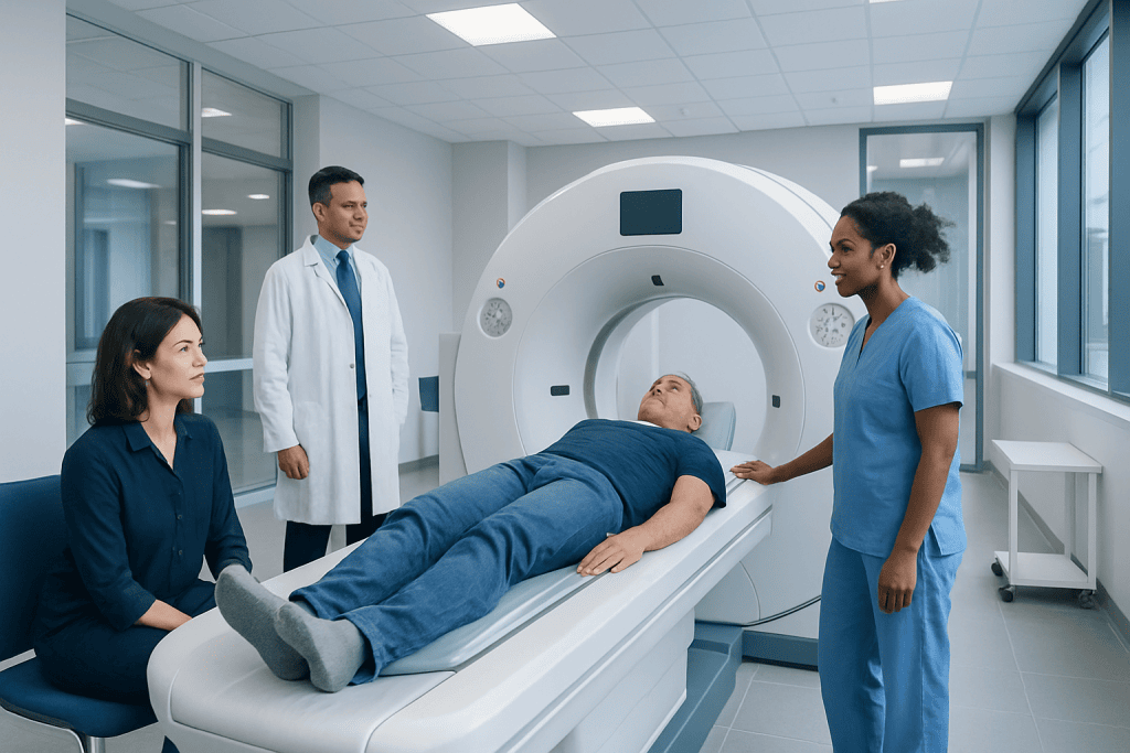
A ct urogram examines the kidneys, ureters, and bladder in detail, often using intravenous contrast. It is commonly ordered for blood in the urine, suspected urinary tract blockage, kidney stones, or masses.
Key Takeaways
- A ct urogram examines the kidneys, ureters, and bladder in detail, often using intravenous contrast.
- It is commonly ordered for blood in the urine, suspected urinary tract blockage, kidney stones, or masses.
- Preparation may include hydration, temporary fasting, and checking kidney function before contrast is given.
- The test is usually quick and noninvasive, but it does involve radiation and may not suit everyone.
- Results help guide whether follow-up, medication, cystoscopy, or other treatment is needed.
A ct urogram is a computed tomography scan that creates detailed images of the kidneys, ureters, and bladder, usually before and after contrast dye. It is commonly used to investigate blood in the urine, pain, recurrent urinary symptoms, or possible blockage, stones, or tumors.
What a CT Urogram Is
A ct urogram is a specialized CT scan that looks closely at the urinary tract: the kidneys, the tubes that carry urine to the bladder (ureters), and the bladder itself. In many cases, contrast dye is injected into a vein so the radiologist can see how the kidneys filter the dye and how urine flows through the system. This helps reveal structural problems that may not be clear on an ordinary X-ray or ultrasound.
Doctors most often request a ct urogram when they need a more complete picture of why urinary symptoms are happening. Typical reasons include blood in the urine, repeated urinary tract symptoms, flank pain, suspected kidney stones, blockage, or concern about a mass. The scan can show both the anatomy and, to some extent, how the urinary tract is functioning during the passage of contrast.
It is helpful to think of a ct urogram as a targeted imaging study rather than a diagnosis by itself. The scan provides detailed visual evidence that a doctor combines with symptoms, urine tests, blood tests, and medical history. This evidence-based approach can help distinguish common conditions such as stones or infection from problems that need closer follow-up, including bladder cancer or kidney cancer.
Why a Doctor May Recommend It
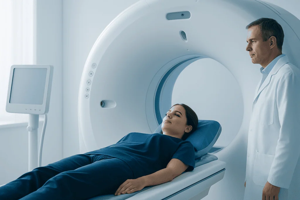
A ct urogram is commonly recommended when a symptom needs more than a routine urine test or basic imaging. One of the main reasons is hematuria, which means blood in the urine. Sometimes the blood is visible, and sometimes it is only found under the microscope. Because blood in the urine can have many causes, a detailed scan can help narrow the possibilities.
Other reasons include suspected kidney stones, repeated urinary tract infections, urinary obstruction, swelling of the kidney, unexplained flank or lower abdominal pain, or concern about an abnormality seen on another scan. It may also be used when a doctor wants to evaluate congenital differences in the urinary tract or look for complications after surgery or other urologic treatment.
In some situations, a ct urogram is chosen because it can detect several potential causes at once. It can identify stones, masses, narrowing, swelling, clots, and changes in the lining of the collecting system. Depending on the symptoms, a doctor may pair imaging findings with other evaluations such as urine cytology or cystoscopy to examine the inside of the bladder directly.
How the Test Is Done and How to Prepare

Before the scan, the imaging team usually reviews medical history, allergies, kidney function, medicines, and whether pregnancy is possible. Patients are often advised to drink water, and some centers ask them not to eat for a few hours beforehand if intravenous contrast will be used. Preparation can vary, so it is important to follow the exact instructions provided by the imaging department.
During the appointment, the patient lies on a table that moves through the CT scanner. Images may be taken before contrast, shortly after contrast, and again after a delay so the dye has time to pass through the urinary tract. The contrast is usually given through a small intravenous line. Some people notice a warm sensation, a metallic taste, or a brief urge to urinate when the dye is injected; these effects are usually short-lived.
The scan itself is generally quick, but the whole visit can take longer because of check-in, IV placement, and delayed imaging phases. Patients are asked to stay still, and at times they may be told to hold their breath for a few seconds. After the study, drinking fluids is often encouraged unless a doctor has advised otherwise.
- Bring a list of current medicines and allergies.
- Ask whether diabetes medicines or blood thinners need special instructions.
- Tell the team about prior reactions to contrast dye.
- Mention any history of kidney disease, asthma, or pregnancy.
What a CT Urogram Can Show
The scan can reveal many different findings within the urinary tract. It may show kidney stones, obstruction caused by a narrowing or clot, swelling of the kidney due to backed-up urine, structural differences present from birth, or signs of inflammation. It can also help detect masses in the kidney or changes affecting the lining of the ureters and bladder.
For blood in the urine, the value of a ct urogram is that it looks beyond one single diagnosis. The cause may be a stone, infection, trauma, an enlarged prostate in men, or a lesion higher in the urinary tract. The images can help a doctor determine whether symptoms are most consistent with a temporary problem or whether additional evaluation is needed.
That said, not every urinary condition is best diagnosed with this test alone. A ct urogram does not replace urine culture for infection, and it may not be the first choice for every person with suspected stones or for children and pregnant patients. If another issue is suspected, the care team may combine imaging with tests or procedures such as kidney stone treatment planning or a referral for urology evaluation.
Benefits, Limits, and Possible Risks
The main benefit of a ct urogram is detail. It can examine the kidneys, ureters, and bladder in a way that is often more comprehensive than ultrasound and more practical than multiple separate tests. This can make it especially useful when symptoms are unclear or when the goal is to rule out several possible causes with one study.
Like all tests, it also has limits. Some very small abnormalities may still be hard to interpret, and imaging findings do not always explain symptoms completely. A normal scan does not automatically mean there is no urinary problem, which is why doctors may still recommend follow-up testing if symptoms continue or if there is ongoing concern based on age, risk factors, or urine results.
The main risks are exposure to ionizing radiation and possible side effects from contrast dye. Most people tolerate contrast well, but reactions can happen, ranging from mild nausea or itching to uncommon allergic-type responses. Contrast can also be a concern in people with reduced kidney function. In selected situations, a doctor may choose a different test, such as ultrasound or MRI, if the risks outweigh the benefits.
Understanding the Results and Next Steps
A radiologist interprets the images and sends a report to the doctor who ordered the test. Results may be described as normal, or they may mention stones, cysts, masses, thickening, dilation, obstruction, or other changes. Some findings are clearly benign, while others need more evaluation to understand what they mean in the context of the patient’s symptoms.
Next steps depend on what the scan shows and why it was ordered. If the issue is a stone, care may range from monitoring and pain control to planning a procedure. If there is a suspicious mass or unexplained thickening, the doctor may recommend more imaging, urine tests, specialist review, or a direct examination of the urinary tract. If the scan suggests a blockage or ongoing urinary retention, treatment may focus on relieving the obstruction and protecting kidney function.
When symptoms persist despite a normal or unclear scan, the doctor may still advise follow-up. This may include repeat urine testing, culture, blood work, or a visit with a specialist. At experienced centers, including Acibadem International, multidisciplinary specialists in JCI-accredited hospitals evaluate urinary tract symptoms and imaging findings for international patients in a coordinated way.
When to Seek Medical Care
Medical care should be sought promptly for visible blood in the urine, severe flank pain, fever with urinary symptoms, difficulty passing urine, or persistent vomiting with pain. These symptoms do not always indicate an emergency, but they do deserve timely evaluation because they can be linked to stones, infection, or obstruction.
It is also sensible to contact a doctor if there is recurrent microscopic blood in the urine, repeated urinary tract infections, unexplained weight loss, worsening urinary frequency, or pain that does not improve. People with a history of smoking, prior urinary tract cancers, kidney disease, or occupational chemical exposure may need a more careful assessment when urinary symptoms occur.
Emergency help may be needed if there is severe pain with fever, confusion, inability to urinate, fainting, or signs of a serious allergic reaction after contrast exposure, such as trouble breathing or swelling of the face or throat. Even when symptoms seem mild, a qualified clinician can advise whether observation is enough or whether imaging and specialist evaluation are appropriate.
Frequently asked questions
Is a ct urogram the same as a regular CT scan?
Not exactly. A ct urogram is a CT study designed specifically to evaluate the urinary tract, often using timed images after intravenous contrast. Those extra phases help show the kidneys, ureters, and bladder in greater detail.
Why is contrast dye often used in a ct urogram?
Contrast helps outline the urinary tract as the kidneys filter it into the urine. This can make it easier to detect blockage, masses, narrowing, and abnormalities in the lining of the collecting system. In some people, contrast may be avoided if kidney function is reduced or if there is a history of significant reaction.
Does a ct urogram hurt?
The scan itself is usually painless. The main discomfort is often the IV insertion, and some people briefly feel warmth, a metallic taste, or a sensation of needing to urinate when contrast is injected. These sensations generally pass quickly.
How long does a ct urogram take?
The actual imaging is often quick, but the total visit may take longer because images are sometimes taken in several phases. Delayed images allow time for contrast to move through the urinary tract. The imaging center can explain the expected timing for that specific protocol.
Can a ct urogram detect cancer?
It can help identify findings that raise concern for cancer in the kidneys, ureters, or bladder, and it is often used when blood in the urine needs evaluation. However, it does not confirm every diagnosis by itself. Further tests, including cystoscopy, laboratory studies, or biopsy, may still be needed.
Who may need a different test instead of a ct urogram?
Pregnant patients, some children, and people with certain contrast allergies or reduced kidney function may need an alternative approach. Depending on the clinical question, a doctor may choose ultrasound, MRI, or a non-contrast CT scan instead. The best test depends on symptoms, age, medical history, and the suspected problem.
References
- American College of Radiology
- Radiological Society of North America
- National Institute of Diabetes and Digestive and Kidney Diseases
- Urology Care Foundation
- National Cancer Institute
This article is for general information only and is not a substitute for professional medical advice. Please consult a qualified doctor about your individual situation.
Explore treatments in Turkey — costs, top hospitals & a free quote
JCI-accredited · board-certified surgeons · reply within 24h