Fetal Echo Ultrasound: Preparation, Procedure and Results

A fetal echo ultrasound provides a detailed assessment of the developing heart and circulation. Most pregnant people do not need to fast or make major changes to their usual routine before the test.
Key Takeaways
- A fetal echo ultrasound provides a detailed assessment of the developing heart and circulation.
- Most pregnant people do not need to fast or make major changes to their usual routine before the test.
- The scan is commonly performed between 18 and 24 weeks of pregnancy, although timing can vary.
- Many fetal echocardiogram results are reassuring; an abnormal result does not always mean a severe heart condition.
- If a concern is found, follow-up may include repeat imaging, genetic counseling, pregnancy monitoring and birth planning.
A fetal echo ultrasound, also called a fetal echocardiogram, is a specialized ultrasound examination that evaluates a baby’s heart structure, rhythm and blood flow before birth. It is painless, uses no radiation and usually needs very little preparation; a fetal cardiology team explains the findings and any next steps.
Fetal Echo Ultrasound Overview
A fetal echo ultrasound is a detailed imaging test that examines a baby’s heart before birth. It uses the same sound-wave technology as a routine pregnancy ultrasound, but it is performed with a focused assessment of the heart chambers, valves, major blood vessels, heartbeat and blood flow. The test is also called a fetal echocardiogram.
The examination may be recommended after a routine prenatal scan shows a possible heart-related finding, or when there is a factor that increases the chance of a congenital heart difference. It does not use ionizing radiation, and it does not involve needles or injections. In most cases, it is an outpatient appointment and the pregnant person can return to normal activities immediately afterward.
A fetal echo ultrasound can provide important information for the pregnancy care team and parents. It may confirm that the heart appears normal, clarify an uncertain routine ultrasound finding, or identify a condition that benefits from closer monitoring and coordinated care before and after delivery.
How a Fetal Echo Ultrasound Works and Who May Need One
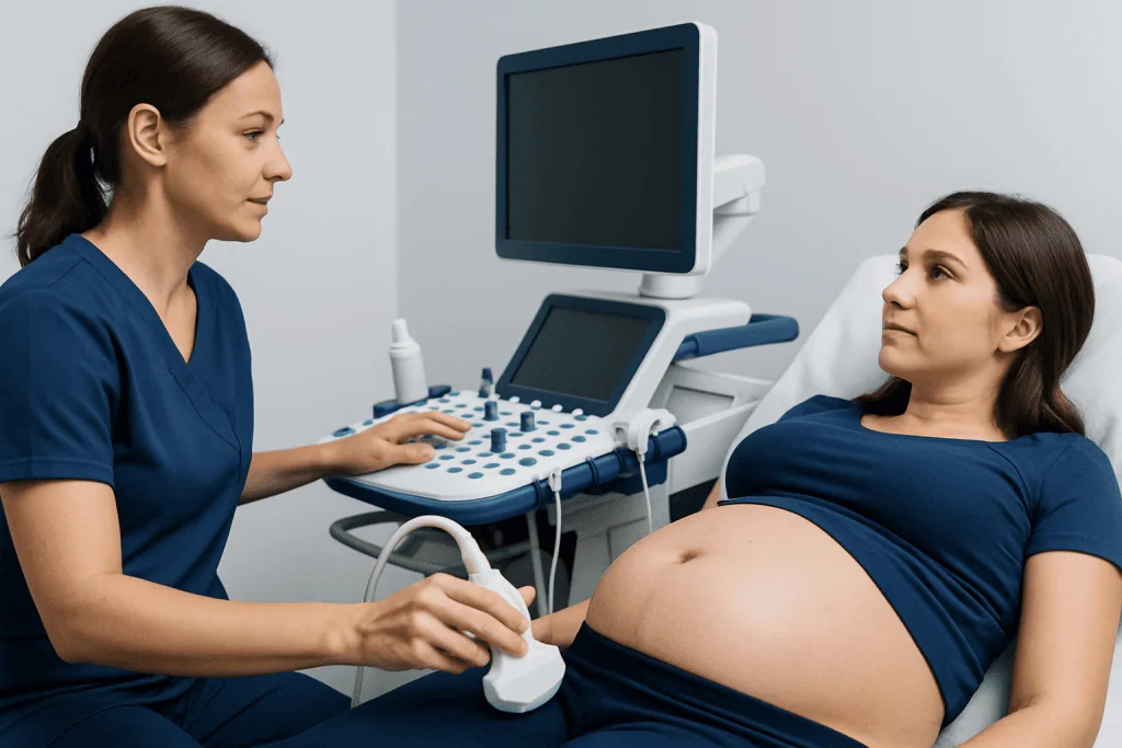
During a fetal echocardiogram, a specially trained clinician uses an ultrasound transducer on the abdomen to create moving images of the fetal heart. Doppler ultrasound may also be used to show the direction and speed of blood flow. The examiner assesses the heart’s four chambers, valves, pumping function, major vessels and rhythm, while taking into account the baby’s position and gestational age.
A referral may be appropriate when a routine anatomy ultrasound suggests a possible heart difference, the heartbeat appears irregular, or the images were limited. A fetal echo may also be advised when there is a parent, sibling or previous child with congenital heart disease; certain maternal medical conditions; exposure to selected medicines early in pregnancy; or a fetal genetic or structural finding associated with heart differences.
Not every pregnancy with a risk factor will reveal a heart condition, and not every suspected finding on a routine scan is confirmed. The purpose of the fetal echo ultrasound is to obtain a more complete and accurate view. The test is often performed around 18 to 24 weeks of pregnancy, though it can be done earlier or later when clinically needed.
What to Do Before a Fetal Echo

Do I need to be on an empty stomach for a fetal echo test? Usually, no. Most people do not need to fast for a fetal echo ultrasound and can eat and drink normally unless their own clinician gives different instructions. Fasting is not generally part of fetal echo prep because the examination is performed through the abdomen and does not require sedation.
What to do before a fetal echo? The most useful fetal echocardiogram preparation is to bring relevant pregnancy records, routine ultrasound reports and information about medicines, health conditions and family heart history. Comfortable two-piece clothing can make the abdominal scan easier. It can also be helpful to allow enough time for the visit, as image collection may take longer when the baby is moving or positioned in a way that limits views.
There is usually no special fetal ultrasound preparation involving a full bladder for a mid-pregnancy fetal echocardiogram. However, individual instructions can differ depending on gestational age, the imaging center and other planned tests. If there is any uncertainty, the pregnant person should contact the referring obstetric team or imaging department before the appointment.
Fetal Echo Ultrasound Protocol: What Happens During the Test
A typical fetal echo ultrasound protocol begins with a review of the reason for referral and relevant medical and family history. The pregnant person lies on an examination couch, often slightly tilted or with support under one side to remain comfortable. A clear gel is applied to the abdomen, and the transducer is moved across the skin to obtain images from different angles.
The clinician records multiple views of the heart and uses Doppler techniques to assess blood flow through the chambers, valves and major vessels. The baby’s position, movement, maternal body shape and pregnancy stage can affect image quality. A short break, a change in position or a repeat appointment may occasionally be needed to obtain the clearest possible views.
The scan commonly lasts about 30 to 60 minutes, though this varies. It should not be painful, but pressure from the transducer and remaining still can be uncomfortable for some people. The procedure is noninvasive, and there is no recovery period. The person may eat, drink, travel and continue usual activities after the appointment unless their care team advises otherwise.
The main benefit is detailed prenatal information that can guide monitoring and delivery planning when needed. Ultrasound has an established safety record in pregnancy when medically indicated and performed by trained professionals. Its limitations are also important: very small defects, some valve abnormalities and conditions that develop later in pregnancy may not always be detected before birth.
Do You Get Fetal Echo Results Right Away?
Do you get fetal echo results right away? Often, the specialist can discuss preliminary findings at the end of the appointment, particularly when the images are clear and reassuring. A formal report is usually sent to the referring obstetrician or maternal-fetal medicine team after the images have been reviewed and documented.
In some situations, interpretation takes longer. Additional image review, a repeat scan or discussion with other specialists may be appropriate if the baby’s position limited the examination or a possible finding needs confirmation. The care team should explain when and how final results will be communicated.
A normal fetal echo is reassuring, but it cannot exclude every possible heart condition. Certain small openings in the heart, mild valve changes, rhythm concerns that come and go, and changes that become more apparent later in pregnancy or after birth may not be visible at the time of scanning. The clinician can explain what the test can and cannot show in the individual situation.
What If a Fetal Echo Is Abnormal?
What if a fetal echo is abnormal? An abnormal result means that the team has identified, or suspects, a difference in heart structure, blood flow or rhythm. It does not automatically indicate a severe condition, and the significance varies widely. Some findings need only observation, while others benefit from specialist follow-up during pregnancy and a planned evaluation after birth.
The next step may include a repeat fetal echocardiogram, consultation with a fetal or pediatric cardiologist, and additional prenatal imaging. Depending on the finding, genetic counseling or testing may be discussed as an option, not a requirement. The obstetric and cardiology teams can describe the expected course, available monitoring and any implications for where and how delivery is planned.
When a significant congenital heart condition is suspected, coordinated care can help families prepare. This may involve maternal-fetal medicine specialists, neonatologists, pediatric cardiologists and cardiac surgeons. Information about related conditions may be useful when a specialist has identified a specific concern, including congenital heart disease.
Parents should feel welcome to ask for clear explanations, written information and time to consider recommendations. A second specialist opinion may also be reasonable when a complex fetal heart finding is suspected.
When to Seek Medical Care
A fetal echo ultrasound is usually arranged through prenatal care rather than as an emergency test. A pregnant person should contact their obstetrician, midwife or maternity assessment service promptly if they have been told that a routine ultrasound showed a possible fetal heart concern, if they have questions about a referral, or if they have not received planned follow-up information.
Urgent maternity advice is important for symptoms such as vaginal bleeding, fluid leakage, severe or persistent abdominal pain, regular painful contractions before term, fever, severe headache, sudden swelling, vision changes or a noticeable reduction in fetal movement after movements have become established. These symptoms are not specific to fetal heart problems, but they require timely assessment in pregnancy.
For people traveling for care, multidisciplinary specialists at Acibadem International and its JCI-accredited hospitals can coordinate fetal cardiology assessment with obstetric, maternal-fetal medicine and neonatal teams for international patients. Individual care plans should always be based on the pregnancy, scan findings and advice from qualified clinicians.
Frequently asked questions
Is a fetal echo ultrasound safe for the baby?
A fetal echo ultrasound uses sound waves, not radiation, and is considered safe when performed for a medical reason by appropriately trained professionals. It is noninvasive and does not involve injections or contrast dye. The team uses the examination time and settings needed to obtain clinically useful images.
How long does a fetal echocardiogram take?
Many fetal echocardiograms take approximately 30 to 60 minutes. The duration can vary because the examiner needs several detailed views and the baby’s position may affect how easily images are obtained. A repeat scan may occasionally be arranged if all views cannot be completed.
Can a fetal echo detect every heart problem?
No prenatal test can detect every heart condition. A fetal echocardiogram is very detailed, but some small defects, mild valve problems, intermittent rhythm changes and conditions that develop later may not be apparent during the scan. Newborn examination and, when indicated, postnatal heart testing remain important.
Why would a routine pregnancy ultrasound lead to a fetal echo referral?
Routine pregnancy ultrasounds provide screening views of the heart, but they are not always designed to evaluate every cardiac structure in depth. A referral may occur when an image is unclear, the heart appears different from expected, or there is a risk factor in the pregnancy or family history. Referral is a careful step to obtain more detailed information and does not by itself confirm a diagnosis.
Can a fetal echocardiogram show an irregular heartbeat?
Yes. The examination can assess fetal heart rhythm and may help identify whether a rhythm appears too fast, too slow or irregular. Some rhythm changes are temporary, while others need monitoring by a fetal and pediatric cardiology team. The recommended follow-up depends on the rhythm pattern and the baby’s overall condition.
What happens after a normal fetal echo?
If the fetal echo is normal, the person usually returns to routine prenatal care unless another pregnancy issue requires monitoring. The referring clinician will explain whether any additional scan is needed. A normal result is reassuring, while recognizing that a small number of heart differences may only become detectable later or after birth.
References
- American College of Obstetricians and Gynecologists
- American Institute of Ultrasound in Medicine
- American Heart Association
- International Society of Ultrasound in Obstetrics and Gynecology
This article is for general information only and is not a substitute for professional medical advice. Please consult a qualified doctor about your individual situation.
Ultrasound in Turkey — costs, top hospitals & a free quote
JCI-accredited · board-certified surgeons · reply within 24h
Add us as a Preferred Source to see more of our trusted health content across Google Search, AI Overviews and Discover.

Check-up & Preventive Medicine
Comprehensive health screenings designed for early detection and prevention.
3 specialists in this unit