Glioma Cancer Treatment: How It Works, Results and What to Expect
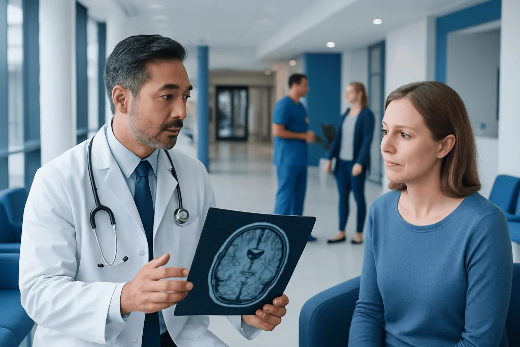
Gliomas are brain and spinal cord tumors that vary greatly in behavior, from slower-growing to aggressive forms. Surgery is often the first treatment when a tumor can be safely reached, both to obtain a diagnosis and reduce tumor volume.
Key Takeaways
- Gliomas are brain and spinal cord tumors that vary greatly in behavior, from slower-growing to aggressive forms.
- Surgery is often the first treatment when a tumor can be safely reached, both to obtain a diagnosis and reduce tumor volume.
- Radiation and drug therapy are commonly used after surgery or when surgery is not appropriate.
- Molecular testing helps clinicians classify gliomas and choose a more personalized treatment approach.
- Outlook varies widely; some gliomas can be controlled for years, while higher-grade gliomas usually require ongoing care.
- New or worsening neurological symptoms, seizures or sudden severe headache need prompt medical assessment.
Glioma cancer treatment is individualized and may include surgery, radiation therapy, chemotherapy or other drug treatments, tumor-treating fields, clinical trials and supportive care. The treatment plan depends on the glioma’s grade, location, molecular features, how much can be safely removed and the person’s overall health and goals.
Overview: How glioma cancer treatment works
Glioma cancer treatment aims to diagnose the tumor accurately, remove or reduce as much tumor as can be done safely, slow remaining tumor cells and protect daily function. Treatment is planned by a multidisciplinary team and commonly combines surgery, radiation therapy and medicines. The exact approach differs substantially between low-grade gliomas and higher-grade gliomas, including glioblastoma.
Gliomas begin in glial cells, which support nerve cells in the brain and spinal cord. They include astrocytomas, oligodendrogliomas and other related tumor types. Modern classification uses the tumor’s appearance under a microscope together with molecular findings, such as changes involving IDH genes and chromosome regions. These features can affect expected behavior and treatment decisions.
A specialist team usually includes neurosurgeons, neuro-oncologists, radiation oncologists, neuroradiologists, neuropathologists, neurologists, rehabilitation professionals and supportive-care clinicians. Care is designed around both tumor control and quality of life, including thinking, movement, speech, mood and independence.
Who may be a candidate for each treatment?
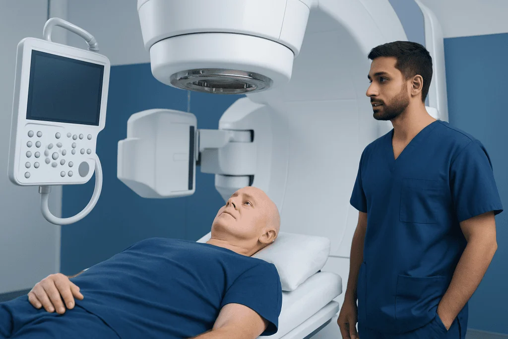
Most people with a newly diagnosed glioma benefit from evaluation at a center experienced in brain tumors. Whether surgery, radiation or medication is advised depends on the tumor’s location and size, imaging findings, symptoms, growth rate, molecular profile and whether it is newly diagnosed or has returned after treatment.
Surgery may be suitable when a neurosurgeon believes tissue can be obtained or tumor can be removed without unacceptable risk to important functions. Tumors close to areas controlling language, movement, vision or memory may require specialized planning, such as functional imaging, awake mapping or intraoperative monitoring. In some cases, a biopsy alone is safer than attempted removal.
Radiation therapy and systemic treatments may be offered after surgery, as primary treatment when surgery is not feasible, or for recurrent disease. A person’s age, general health, neurological function, prior treatments and preferences are all important. For people with symptoms from swelling, seizures or reduced function, supportive medicines and rehabilitation can begin alongside tumor-directed care.
The treatment pathway: step by step
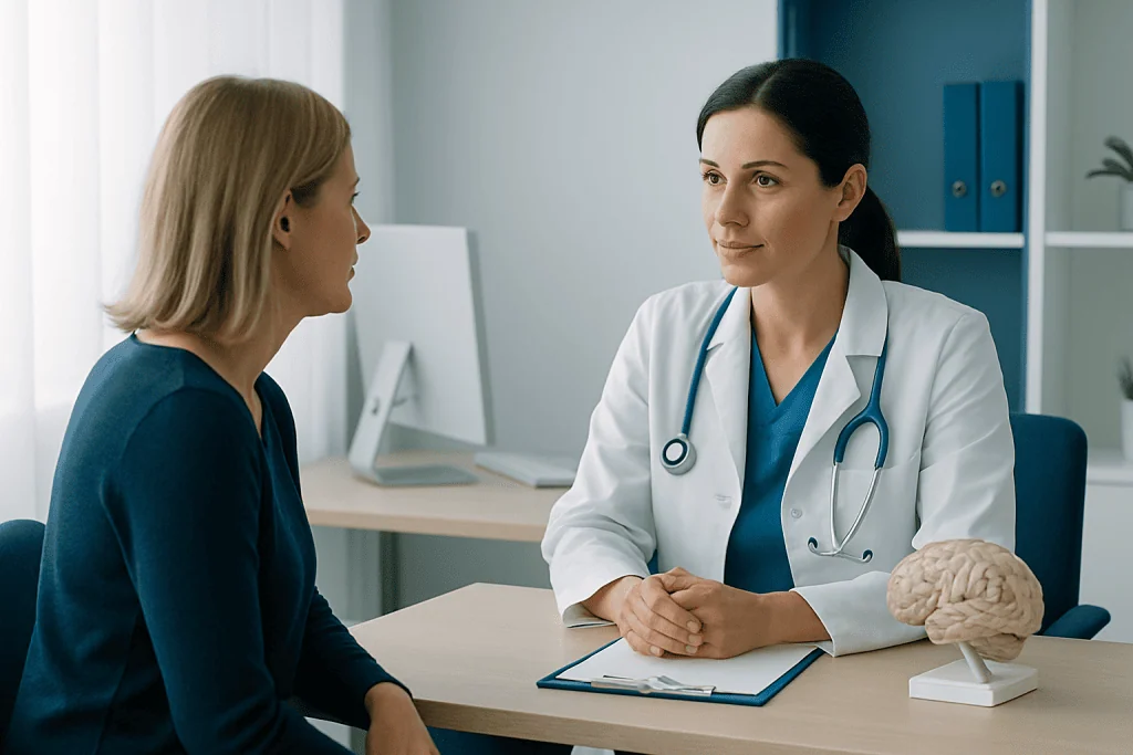
The process usually starts with magnetic resonance imaging (MRI) and a detailed neurological examination. MRI helps show the tumor’s size, location and relationship to sensitive brain structures, but imaging alone cannot always identify the precise tumor type. A biopsy or surgical removal provides tissue for pathology and molecular testing.
When surgery is recommended, the neurosurgical team plans the safest route to the tumor using detailed scans and navigation technology. The operation is performed under anesthesia in most cases. For selected tumors near language or movement areas, awake surgery may be used so the team can monitor important functions while removing tissue. The goal is maximal safe resection, not removal at the expense of major neurological harm.
After surgery, pathology and molecular results are reviewed by the multidisciplinary team. Radiation therapy may be delivered externally in carefully planned daily sessions over several weeks. Drug treatment may be given during or after radiation, or in other schedules depending on the glioma type. Brain tumor treatment may also involve therapies for recurrent disease, symptom management, rehabilitation and consideration of suitable clinical trials.
Follow-up MRI scans are essential because gliomas can change over time. The team compares scans, symptoms and treatment effects to decide whether surveillance, continued treatment or a different strategy is appropriate.
Benefits, limits and possible risks
The potential benefit of surgery is obtaining a reliable diagnosis, relieving pressure from the tumor and reducing the amount of tumor that needs further treatment. In many gliomas, removing more tumor safely is associated with better disease control, but the safest achievable extent of surgery differs from person to person.
Radiation therapy can slow tumor growth and reduce the chance of progression after surgery. Drug therapies can also improve tumor control in particular glioma types. However, treatment cannot always eliminate every tumor cell, especially where cells have spread microscopically into surrounding brain tissue. Some gliomas return despite appropriate care.
Possible surgical risks include bleeding, infection, seizures, stroke-like injury, fluid leakage and changes in speech, strength, sensation, vision, memory or personality. Radiation can cause fatigue, scalp irritation, temporary worsening from swelling and, less commonly, delayed effects on cognition or hormone function. Drug-treatment effects vary, but may include fatigue, nausea, lowered blood counts or infection risk. The care team monitors for complications and discusses ways to reduce them.
- Antiseizure medicines may be used when a person has had seizures.
- Corticosteroids may temporarily reduce swelling and related symptoms.
- Physical, occupational and speech therapy can support recovery and daily function.
- Psychological, nutritional and palliative-care support can be helpful at any stage of illness.
Recovery timeline and follow-up care
Recovery after a biopsy or craniotomy varies with the tumor location, extent of surgery and a person’s baseline health. Many people stay in hospital for several days after surgery, while some need a longer stay or inpatient rehabilitation. Tiredness, headaches and temporary neurological symptoms can occur in the early recovery period.
The pathology report and molecular testing may take days to a few weeks, depending on the analyses required. A post-operative MRI is often performed soon after surgery to assess the extent of resection. Once the wound has healed and treatment planning is complete, radiation and medication may begin if indicated.
During the following months, care focuses on surveillance imaging, managing symptoms and rebuilding function. Some changes improve as swelling settles and rehabilitation progresses; others may be longer lasting. Patients should tell their team about new headaches, seizures, weakness, confusion, personality changes, problems with speech or worsening balance, rather than waiting for the next scheduled visit.
Acibadem International’s multidisciplinary specialists and JCI-accredited hospitals assess and treat gliomas for international patients, coordinating neurosurgery, oncology, radiation treatment and rehabilitation when needed.
How long do people live with gliomas?
How long a person lives with a glioma depends on factors that are specific to the individual and tumor. These include the glioma type and grade, molecular characteristics, age, neurological function, tumor location, how much tumor can be safely removed and response to treatment. It is not possible to estimate an individual outcome accurately from the word “glioma” alone.
Some lower-grade gliomas may grow slowly and can be managed for many years, although they still need regular monitoring and may require treatment over time. Higher-grade gliomas generally grow faster and have a more serious outlook, but people’s experiences and treatment responses differ. A neuro-oncology team can discuss prognosis using the complete pathology and imaging information.
It can be helpful for patients and families to ask what the goals of treatment are now, what changes may be expected and how follow-up results will guide future decisions. Supportive and palliative care can improve comfort, function and communication throughout treatment; it is not limited to end-of-life care.
Can gliomas be completely cured?
Whether a glioma can be completely cured depends largely on its type, grade, molecular features and location. Some slow-growing tumors can be fully removed when they are small and located in an area where surgery can be performed safely. Even then, ongoing MRI follow-up is important because some gliomas can recur after a long interval.
Many gliomas are difficult to cure completely because tumor cells may extend beyond what is visible on scans or what can be safely removed from brain tissue. For these tumors, treatment is often focused on long-term control, delaying progression, preserving neurological abilities and managing symptoms.
Patients may hear terms such as “no evidence of disease,” “stable disease,” “response” or “progression.” These terms describe the current status rather than guaranteeing a permanent cure. Discussing the meaning of scan results with the treating team can prevent misunderstanding and help guide realistic planning.
How serious is a glioma tumor? Can people survive gliomas?
A glioma tumor should always be assessed by qualified clinicians because it occurs in the brain or spinal cord, where even a small tumor can affect important functions. Seriousness ranges widely: some gliomas are slower growing and manageable over extended periods, while others are aggressive and need prompt, intensive treatment. Symptoms and scans alone do not determine seriousness; tissue diagnosis and molecular testing are central.
People can survive gliomas, and many live meaningful lives during and after treatment. Survival differs considerably according to the features of the tumor and the person’s health, so broad predictions can be misleading. Advances in surgical techniques, precision radiation planning, molecular diagnosis, rehabilitation and supportive care continue to improve how treatment is tailored.
Families should consider asking the team about the tumor’s integrated diagnosis, treatment goal, possible impact on daily activities and the support available. Related conditions that may be evaluated within neuro-oncology include brain tumors, although each tumor diagnosis requires its own management plan.
When to seek medical care
New neurological symptoms should be assessed by a doctor, particularly if they are persistent, progressive or interfere with everyday activities. Symptoms that can occur with a glioma include a first seizure, worsening headaches, nausea or vomiting with headache, weakness or numbness on one side, balance difficulty, speech problems, vision changes, confusion or noticeable changes in personality or behavior. These symptoms can also have causes other than a tumor, but they should not be ignored.
Emergency medical care is appropriate for a seizure lasting more than five minutes, repeated seizures without full recovery, sudden severe headache, new facial drooping, sudden weakness, loss of consciousness or severe confusion. Someone having a seizure should be kept safe from injury, placed on their side if possible and should not be restrained or given food or drink until fully alert.
For people already diagnosed with a glioma, prompt contact with the care team is important for new seizures, rapidly worsening headache, fever after surgery, wound redness or drainage, increasing drowsiness or a sudden decline in neurological function. Timely review can help identify treatable causes such as swelling, infection, medication effects or tumor progression.
Frequently asked questions
What is the first-line glioma cancer treatment?
Surgery is often the first treatment when it can be performed safely, because it provides tissue for diagnosis and can reduce tumor volume. However, some tumors are best approached with a biopsy, radiation therapy, drug treatment or a combination of these options. The choice depends on the tumor’s location, type and molecular findings.
Does every glioma require surgery?
No. Surgery is not always the safest or most appropriate option, especially when a tumor is deep in the brain or close to critical areas controlling movement, speech or other functions. A stereotactic biopsy may be used to obtain a diagnosis, followed by radiation and/or medication when appropriate.
What happens after glioma surgery?
After surgery, patients are monitored in hospital and usually have follow-up brain imaging to assess the operation. Pathology and molecular testing help determine whether radiation, chemotherapy or another treatment is needed. Rehabilitation and symptom management may be part of recovery.
Can radiation therapy cure a glioma?
Radiation therapy can be an important treatment for controlling glioma growth and reducing the risk of progression, particularly after surgery. Whether it can provide a cure depends on the individual tumor type and other factors. It is often used as part of a combined treatment plan rather than as a stand-alone cure.
How often are MRI scans needed after glioma treatment?
MRI schedules vary according to tumor grade, treatment stage, scan results and symptoms. Scans are often more frequent soon after treatment and may become less frequent when the tumor is stable. The treating neuro-oncology team sets an individual surveillance plan.
Can a glioma come back after treatment?
Yes, some gliomas can recur or progress after treatment, including tumors that were initially controlled. Follow-up MRI scans help detect changes early. If recurrence occurs, options may include further surgery, radiation in selected cases, medicines, clinical trials and supportive care.
References
- World Health Organization
- National Cancer Institute
- National Institute of Neurological Disorders and Stroke
- European Association of Neuro-Oncology
- American Cancer Society
This article is for general information only and is not a substitute for professional medical advice. Please consult a qualified doctor about your individual situation.
Oncology care in Turkey — second opinion and treatment plan
JCI-accredited · board-certified surgeons · reply within 24h
Add us as a Preferred Source to see more of our trusted health content across Google Search, AI Overviews and Discover.