Hilum: What Patients Need to Know

A hilum is a normal anatomical gateway in an organ, not a disease by itself. The term is commonly used for the lungs, but hila also exist in organs such as the kidneys, liver, spleen, and lymph nodes.
Key Takeaways
- A hilum is a normal anatomical gateway in an organ, not a disease by itself.
- The term is commonly used for the lungs, but hila also exist in organs such as the kidneys, liver, spleen, and lymph nodes.
- Doctors often assess the hilar region on imaging to look for enlarged lymph nodes, blood vessel changes, or masses.
- Abnormal findings in the hilum may be caused by infection, inflammation, vascular changes, or cancer, but imaging findings alone do not confirm a diagnosis.
- Further evaluation may include CT scans, blood tests, bronchoscopy, biopsy, or follow-up imaging depending on the suspected cause.
The hilum is the area of an organ where important structures such as blood vessels, nerves, lymphatics, or ducts enter and leave. Most often, patients hear the term when discussing the lungs on a chest X-ray or CT scan, where the hila can provide clues about infection, inflammation, enlarged lymph nodes, or other conditions.
Overview: What the Hilum Is
The hilum is the point where structures enter and leave an organ. It can include blood vessels, lymphatic vessels, nerves, ducts, or airways, depending on the organ involved. In simple terms, it is the organ’s “gateway” or connection point to the rest of the body.
Patients most often encounter this term in reports about the lungs. Each lung has a hilum where the main bronchus, pulmonary arteries, pulmonary veins, lymphatic channels, and nerves pass in and out. On a chest X-ray or CT scan, doctors examine the right and left hila because changes in this area may suggest infection, enlarged lymph nodes, vascular enlargement, or a mass.
The word may also appear in discussions of other organs. The kidneys have a hilum where the renal artery, renal vein, and ureter connect. The liver and spleen also have hilar regions. So, if a radiology report mentions the hilum, the meaning depends on which organ is being described.
Why the Hilum Matters in Medical Care
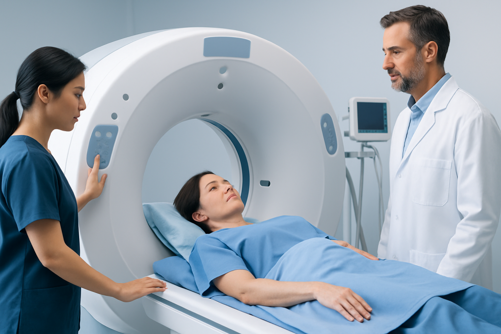
The hilum matters because many important pathways pass through it. If there is swelling, blockage, inflammation, or abnormal tissue growth in this area, the organ’s function may be affected. In the lungs, for example, a hilar abnormality may influence breathing, blood flow, or lymphatic drainage.
For clinicians, the hilum is also valuable because it can provide early clues on imaging. A chest X-ray may show that one hilum looks larger than expected, denser, or asymmetric compared with the other side. That does not automatically mean a serious disease is present, but it does tell the doctor that a closer look may be needed.
Radiologists evaluate the shape, size, and position of the hila together with the patient’s symptoms, medical history, and other scan findings. This broader picture helps distinguish normal variation from conditions that require more investigation.
The Hilum in the Lungs: A Common Focus

When people search for “hilum,” they are usually asking about the lung hilum. The right and left lung hila are not perfectly identical. Small differences in height or contour can be normal because the blood vessels and bronchial anatomy are arranged slightly differently on each side.
On imaging, the hilar shadows seen on a chest X-ray are created mainly by the pulmonary arteries, along with bronchi and nearby soft tissues. Because this is a busy anatomical area, several different problems can change how the hilum looks. These changes may include hilar enlargement, prominent vessels, enlarged lymph nodes, or a visible mass.
Doctors may mention hilar findings when assessing conditions such as pneumonia, sarcoidosis, tuberculosis, <a href="https://acibademinternational.com/diseases/pulmonary-hypertension/”>pulmonary hypertension, or suspected lung cancer. In many cases, additional tests are needed because more than one condition can produce similar imaging appearances.
Another term patients may see is hilar lymph nodes. These are lymph nodes near the lung hila. They can become enlarged in response to infection, inflammatory disease, or cancer. Enlarged hilar lymph nodes are a finding, not a final diagnosis.
Symptoms: Can a Hilar Problem Cause Noticeable Signs?
The hilum itself does not cause symptoms simply by existing, because it is a normal part of anatomy. Symptoms appear only if there is an underlying condition affecting the hilar region. Some patients have no symptoms at all, and the abnormality is found incidentally on imaging done for another reason.
When symptoms do occur, they depend on the cause. In the lungs, a person may experience:
- Cough
- Shortness of breath
- Chest discomfort
- Wheezing
- Fever or chills if infection is present
- Unexplained weight loss or fatigue in some chronic conditions
Symptoms can also come from related structures. For example, enlarged lymph nodes near the hilum may irritate nearby airways, and vascular changes may be associated with breathlessness or reduced exercise tolerance. Because the same symptoms can occur in many common conditions, a medical assessment is needed to determine the cause.
What Can Affect the Hilum?
Several different types of conditions can affect the hilum. In the lungs, one broad group involves infection. Pneumonia, tuberculosis, and some fungal infections can lead to enlarged lymph nodes or inflammation around the hilar structures. Another group includes inflammatory or immune-related disorders, such as sarcoidosis, which often causes enlargement of hilar lymph nodes on both sides of the chest.
Vascular conditions can also change the appearance of the hilum. The hilar region contains major pulmonary blood vessels, so enlargement of these vessels may be seen in pulmonary hypertension or certain heart and lung disorders. This is one reason the hilum is important in chest imaging interpretation.
Benign and malignant growths are another possibility. A mass near the hilum may arise from the airway, lymph nodes, or surrounding tissues. A doctor may consider tumors, metastases, or lymphoma depending on the pattern seen on scans and the person’s medical history.
Not every apparent hilar abnormality represents disease. Normal anatomical variation, overlapping shadows on chest X-ray, and technical imaging factors can sometimes make the region look unusual. That is why a report often recommends correlation with symptoms or further imaging rather than making a conclusion from one picture alone.
How Doctors Evaluate a Hilar Finding
Evaluation usually begins with a clinical review and imaging. If the hilum looks enlarged or unusual on a chest X-ray, the next step is often a more detailed scan such as a computed tomography (CT) scan. CT can help show whether the change is related to blood vessels, lymph nodes, airway structures, or a mass.
Doctors may also use other tests based on the suspected cause. Blood tests can help look for infection, inflammation, or other systemic disease. In some cases, breathing tests or heart evaluation are helpful if vascular or cardiopulmonary causes are being considered.
If a tissue diagnosis is needed, procedures such as bronchoscopy may allow specialists to examine the airways and sample nearby tissue. Some patients may also need biopsy of a lymph node or mass to clarify whether the finding is inflammatory, infectious, or malignant.
Importantly, the evaluation is guided by the whole clinical picture. Age, smoking history, immune status, travel history, prior infections, cancer history, and current symptoms all influence how urgently a hilar finding should be investigated and which tests are most appropriate.
Treatment and Follow-Up
There is no single treatment for the hilum because the hilum is an anatomical region, not a disease. Treatment is directed at the underlying cause. For example, bacterial infections may be treated with antibiotics, inflammatory conditions may require specialist-guided medications, and vascular causes are managed according to the specific heart or lung disorder involved.
If imaging suggests a tumor or enlarged lymph nodes that need further clarification, treatment planning may involve pulmonology, radiology, pathology, thoracic surgery, oncology, or infectious disease specialists. Some findings require only repeat imaging after a period of observation, while others need prompt investigation.
Follow-up is often important even when symptoms are mild. A doctor may recommend repeat chest imaging to check whether a hilar abnormality resolves, stays stable, or changes over time. This helps avoid both unnecessary procedures and delays in diagnosing a more significant condition.
For international patients who need coordinated assessment, Acibadem International’s multidisciplinary specialists and JCI-accredited hospitals diagnose and treat conditions involving the chest, lungs, and related structures using imaging, endoscopic evaluation, and pathology review.
When to Seek Medical Care
A patient should seek medical care if a scan report mentions a hilar abnormality and there are symptoms such as ongoing cough, shortness of breath, chest pain, fever, coughing up blood, or unexplained weight loss. These symptoms do not always signal a serious condition, but they do deserve professional evaluation.
Urgent medical attention is important for severe breathing difficulty, blue lips, confusion, significant chest pain, or rapid worsening of symptoms. These signs may indicate a condition that needs immediate treatment.
If the hilar finding was discovered incidentally and there are no symptoms, follow-up is still worthwhile. A clinician can explain what the report means, compare it with prior imaging, and decide whether watchful waiting, repeat scanning, or specialist referral is the safest next step.
Frequently asked questions
Is the hilum a disease?
No. The hilum is a normal anatomical area where structures such as blood vessels, nerves, or airways enter and leave an organ. A problem in the hilar region may point to an underlying condition, but the hilum itself is not a disease.
What does it mean if a report says hilar enlargement?
Hilar enlargement means the area looks larger or more prominent than expected on imaging. This can happen for different reasons, including enlarged blood vessels, swollen lymph nodes, infection, inflammation, or a mass. More detailed imaging or follow-up may be needed to determine the cause.
Are hilar lymph nodes always cancer?
No. Hilar lymph nodes can enlarge for many non-cancerous reasons, especially infections and inflammatory conditions such as sarcoidosis. Cancer is only one possible explanation, and a diagnosis usually requires imaging review and sometimes tissue sampling.
Can a chest X-ray diagnose the cause of a hilar abnormality?
Not always. A chest X-ray can show that the hilar area looks unusual, but it may not clearly identify why. CT scanning and, in some cases, bronchoscopy or biopsy are often used to clarify the finding.
Does a hilar finding always cause symptoms?
No. Some people have no symptoms, and the finding is discovered incidentally on imaging. Symptoms, when present, usually come from the underlying condition affecting the hilum rather than from the hilum itself.
Which organs have a hilum?
Several organs have a hilum, including the lungs, kidneys, spleen, and liver. In each organ, the hilum serves as the connection point for important vessels, ducts, nerves, or airways. The exact structures differ depending on the organ.
References
- American College of Radiology
- National Heart, Lung, and Blood Institute
- Radiological Society of North America
- American Thoracic Society
- MedlinePlus
This article is for general information only and is not a substitute for professional medical advice. Please consult a qualified doctor about your individual situation.
Explore treatments in Turkey — costs, top hospitals & a free quote
JCI-accredited · board-certified surgeons · reply within 24h

More from the Health Library



Related Specialists

Assoc. Prof. Dr. Bertan Cengiz
Orthopedic Surgery & Traumatology
Dt. Gülçin Doğusal İşgüder
Dentistry
Prof. Dr. K. Latif Abbasoğlu
Pediatric Surgery
Dr. Aynur Göksel
Physical Medicine and Rehabilitation