Lower Leg Anatomy: A Complete Medical Overview
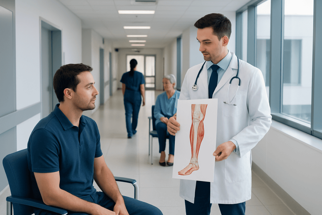
The lower leg is mainly made up of the tibia, fibula, muscle compartments, nerves, and blood vessels. Its structures work together to allow walking, running, balance, and foot and ankle movement.
Key Takeaways
- The lower leg is mainly made up of the tibia, fibula, muscle compartments, nerves, and blood vessels.
- Its structures work together to allow walking, running, balance, and foot and ankle movement.
- Pain, swelling, numbness, weakness, or skin color changes can point to injury or an underlying medical problem.
- Diagnosis may involve a physical exam, imaging, and circulation or nerve tests depending on symptoms.
- Prompt medical assessment is important if symptoms follow trauma or include severe swelling, deformity, or loss of sensation.
Lower leg anatomy refers to the bones, muscles, tendons, nerves, and blood vessels between the knee and ankle. Together, these structures support walking, standing, balance, and blood flow back to the heart.
Overview of Lower Leg Anatomy
Lower leg anatomy includes all of the important structures located between the knee and the ankle. In practical terms, this area contains two long bones, several muscle groups, tendons that connect muscle to bone, ligaments that support nearby joints, major nerves, and blood vessels. These parts function as one coordinated system to help the body stand upright, absorb impact, and move efficiently.
The lower leg has a demanding role in daily life. Each step requires the bones to bear weight, the muscles to control motion, and the vessels and nerves to keep tissues nourished and responsive. Even simple actions such as rising onto the toes, climbing stairs, or keeping balance on uneven ground depend on the health of the lower leg.
Clinicians often describe the region by its compartments rather than by a simple front-and-back view. The muscles are grouped into anterior, lateral, superficial posterior, and deep posterior compartments, each with distinct functions and nerve supplies. Understanding this organization helps explain why certain injuries or conditions cause very specific patterns of pain, weakness, or numbness.
Bones and Joints of the Lower Leg
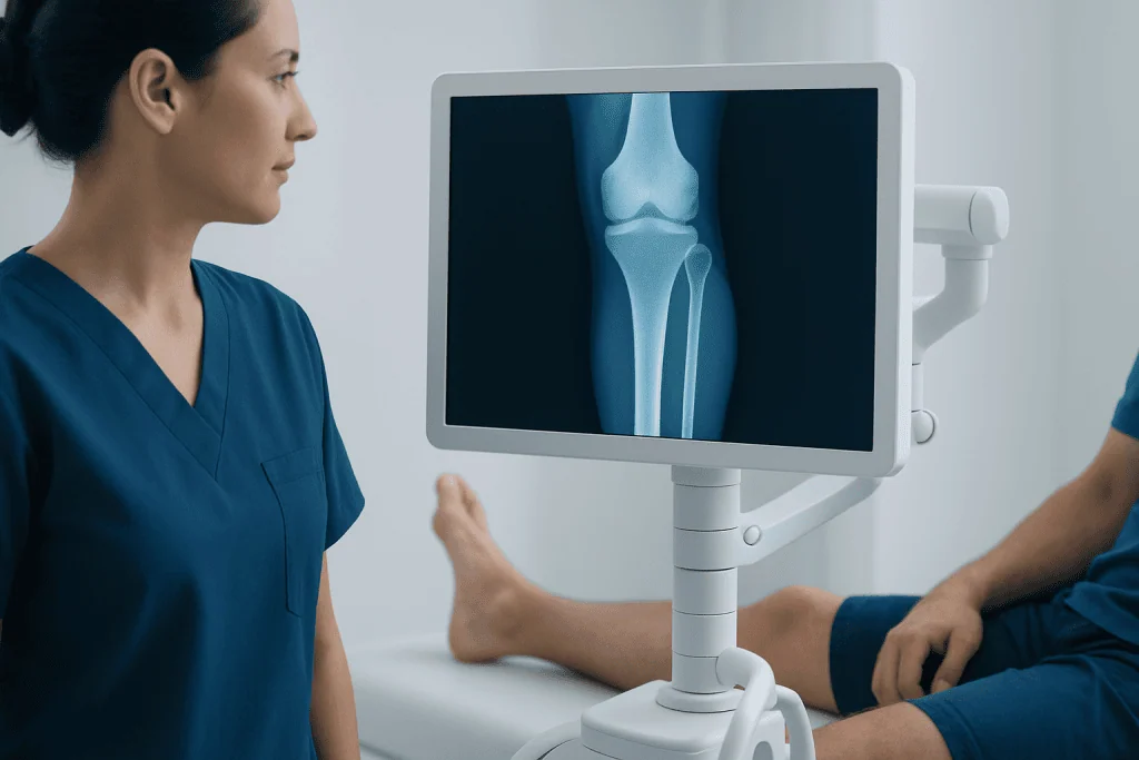
The two main bones of the lower leg are the tibia and the fibula. The tibia, or shinbone, is the larger and stronger of the two. It carries most of the body’s weight and forms major parts of both the knee joint above and the ankle joint below. Because it bears so much load, it is especially important in standing, walking, and recovery after injury.
The fibula is thinner and sits on the outer side of the leg. Although it carries much less body weight than the tibia, it is still essential. It provides stability at the ankle, serves as an attachment point for muscles and ligaments, and contributes to the overall structure of the leg. The tibia and fibula are connected along their length by a strong sheet of tissue called the interosseous membrane.
At the top of the lower leg, the tibia meets the femur at the knee. At the bottom, the tibia and fibula form the ankle mortise, which surrounds the upper part of the talus. Stable alignment of these bones is important for smooth movement and shock absorption. Fractures, ligament injuries, or joint wear can disrupt this alignment and may lead to pain or difficulty walking. In some cases, imaging and specialist assessment are needed to evaluate ankle fracture or other bone injuries.
Muscles, Tendons, and Compartments
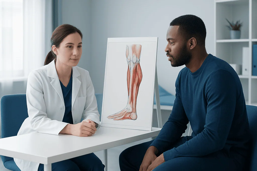
The lower leg muscles are usually described by compartment. The anterior compartment lies along the front of the leg and helps lift the foot and toes upward, a motion called dorsiflexion. This action is important for clearing the foot during walking. The lateral compartment, on the outer side, helps turn the sole outward and supports side-to-side ankle stability.
The posterior part of the leg is divided into superficial and deep layers. The superficial posterior compartment includes the gastrocnemius and soleus, which form most of the calf. These muscles are responsible for plantarflexion, or pointing the foot downward, and they are essential for pushing off during walking, running, and jumping. The deep posterior muscles assist with toe movement, foot inversion, and support of the arch.
Tendons extend these muscle functions to the foot and ankle. The Achilles tendon, which connects the calf muscles to the heel bone, is the largest and strongest tendon in the body. Other tendons travel behind the ankle or across the front of the foot to control fine movement and stability. Repetitive strain, sudden sports activity, or poor biomechanics can contribute to inflammation, overuse injuries, or tears such as Achilles tendon rupture.
Because these muscle groups are enclosed within fascial compartments, swelling inside the leg can sometimes create dangerous pressure. This is one reason severe pain after trauma or intense exercise should not be ignored. Compartment-related problems need urgent evaluation to protect muscles, nerves, and circulation.
Nerves and Blood Vessels in the Lower Leg
The lower leg depends on an organized nerve network to provide movement and sensation. Branches of the sciatic nerve divide higher in the leg into the tibial nerve and common fibular nerve. These then give rise to smaller branches that supply different compartments. For example, the deep fibular nerve helps control the muscles that lift the foot, while the superficial fibular nerve contributes to foot eversion and sensation over parts of the foot.
The tibial nerve travels through the back of the leg and supports many of the calf and deep posterior muscles. It also continues into the foot, where it helps with sensation and intrinsic foot muscle function. Nerve compression, trauma, diabetes-related nerve damage, or spine-related problems may all cause symptoms in the lower leg, including numbness, tingling, burning, or weakness.
Blood reaches the lower leg mainly through branches of the popliteal artery, which divide into the anterior tibial, posterior tibial, and fibular arteries. These vessels carry oxygen and nutrients to muscles, bones, skin, and nerves. Veins return blood to the heart, and the calf muscles help pump venous blood upward with each step. Problems in either arteries or veins can cause pain, swelling, skin color changes, or poor healing. In some cases, doctors may assess for varicose veins or other vascular conditions affecting comfort and circulation.
How the Lower Leg Works in Movement and Balance
The lower leg is not only a structural region; it is also a dynamic control center for gait. During walking, the front muscles help lift the foot during the swing phase, the calf muscles slow and then propel the body during stance, and the smaller stabilizing muscles keep the ankle aligned. This coordinated sequence allows movement to remain efficient and balanced.
Shock absorption is another key function. As the foot strikes the ground, the lower leg helps distribute force through the ankle and up the body. Tendons store and release energy, especially during faster activities such as jogging or climbing stairs. This spring-like role is one reason calf tightness or tendon dysfunction can affect overall comfort and performance.
The lower leg also helps maintain posture. Subtle muscle activity constantly adjusts the body’s position over the feet, especially when standing for long periods or walking on uneven surfaces. If one structure is weakened or injured, nearby muscles often compensate. Over time, this can lead to overuse pain, altered gait, and secondary problems in the knee, hip, or back.
Common Problems That Affect the Lower Leg
Several conditions can affect lower leg anatomy. Acute injuries include fractures, muscle strains, tendon tears, and ligament damage around the ankle. Overuse conditions are also common, especially in runners and physically active adults. These may include shin splints, tendinopathy, stress fractures, and calf muscle strain. Symptoms often depend on which tissue is involved.
Vascular and nerve-related issues can also develop in this area. Swelling may result from venous insufficiency, injury, infection, or prolonged immobility. Cramping or exercise-related pain may sometimes suggest reduced blood flow. Numbness, weakness, or foot drop can occur with nerve compression or injury. Skin warmth, redness, or tenderness may indicate inflammation or another medical issue that needs assessment.
Some symptoms overlap across different conditions, so evaluation is important when pain persists or function changes. Doctors may also consider referred pain from the back, knee, or foot. Depending on the cause, treatment may range from activity modification and physical therapy to bracing, medication, or orthopedic rehabilitation. More complex injuries may require care from specialists in orthopedics and traumatology.
How Lower Leg Problems Are Diagnosed and Treated
Diagnosis starts with a careful history and physical examination. A clinician will usually ask where the symptoms are located, when they began, whether they followed exercise or injury, and whether walking, rest, or certain positions make them better or worse. The exam may include checking tenderness, swelling, muscle strength, range of motion, pulses, reflexes, and skin temperature or color.
Imaging and other tests may be helpful depending on the symptoms. X-rays are often used when a fracture is suspected. Ultrasound can help assess soft tissues or blood flow, while MRI may provide a clearer view of muscles, tendons, stress injuries, or compartment-related concerns. If circulation is a concern, doctors may request vascular testing; if numbness or weakness is present, nerve studies can sometimes help.
Treatment depends on the underlying cause. Minor strains or overuse injuries may improve with rest, gradual return to activity, stretching, supportive footwear, and guided exercise. More significant injuries may require immobilization, physical therapy, or surgery. Vascular and nerve conditions are treated according to their cause and severity. If surgery is needed for bone or tendon injury, it may be planned alongside physical therapy and rehabilitation to restore safe movement and strength.
Near the end of the care pathway, some patients benefit from coordinated specialist input, especially if the issue affects mobility, circulation, or recovery after trauma. Acibadem International’s multidisciplinary specialists and JCI-accredited hospitals diagnose and treat lower leg conditions for international patients when advanced evaluation or treatment is required.
Self-care and When to Seek Medical Care
Simple self-care can support lower leg health and reduce strain. Helpful measures include warming up before exercise, increasing activity gradually, wearing well-fitting footwear, stretching the calf muscles, and allowing recovery time after repetitive impact. People who stand or sit for long periods may also benefit from regular movement, ankle pumps, and leg elevation when appropriate.
It is sensible to seek medical advice if lower leg pain lasts more than a few days, returns repeatedly, or interferes with walking, sport, or sleep. Evaluation is also appropriate for visible swelling, persistent cramping, skin discoloration, warmth, numbness, tingling, or new weakness. These symptoms do not always mean a serious problem, but they should be assessed in context.
Urgent medical care is important after significant trauma, obvious deformity, inability to bear weight, severe calf swelling, sudden loss of sensation, a cold or pale foot, or rapidly worsening pain. Prompt treatment can help protect function and reduce the risk of complications. If symptoms are severe or unclear, a qualified doctor can determine whether the cause is musculoskeletal, vascular, or neurologic.
Frequently asked questions
What structures are included in lower leg anatomy?
Lower leg anatomy includes the tibia, fibula, muscles, tendons, ligaments, nerves, arteries, and veins between the knee and ankle. These structures work together to support movement, balance, and circulation.
What is the difference between the tibia and fibula?
The tibia is the larger shinbone and carries most of the body's weight. The fibula is thinner and sits on the outer side of the leg, helping with ankle stability and muscle attachment.
Which muscles make up the calf?
The calf is mainly formed by the gastrocnemius and soleus muscles in the superficial back part of the lower leg. These muscles help point the foot downward and are essential for walking, climbing stairs, and pushing off the ground.
Why does the lower leg hurt during exercise?
Exercise-related lower leg pain can come from muscle strain, shin splints, tendon irritation, stress injury, or circulation-related causes. If pain keeps returning, becomes severe, or limits activity, a medical assessment is a good idea.
Can lower leg symptoms come from nerves or blood vessels?
Yes. Numbness, tingling, burning, weakness, swelling, skin color change, or coldness in the foot can sometimes be related to nerve or vascular problems rather than only muscle or bone injury. These symptoms should be evaluated by a clinician, especially if they are new or worsening.
When should someone seek urgent care for lower leg symptoms?
Urgent care is important after major injury, if there is obvious deformity, severe swelling, inability to walk, or rapidly worsening pain. Immediate assessment is also needed for a pale, cold foot or sudden loss of feeling, as these may suggest impaired circulation or nerve function.
References
- American Academy of Orthopaedic Surgeons
- MedlinePlus
- Merck Manual Consumer Version
- National Institute of Arthritis and Musculoskeletal and Skin Diseases
- Cleveland Clinic
This article is for general information only and is not a substitute for professional medical advice. Please consult a qualified doctor about your individual situation.
Joint and spine care in Turkey — expert assessment & treatment
JCI-accredited · board-certified surgeons · reply within 24h