Lungs Xray Smoker: Preparation, Procedure and Results

A chest X-ray uses a small amount of radiation to create images of the lungs, heart, ribs, and nearby structures. Smoking-related changes may not be visible on an X-ray, particularly in early disease.
Key Takeaways
- A chest X-ray uses a small amount of radiation to create images of the lungs, heart, ribs, and nearby structures.
- Smoking-related changes may not be visible on an X-ray, particularly in early disease.
- For people who meet risk criteria, yearly low-dose CT is the recommended screening test for lung cancer rather than a routine chest X-ray.
- An abnormal X-ray does not automatically mean cancer; infection, scarring, and other non-cancerous conditions can cause changes.
- Stopping smoking benefits lung health at any age and is the most effective step to reduce future smoking-related harm.
A lungs X-ray for a smoker, also called a chest X-ray, is a quick imaging test that can show some infections, fluid buildup, major lung changes, or suspicious findings. It cannot reliably rule out early lung cancer or all smoking-related lung disease, so the next step depends on symptoms, personal risk factors, and the X-ray result.
Overview: What a Lungs X-Ray Can Show in Smokers
A lungs X-ray for a smoker is usually a chest X-ray, an imaging examination that produces a picture of the lungs, airways, heart, ribs, and diaphragm. It may be requested when a person has symptoms such as a persistent cough, shortness of breath, chest discomfort, fever, or coughing up blood. It can also be used to assess a possible infection or to follow up a finding from another test.
A lungs smoker X-ray can sometimes show signs such as pneumonia, fluid around the lung, a collapsed lung, enlarged heart, substantial scarring, or a lung mass. However, many early smoking-related problems are too subtle to appear on a plain X-ray. A normal image therefore does not prove that the lungs are free of disease.
Chest X-rays are not recommended as a stand-alone screening test for lung cancer. For eligible adults with a substantial smoking history, clinicians may recommend low-dose CT lung cancer screening, which can detect smaller lung abnormalities than a standard X-ray. The best test is selected according to the person’s symptoms, age, smoking exposure, and medical history.
How a Chest X-Ray Works and Who May Need One
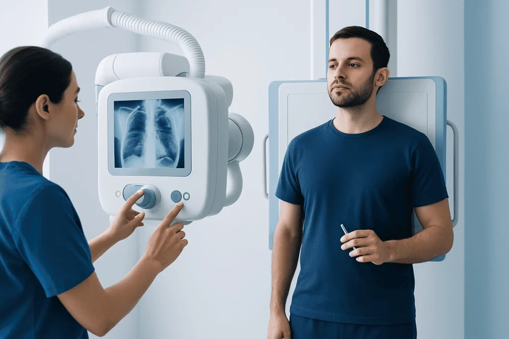
Chest X-rays use low-dose ionizing radiation to pass through the chest and create a black-and-white image. Air-filled lungs generally look darker, while denser structures such as bones, the heart, fluid, or some abnormalities appear lighter. A radiologist interprets the images and sends a report to the clinician who ordered the examination.
A clinician may request a chest X-ray for a current or former smoker with a new or worsening cough, unexplained breathlessness, recurrent chest infections, fever, wheeze, chest pain, or unexplained weight loss. It may also help assess chronic respiratory symptoms in people with conditions such as chronic obstructive pulmonary disease (COPD), although breathing tests are often needed to confirm and measure COPD.
Most people can have a chest X-ray safely. It is important to tell the imaging team about pregnancy or the possibility of pregnancy, previous chest imaging, implanted devices, and current symptoms. A chest X-ray is not routinely needed simply because someone smokes if they have no symptoms and do not meet criteria for CT-based screening.
Step-by-Step: Preparation, Procedure and Recovery
Preparation is usually simple. A person may eat, drink, and take regular medicines unless their clinician gives different instructions. They may be asked to remove jewelry, clothing with metal fasteners, or other objects from the chest area and to wear a hospital gown. Bringing prior imaging information can help the radiologist compare changes over time.
During the procedure, the radiographer positions the person standing against an image detector whenever possible. Most chest X-rays include a front view and a side view. The person will be asked to hold still, take a deep breath, and briefly hold that breath while each image is taken. The imaging itself lasts only moments, and the appointment is commonly completed within a short time.
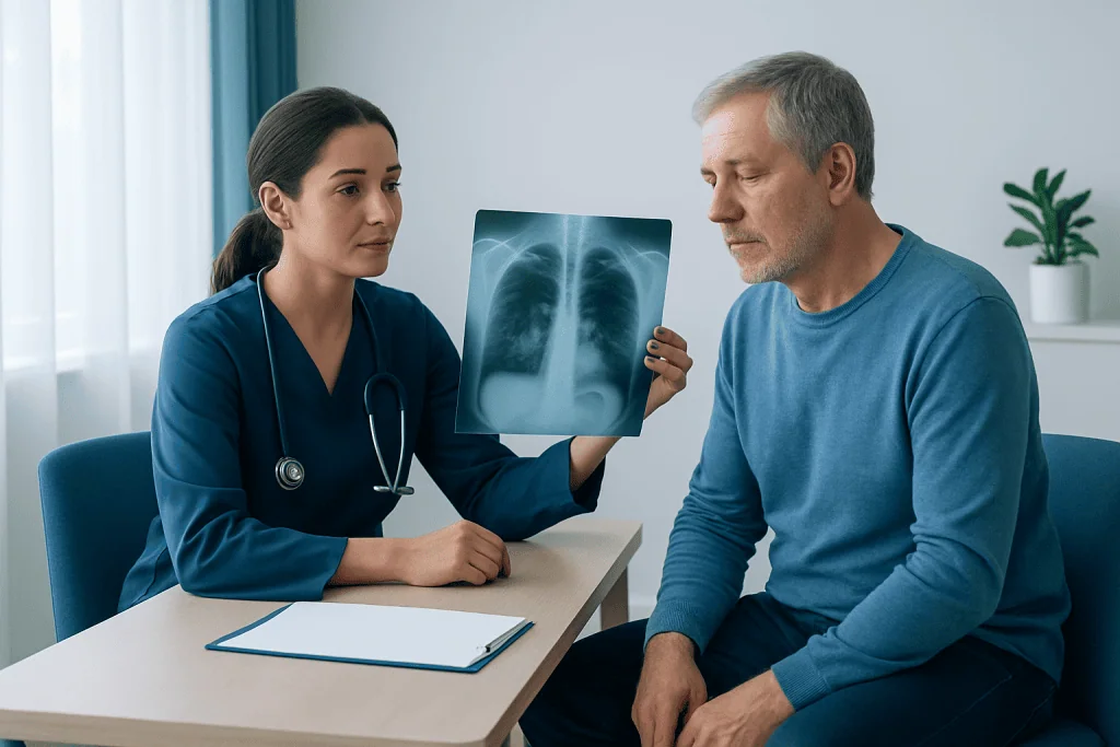
There is no recovery period after a routine chest X-ray. The person can return to normal activities immediately. Results may be available the same day or after a radiologist has reviewed the images, depending on the care setting. If an X-ray identifies an unclear shadow or other change, the clinician may recommend repeat imaging, a CT scan, breathing tests, or referral to a respiratory or lung specialist.
Benefits, Limits and Risks of Lungs X-Ray on Smoking
The main benefit of a chest X-ray is that it is fast, widely available, painless, and useful for evaluating several common causes of respiratory symptoms. It can help identify urgent conditions, including some infections, fluid around the lungs, or a collapsed lung. It may also show changes that need further assessment.
Its most important limitation is that it does not detect every lung condition. Small cancers, early emphysema, early airway disease, small blood clots, and some infections can be missed. For this reason, persistent symptoms deserve medical review even if a lungs X-ray on smoking is reported as normal.
The radiation dose from a chest X-ray is low, but radiation exposure is not zero. The imaging team uses the lowest practical dose and avoids unnecessary testing. An abnormal finding can also lead to additional scans or procedures, which may cause understandable worry; however, many X-ray abnormalities have non-cancerous explanations, including infection, old inflammation, or benign scarring.
What Are the First Signs of Lung Problems?
Early signs of lung problems can be mild and may develop gradually. Common examples include a cough that lasts longer than expected, increasing mucus, wheezing, breathlessness during everyday activity, reduced exercise tolerance, frequent chest infections, or tiredness. Smokers may mistakenly consider a regular cough normal, but a change in a long-standing cough should be assessed.
Symptoms alone cannot identify the cause. They may be linked to asthma, infection, allergies, reflux, heart conditions, COPD, or other concerns. A clinician may ask about smoking and occupational exposure, listen to the chest, check oxygen levels, and arrange tests such as a chest X-ray, spirometry, or CT when appropriate.
Keeping a brief record of when symptoms began, what makes them worse, and whether there is fever, weight loss, or blood in mucus can be useful at an appointment. Seeking advice early often makes it easier to identify treatable causes and plan appropriate follow-up.
What Are Three Warning Signs of Lung Cancer?
Three warning signs that should be discussed promptly with a clinician are a cough that persists or changes, coughing up blood, and unexplained shortness of breath or chest pain. Other possible symptoms include repeated chest infections, hoarseness, unexplained weight loss, marked fatigue, or pain in the shoulder or upper back. These symptoms are common in conditions other than cancer, but they should not be ignored.
Lung cancer may not cause symptoms in its earliest stages. This is why people who meet local eligibility criteria for annual low-dose CT screening should discuss screening with a qualified clinician. Screening is intended for people at increased risk who do not have symptoms; people with symptoms need diagnostic assessment instead.
If imaging shows a concerning area, the care team may recommend CT imaging and, in selected cases, further tests such as PET imaging, bronchoscopy, or a biopsy. Evaluation is individualized, and an X-ray finding by itself cannot confirm a diagnosis of lung cancer.
Where Would You Feel Lung Pain?
The lung tissue itself has few pain-sensitive nerves, so people do not always feel pain directly from the lungs. Pain related to the chest may be felt in the center or side of the chest, under the ribs, in the upper back, or sometimes in the shoulder. It may become sharper with deep breathing or coughing when the lining around the lungs is irritated.
Chest discomfort has many possible causes, including muscle strain, rib injury, reflux, infection, inflammation of the lung lining, heart disease, and anxiety. Its location or character cannot reliably establish the cause. New, persistent, severe, or worsening chest pain should be evaluated rather than self-diagnosed.
Urgent assessment is important for chest pain accompanied by severe breathlessness, fainting, sweating, nausea, blue or gray lips, confusion, or coughing up more than a small streak of blood. Emergency services should be contacted for symptoms that may indicate a serious heart or lung problem.
How Can I Get My Lungs Healthy Again?
Stopping smoking is the most important action a smoker can take for lung health. The body begins to recover after quitting, and the risks of smoking-related disease decrease over time, although existing scarring or chronic lung damage may not fully reverse. Support can include behavioral counseling, nicotine replacement therapy, and prescription medicines when suitable; a clinician can help create a personalized quit plan.
Regular physical activity within a person’s abilities, balanced nutrition, recommended vaccinations, good sleep, and avoiding secondhand smoke and workplace irritants also support respiratory health. People with diagnosed lung disease may benefit from pulmonary rehabilitation, inhaler treatment, and follow-up care tailored to their condition. It is best not to start supplements or unproven “lung cleanse” products as a substitute for evidence-based care.
Acibadem International’s multidisciplinary specialists and JCI-accredited hospitals assess respiratory symptoms and coordinate diagnostic imaging, smoking cessation support, and treatment planning for international patients. A clinician can explain whether a chest X-ray, CT screening, or another evaluation is appropriate for the individual.
Frequently asked questions
Can a chest X-ray show smoking damage to the lungs?
A chest X-ray can show some advanced or indirect changes, such as lung overexpansion, scarring, infection, or a suspicious mass. However, it may be normal in early COPD, emphysema, or early lung cancer. Further testing may be needed when symptoms or risk factors are present.
Should every smoker have a chest X-ray?
No. Routine chest X-rays have not been shown to be an effective lung cancer screening method for all smokers. People with substantial smoking exposure may qualify for annual low-dose CT screening, while those with symptoms should see a clinician for diagnostic evaluation.
What does a normal chest X-ray mean for a smoker?
A normal result means no clear abnormality was seen on that image. It does not exclude all lung disease, especially early airway disease, small cancers, or conditions that are better assessed with CT or lung function tests. Ongoing or worsening symptoms should still be reviewed.
How long does a chest X-ray take?
The actual images are usually taken in a few minutes. Allowing time for registration, positioning, and instructions, the visit is often brief. There is no special recovery time afterward.
Can a chest X-ray diagnose COPD?
A chest X-ray may reveal changes that suggest COPD or rule out other causes of symptoms, but it cannot diagnose COPD on its own. Spirometry, a breathing test performed before and after bronchodilator medicine when appropriate, is central to confirming the diagnosis.
When should a smoker seek urgent medical care?
Urgent medical care is needed for severe or sudden breathlessness, chest pain with sweating or fainting, blue or gray lips, confusion, or significant coughing up of blood. A prompt non-emergency appointment is appropriate for a persistent or changing cough, repeated chest infections, unexplained weight loss, or new breathlessness.
References
- American Cancer Society
- Centers for Disease Control and Prevention
- National Cancer Institute
- U.S. Preventive Services Task Force
- Radiological Society of North America
This article is for general information only and is not a substitute for professional medical advice. Please consult a qualified doctor about your individual situation.
Oncology care in Turkey — second opinion and treatment plan
JCI-accredited · board-certified surgeons · reply within 24h
Add us as a Preferred Source to see more of our trusted health content across Google Search, AI Overviews and Discover.

More from the Health Library



Related Specialists

Fzt. Melis Eda Erkanlı
Physical Medicine and Rehabilitation
Dr. Sinem Altunyuva Usta
Pediatric Cardiology
Prof. Dr. Arzu Ertürk
Pulmonary Medicine
Prof. Dr. Faysal Dane
Medical Oncology