Myocardial Infusion: Procedure, Recovery and Results

Myocardial perfusion imaging evaluates blood flow to the heart muscle and can help identify possible coronary artery disease. The test may involve exercise on a treadmill or medication that temporarily simulates the effects of exercise.
Key Takeaways
- Myocardial perfusion imaging evaluates blood flow to the heart muscle and can help identify possible coronary artery disease.
- The test may involve exercise on a treadmill or medication that temporarily simulates the effects of exercise.
- Most people return to usual activities the same day, although individual instructions may differ.
- An abnormal result does not by itself confirm a blockage; clinicians interpret it alongside symptoms, history and other tests.
- Urgent chest pain, severe breathlessness, fainting or new concerning symptoms require prompt medical assessment.
Myocardial infusion is commonly used to describe myocardial perfusion imaging, also called a nuclear stress test. This noninvasive test uses a small amount of radioactive tracer and imaging to show how well blood reaches the heart muscle at rest and during stress.
Overview: What Is Myocardial Infusion?
The term myocardial infusion is often used when referring to myocardial perfusion imaging (MPI), a type of nuclear stress test. It is not an infusion treatment into the heart. Instead, it is an imaging examination that assesses whether the heart muscle receives enough blood, particularly when the heart is working harder.
During the scan, a healthcare professional injects a small amount of radioactive tracer into a vein. A special camera detects the tracer as it travels through the bloodstream to the heart. Areas of heart muscle with good blood supply take up more tracer than areas receiving reduced blood flow.
Images are usually taken while the patient is at rest and again after exercise or medication-induced stress. Comparing these images can help the cardiology team assess possible narrowing in the coronary arteries, previous heart muscle injury, and the heart’s pumping function. It is an important diagnostic tool, but its findings are considered together with symptoms, medical history and other investigations.
How Myocardial Perfusion Imaging Works and Who May Need It
The coronary arteries supply oxygen-rich blood to the heart muscle. If an artery is narrowed by coronary artery disease, blood flow may be adequate at rest but insufficient during physical exertion or another form of stress. Myocardial perfusion imaging can reveal this difference by showing the distribution of tracer in the heart under both conditions.
A doctor may recommend this test for people with chest discomfort, unexplained shortness of breath, reduced exercise tolerance, an abnormal electrocardiogram, or a known history of coronary artery disease. It may also be used to evaluate symptoms after certain heart treatments or to help guide further investigation.
Not everyone with chest symptoms needs a nuclear stress test. The most appropriate test depends on the person’s age, symptoms, ability to exercise, heart risk factors, previous test results and medical conditions. People with possible coronary disease may also benefit from information about coronary artery disease and its assessment.
- Possible candidates include people with suspected reduced blood flow to the heart.
- It can help assess known coronary artery disease or prior heart attack.
- Pregnancy, breastfeeding, allergies, lung disease and certain medicines should be discussed before scheduling.
What Happens During a Myocardial Perfusion Scan?

Preparation instructions vary by facility and by the medicines a person takes. Patients may be asked to avoid caffeine for a period before the test, because caffeine can interfere with some stress medications. They may also need to avoid eating for several hours, wear comfortable clothing and bring a current medication list. A clinician should advise whether any prescribed medicine needs to be temporarily withheld; patients should not stop medications on their own.
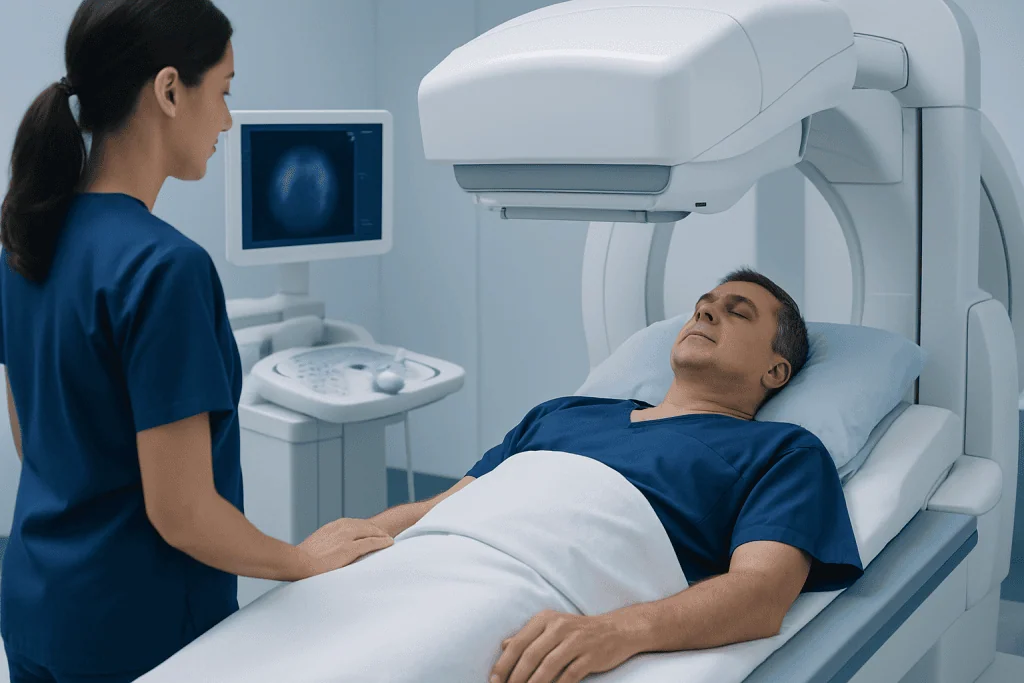
For the resting part of the test, the tracer is injected through an intravenous line. After allowing time for it to circulate, the patient lies still on an imaging table while a gamma camera obtains images. The camera does not cause pain, although remaining still can be uncomfortable for some people.
For the stress portion, the patient may walk on a treadmill with gradually increasing intensity. Heart rhythm, blood pressure and symptoms are monitored. If exercise is not suitable, medication may be given to increase blood flow through the coronary arteries or simulate the heart’s response to exercise. Another tracer injection and imaging session follow.
The appointment may take several hours because there can be waiting periods between injection and imaging. The scan itself is noninvasive apart from the IV line. The care team remains available throughout the examination and can stop the stress phase if concerning symptoms or changes arise.
Recovery Timeline, Benefits and Possible Risks
Most people can go home soon after a myocardial perfusion scan and resume normal routine activities that day. Drinking water, unless fluid intake has been restricted for another medical reason, can help the body eliminate the tracer through urine. The tracer amount is small and typically leaves the body over a short period.
After treadmill exercise, temporary tiredness, muscle fatigue or mild breathlessness may occur, much like after exertion. When stress medication is used, some people briefly experience flushing, headache, chest pressure, dizziness, nausea or shortness of breath. These symptoms usually settle quickly once the medication effect passes, and the medical team monitors the person during recovery.
The main benefit is that the test can provide useful information about heart blood flow without placing a catheter into an artery. It can help a clinician estimate the likelihood of clinically important reduced blood flow and decide whether medication, lifestyle measures, another test or a procedure may be appropriate.
Risks are generally low but include reactions to the stress medicine, abnormal heart rhythms, a small amount of radiation exposure, and rare allergic reactions. Serious complications are uncommon, and the test is performed in a setting prepared to respond to symptoms. A cardiologist can explain why the expected benefit outweighs any risks for an individual patient.
How Long Do You Feel Bad After a Nuclear Stress Test?
Many people do not feel unwell after a nuclear stress test. If symptoms occur, they are usually mild and short-lived. Tiredness after walking on the treadmill may last for the rest of the day, while effects such as flushing, headache, lightheadedness or nausea from stress medication commonly improve within minutes to a few hours.
The recovery experience depends on the person’s general health, whether exercise or medication was used, and whether they had symptoms before testing. Rest, hydration and following the care team’s discharge instructions are often sufficient. Caffeine should only be resumed according to the specific instructions given by the testing facility.
Persistent or worsening chest pain, severe shortness of breath, fainting, a racing or irregular heartbeat, or other concerning symptoms after leaving the facility should not be assumed to be a normal effect of the test. Medical advice should be sought urgently in these situations.
Is a Myocardial Perfusion Scan Serious?
A myocardial perfusion scan is a commonly used diagnostic test and is generally considered safe when it is clinically appropriate and carried out by trained professionals. It does involve a radioactive tracer and sometimes a medicine that stresses the heart, so it should be ordered and supervised carefully. The radiation exposure is kept as low as reasonably achievable while still obtaining useful images.
The seriousness of the result depends on what the images show, not on the test itself. A normal scan is reassuring because it suggests that blood flow is adequate under the conditions tested. An abnormal scan may indicate reduced blood flow, a previous area of heart damage, or occasionally a technical imaging issue that needs clarification.
An abnormal result is not a diagnosis on its own. The cardiologist considers the location and size of any perfusion change, the person’s symptoms, electrocardiogram findings, risk factors and other imaging or blood tests before making recommendations.
Can You Drive After a Myocardial Perfusion Scan?
Many patients can drive after a myocardial perfusion scan, especially if they feel well and did not receive sedation. However, the correct answer depends on the stress method used, the person’s symptoms, local policy and the instructions from the testing team. Patients should arrange transport in advance if they have been advised not to drive or are concerned about feeling unwell.
A person should not drive if they remain dizzy, weak, unusually short of breath, have chest discomfort, or have been given a medicine that affects alertness. Those who experienced significant symptoms or abnormal heart rhythm during testing may require additional observation and individual guidance before leaving.
It is sensible to ask the facility directly about driving before the appointment, particularly when travelling alone or when medication stress testing is planned. The team’s discharge instructions should take priority over general advice.
What Happens if They Find a Blockage During a Nuclear Stress Test?
A nuclear stress test does not directly show the inside of coronary arteries in the same way as coronary angiography. Instead, it can show a pattern of reduced blood flow that may suggest a significant narrowing or blockage. The clinician will explain whether the finding appears mild, moderate or more extensive and how well it matches the person’s symptoms.
Next steps may include optimizing medicines and lifestyle measures, arranging additional noninvasive testing, or considering invasive coronary angiography to look directly at the arteries. If a significant narrowing is confirmed and treatment is needed, options can include coronary angiography and, in selected circumstances, coronary angioplasty and stenting. The best approach depends on the arteries involved, symptom burden, heart function and overall health.
Some findings can be managed without an immediate procedure. Treatments for coronary disease often include risk-factor management, such as stopping smoking, being physically active when medically cleared, following a heart-healthy eating pattern, and taking prescribed medicines consistently. Decisions should be made with a cardiologist after a full review.
When to Seek Medical Care
Medical care should be sought promptly for new, severe or persistent chest pressure or pain, especially if it spreads to the arm, shoulder, jaw, back or upper abdomen. Sudden breathlessness, cold sweating, nausea, fainting or a new irregular heartbeat can also require urgent assessment, particularly in someone with heart disease risk factors.
After a myocardial perfusion scan, patients should contact the test center or their doctor if they have ongoing symptoms that do not improve as expected. Emergency services should be contacted for severe symptoms rather than waiting for a routine appointment.
For planned assessment and ongoing care, Acibadem International’s multidisciplinary specialists and JCI-accredited hospitals diagnose and treat heart conditions for international patients. A cardiology team can explain scan findings in context and discuss whether further testing, preventive care or treatment is appropriate.
Frequently asked questions
What is the difference between myocardial infusion and myocardial perfusion imaging?
Myocardial infusion is not the standard medical name for a heart scan. In many searches, it refers to myocardial perfusion imaging, also called a nuclear stress test, which evaluates blood flow to the heart muscle. The test involves an injected tracer, but it is not an infusion treatment for the heart.
How should a patient prepare for a myocardial perfusion scan?
Preparation commonly includes avoiding caffeine for a specified time, following instructions about food and drink, and wearing comfortable clothing for possible treadmill exercise. Some medicines may affect the test, so the patient should provide a full medication list and follow the clinician’s instructions. Prescribed medication should not be stopped without medical advice.
Does a myocardial perfusion scan hurt?
The scan itself is painless, although there is a brief needle stick for the IV injection. Lying still for images may be uncomfortable for some people. Exercise or stress medication can cause temporary sensations such as tiredness, flushing, breathlessness or chest pressure, which are monitored by the care team.
How long does it take to receive nuclear stress test results?
A specialist must review the images along with the stress-test data and the patient’s clinical history. Some facilities provide preliminary information the same day, while final reporting may take longer. The ordering doctor or cardiologist can explain when and how results will be communicated.
Can a normal myocardial perfusion scan rule out all heart problems?
A normal scan is reassuring about significant blood-flow reduction under the conditions tested, but it does not exclude every heart condition. Some symptoms may come from rhythm disorders, valve disease, microvascular problems, lung conditions or non-heart causes. Persistent symptoms should be reviewed by a qualified clinician.
What does an abnormal myocardial perfusion scan mean?
An abnormal scan can suggest an area of reduced blood flow during stress, prior heart muscle damage, or a result that requires clarification because of imaging artifacts. It does not automatically mean that a person needs a stent or surgery. The cardiologist will assess the complete clinical picture and may recommend medication, further testing or another treatment approach.
References
- American Heart Association
- American College of Cardiology
- Society of Nuclear Medicine and Molecular Imaging
- National Heart, Lung, and Blood Institute
This article is for general information only and is not a substitute for professional medical advice. Please consult a qualified doctor about your individual situation.
Explore treatments in Turkey — costs, top hospitals & a free quote
JCI-accredited · board-certified surgeons · reply within 24h
Check your numbers in seconds
BMI, calories, due date, blood pressure and 30+ more clinical calculators — free, instant, doctor-reviewed ranges.