Scfe Xray: Preparation, Procedure and Results
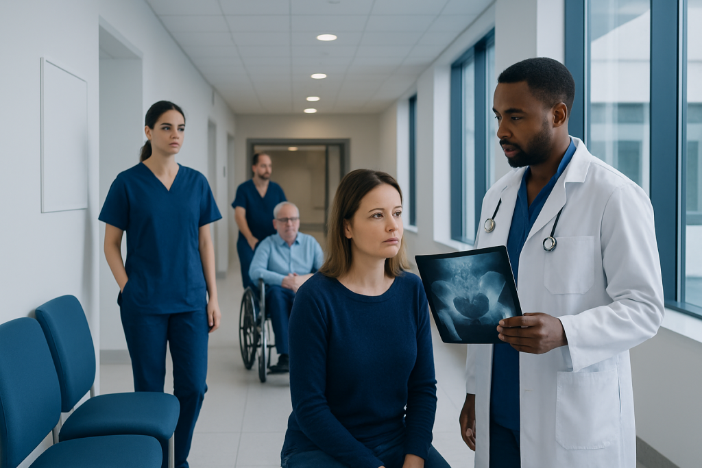
SCFE X-rays usually include front and side views of both hips to compare alignment and detect a slip. Hip or knee pain, limping, and reduced ability to turn the leg inward can be signs of SCFE in adolescents.
Key Takeaways
- SCFE X-rays usually include front and side views of both hips to compare alignment and detect a slip.
- Hip or knee pain, limping, and reduced ability to turn the leg inward can be signs of SCFE in adolescents.
- SCFE is commonly treated urgently with surgery to stabilize the growth plate and reduce the risk of further slipping.
- Recovery after SCFE surgery varies, but protected weight-bearing and follow-up imaging are often needed for several weeks to months.
- A child or teenager with sudden inability to walk, severe hip pain, or worsening limp should receive urgent medical assessment.
An SCFE X-ray is a set of hip radiographs used to assess slipped capital femoral epiphysis (SCFE), a condition in which the growth plate at the top of the thigh bone becomes unstable. It is a quick, noninvasive examination that helps orthopedic specialists confirm the diagnosis, judge severity, and guide timely treatment.
Overview: What Is an SCFE X-Ray?
An SCFE X-ray is an imaging examination used to look for slipped capital femoral epiphysis (SCFE), also called a slipped upper femoral epiphysis. In SCFE, the rounded upper end of the thigh bone remains in the hip socket while the neck of the femur gradually shifts in relation to the growth plate. The condition occurs during adolescence, when the growth plate is naturally weaker than the surrounding bone.
The X-ray is usually the first imaging test when a clinician suspects SCFE. It can show whether the growth plate has widened or become displaced, whether one or both hips are involved, and how much slipping has occurred. Early recognition matters because continued weight-bearing may allow an unstable slip to worsen.
Although symptoms are often felt in the hip or groin, some young people experience pain mainly in the thigh or knee. For this reason, a clinician may examine the hips even when knee pain is the main concern. X-rays are interpreted alongside symptoms, physical examination findings, and the child’s ability to bear weight.
How the X-Ray Works and Who May Need It
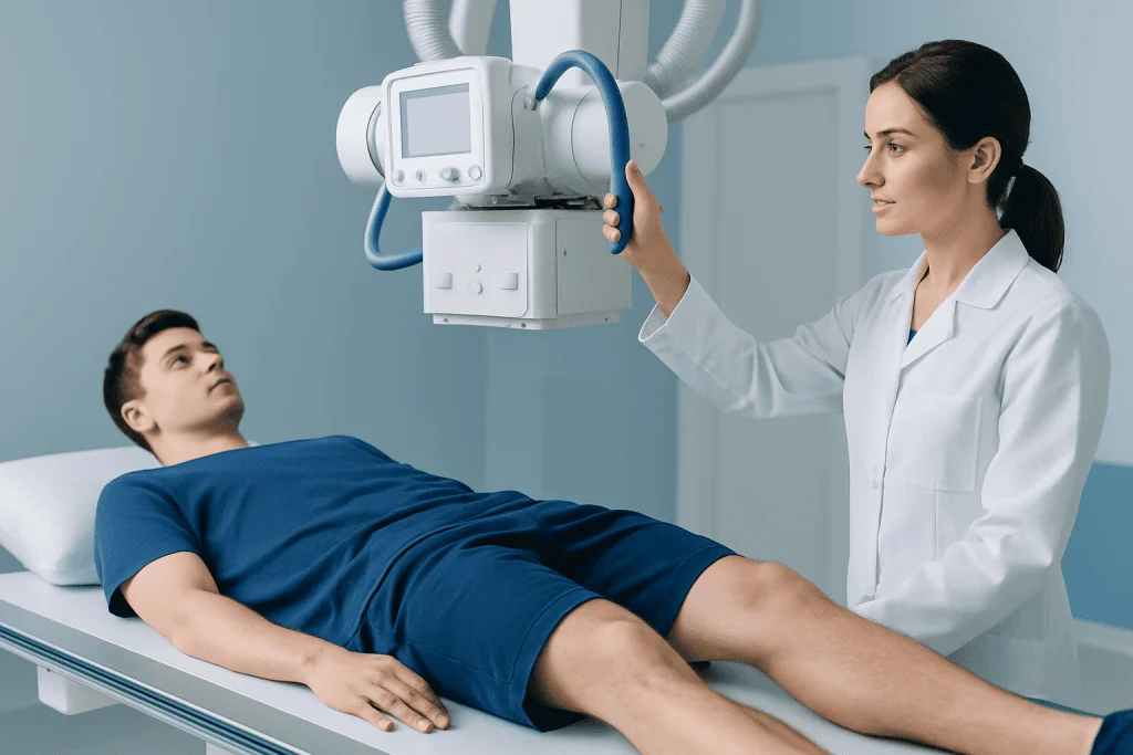
X-rays use a small, controlled amount of ionizing radiation to create images of bone. Bone absorbs more radiation than soft tissues, so its outline and alignment can be assessed clearly. For suspected SCFE, images are commonly taken of both hips because the other hip may show early changes or may be at future risk.
An SCFE X-ray may be recommended for an adolescent with a new limp, hip or groin pain, thigh or knee pain without a clear injury, outward turning of the foot, or reduced hip movement. It is especially important when pain is persistent or worsening, or when the child cannot comfortably put weight through the leg.
SCFE is more likely during periods of rapid growth. Some affected young people have excess body weight, endocrine conditions, kidney disease, or prior radiation treatment, although it can also occur without these factors. A specialist may consider additional evaluation when SCFE occurs at an unusually young age, in a child with low body weight, or on both sides.
- Stable SCFE: the person can still walk or bear weight, with or without crutches.
- Unstable SCFE: the person cannot bear weight, even with support; this requires urgent orthopedic assessment.
Preparing for an SCFE X-Ray and What Happens During the Procedure
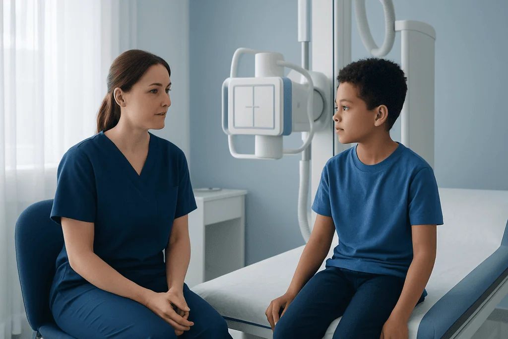
In most cases, no special preparation is needed for an SCFE X-ray. The child or teenager may be asked to remove metal objects around the pelvis, such as belts, zips, jewellery, or clothing with metal fasteners, and may change into an imaging gown. A parent or caregiver can often remain nearby, following the radiology department’s safety guidance.
The radiographer positions the patient on the X-ray table or standing imaging unit, depending on comfort and the requested views. Standard images often include an anteroposterior view, taken from the front, and a side view of the hip. In suspected unstable SCFE, the team avoids movements that could increase pain or potentially worsen the slip; a cross-table lateral view may be used instead of positioning the leg outward.
Each image takes only seconds, although the appointment may take around 15 to 30 minutes including positioning. The child needs to remain still briefly while the images are taken. The examination itself is painless, but positioning may be uncomfortable if the hip is painful, so the imaging team should be told about any severe pain or inability to move the leg.
After the images are complete, normal activity can usually resume unless the clinician has advised avoiding weight-bearing. When SCFE is strongly suspected, the child should not walk on the affected leg until an orthopedic team has assessed them, even if the final report is still pending.
What Are the Radiological Findings of a Slipped Capital Femoral Epiphysis?
On an SCFE X-ray, clinicians look for changes in the relationship between the femoral head and neck at the growth plate. A common early feature is widening or irregularity of the physis, the growth plate. The femoral neck and shaft may appear displaced forward and upward relative to the femoral head, while the head remains seated in the acetabulum, or hip socket.
On the front-view image, a clinician may use Klein’s line as a screening tool. This line is drawn along the upper edge of the femoral neck; normally it intersects part of the femoral head. If it does not intersect the head, or does so less than on the other side, SCFE may be present. Comparison with the unaffected hip can be helpful.
Side-view images are particularly important because mild slips can be subtle on a front view. Other possible findings include loss of the normal smooth contour at the head-neck junction, a visible step at the growth plate, and remodeling in longer-standing cases. X-rays may be normal in very early or subtle cases, so further imaging such as MRI may be considered if symptoms and examination still strongly suggest SCFE.
The radiology report supports, but does not replace, urgent clinical decision-making. If imaging confirms a slip, prompt referral to pediatric orthopedics or an orthopedic surgeon is generally needed to protect the hip and plan stabilization.
What Are the Different Grades of SCFE?
SCFE can be described in more than one way. Clinically, it is classified as stable or unstable according to whether the patient can bear weight. This classification is important because unstable SCFE has a higher risk of complications affecting the blood supply to the femoral head and is treated as an orthopedic urgency.
Radiographic severity is often graded by the degree of slipping seen on X-ray. Mild slips are commonly described as less than one-third displacement, moderate slips as roughly one-third to one-half, and severe slips as more than one-half. Specialists may also measure the slip angle on side-view images to help document severity and guide surgical planning.
SCFE may also be described by the duration of symptoms. Acute symptoms have been present for a short period, chronic symptoms develop gradually over weeks or months, and acute-on-chronic SCFE describes a sudden worsening in someone with prior symptoms. These terms do not reliably show how severe the X-ray changes are, so the clinical and radiological descriptions are considered together.
Grading helps the orthopedic team communicate clearly and monitor the hip over time. It does not determine care on its own, since weight-bearing ability, symptoms, hip stability, skeletal maturity, and the shape of the joint all influence management.
Is SCFE Considered a Fracture?
SCFE is not usually described as a typical traumatic fracture. Instead, it is a growth-plate injury in which the upper femoral growth plate becomes weak and the femoral neck shifts relative to the femoral head. It may develop gradually and can occur without a fall, collision, or other major injury.
However, SCFE is a serious orthopedic condition and is managed with similar caution to an urgent bone injury. Continued walking can increase displacement, particularly when the slip is unstable. For this reason, a child with suspected SCFE may be advised to stop weight-bearing and use a wheelchair, stretcher, or crutches as directed by the treating team.
Trauma can sometimes reveal or worsen an already vulnerable growth plate, but it is not required for SCFE to occur. The condition should not be dismissed as a simple muscle strain, especially when a young person has persistent limping, hip stiffness, or knee pain with no clear explanation.
Treatment, Benefits and Recovery After SCFE Surgery
Once SCFE is diagnosed, treatment usually involves surgery to stabilize the growth plate and prevent additional slipping. The most common approach is in-situ fixation, in which one or more screws are placed across the growth plate to hold the femoral head in position. The aim is generally to prevent progression rather than forcefully return the hip to its previous alignment.
In more complex cases, including severe deformity or selected unstable slips, the orthopedic surgeon may discuss other procedures. Decisions are individualized and take account of X-ray findings, hip stability, symptom duration, and the patient’s growth remaining. Treatment may also include assessment of the opposite hip, because SCFE can later occur on both sides.
The benefits of timely stabilization include reducing the chance of further slip and helping preserve hip function. Potential surgical risks include infection, bleeding, anesthesia-related complications, hardware problems, continued deformity, avascular necrosis, chondrolysis, and later hip arthritis. The treating surgeon explains the relevant risks and expected benefits for the individual child.
Specialists in pediatric orthopedics, radiology, anesthesia, rehabilitation, and endocrinology may be involved when needed. Acibadem International’s multidisciplinary specialists and JCI-accredited hospitals diagnose and treat SCFE for international patients, with care plans based on each patient’s clinical assessment and imaging findings.
How Long Does It Take to Recover From SCFE Surgery?
Recovery from SCFE surgery differs according to whether the slip was stable or unstable, the type of procedure performed, the condition of the other hip, and the child’s overall health. Many patients need crutches or another mobility aid and are asked to limit weight-bearing for a period determined by their surgeon. Follow-up visits and X-rays are used to confirm that the hip is healing and the fixation remains in the intended position.
For uncomplicated stable SCFE treated with in-situ fixation, return to comfortable everyday mobility may take several weeks, while return to running, sports, and high-impact activities usually takes longer and should happen only after orthopedic clearance. Some children need physiotherapy to restore hip movement, strength, gait, and confidence after time spent protecting the leg.
Recovery may be longer after unstable SCFE or more extensive surgery. Ongoing monitoring is important because hip shape and function can change as the child grows. The orthopedic team may continue follow-up until growth is complete, particularly when there is concern about future impingement, uneven leg length, or disease in the other hip.
Families should follow instructions about wound care, medicines, school attendance, weight-bearing, and activity. New fever, wound drainage, increasing pain, sudden loss of function, or a change in the ability to bear weight should be discussed promptly with the surgical team.
When to Seek Medical Care
A child or teenager with a new limp, persistent hip or groin pain, unexplained thigh pain, or knee pain should be assessed by a qualified clinician, particularly if symptoms are worsening or movement of the hip is limited. Early assessment is helpful because SCFE can sometimes present gradually and may be mistaken for a minor strain.
Urgent medical care is needed if the young person cannot bear weight, has severe hip or thigh pain, develops a sudden worsening of a limp, or the leg appears turned outward. They should avoid walking on the affected leg while awaiting assessment unless a clinician advises otherwise.
After treatment, scheduled reviews are important even when symptoms improve. Repeat imaging helps the orthopedic team assess healing, check the fixation, monitor the opposite hip where appropriate, and support a safe return to normal activities.
Frequently asked questions
Does an SCFE X-ray hurt?
The X-ray itself does not hurt. However, moving or positioning a painful hip may be uncomfortable. The radiographer can adapt positioning as safely as possible, especially if the patient cannot bear weight or has severe pain.
Can SCFE be missed on an X-ray?
Mild or early SCFE can be subtle, particularly if only one view is obtained or if the side-view image is limited. When symptoms and examination strongly suggest SCFE despite normal or uncertain X-rays, clinicians may arrange additional imaging, often MRI, or seek urgent orthopedic advice.
Why are both hips X-rayed for suspected SCFE?
Images of both hips allow the clinician to compare growth plates and alignment. SCFE can affect both sides, either at the same time or at different times, so the opposite hip is clinically relevant.
Is SCFE an emergency?
SCFE requires prompt medical assessment because further slipping can harm hip function. Inability to bear weight may indicate unstable SCFE, which needs urgent orthopedic evaluation.
Can a child walk after SCFE surgery?
Many children can walk again after surgery, but they may need crutches or other support while the hip heals. The timing and amount of weight-bearing should follow the orthopedic surgeon’s specific instructions.
Will SCFE cause arthritis later in life?
Some people with SCFE have a higher risk of hip impingement or osteoarthritis later in life, particularly after larger slips or complications. Regular orthopedic follow-up helps identify changes early and supports planning for long-term hip health.
References
- American Academy of Orthopaedic Surgeons
- Pediatric Orthopaedic Society of North America
- National Institute of Arthritis and Musculoskeletal and Skin Diseases
- Radiopaedia
- American College of Radiology
This article is for general information only and is not a substitute for professional medical advice. Please consult a qualified doctor about your individual situation.
Joint and spine care in Turkey — expert assessment & treatment
JCI-accredited · board-certified surgeons · reply within 24h
Add us as a Preferred Source to see more of our trusted health content across Google Search, AI Overviews and Discover.

More from the Health Library



Related Specialists

Dr. Ayten Güner Akbıyık
Thoracic Surgery
Assoc. Prof. Dr. Cüneyt Narin
Cardiovascular Surgery
Dr. Abdurrahman Gölbaşı
Radiology
Assoc. Prof. Dr. Mirza Zafer Dağtaş
Orthopedic Surgery & Traumatology