Skin Cancer Melanoma Surgery: Procedure, Recovery and Results
Wide local excision is the most common operation for melanoma and removes the tumor scar plus surrounding normal-looking skin. Some people with higher-risk melanoma may be offered sentinel lymph node biopsy to check whether cancer cells have reached nearby lymph nodes.
Key Takeaways
- Wide local excision is the most common operation for melanoma and removes the tumor scar plus surrounding normal-looking skin.
- Some people with higher-risk melanoma may be offered sentinel lymph node biopsy to check whether cancer cells have reached nearby lymph nodes.
- Recovery varies with the size and location of surgery, the type of wound closure and whether lymph node surgery or reconstruction is needed.
- Melanoma can return after surgery, so scheduled skin and lymph node checks remain important even after successful treatment.
- Protecting the skin from ultraviolet exposure and learning regular skin self-examination support long-term care.
Skin cancer melanoma surgery is the main treatment for most melanomas that are confined to the skin or nearby tissue. The procedure aims to remove the complete tumor with an appropriate safety margin, while follow-up care helps detect recurrence or a new melanoma early.
Overview: how skin cancer melanoma surgery works
Skin cancer melanoma surgery is usually performed to remove melanoma completely before it can spread or, when melanoma has already spread locally, to manage the affected area. For most early melanomas, surgery is the principal treatment and may be all that is needed. The surgeon removes the melanoma or biopsy scar together with a measured border of surrounding skin, called a surgical margin.
The amount of normal-looking skin removed depends mainly on the melanoma thickness and its location. The specimen is examined by a pathologist, who checks whether the melanoma has been fully removed and reports features that help guide the next steps. A specialist may recommend additional surgery, lymph node assessment, imaging or systemic treatment when the pathology suggests a higher risk of spread.
Melanoma care is planned individually because a small melanoma on the arm is very different from one on the face, scalp, hand, foot or near the eye. Dermatologists, surgical oncologists, plastic and reconstructive surgeons, pathologists and medical oncologists may work together to balance effective cancer treatment with function and appearance.
Who may be a candidate for melanoma surgery?
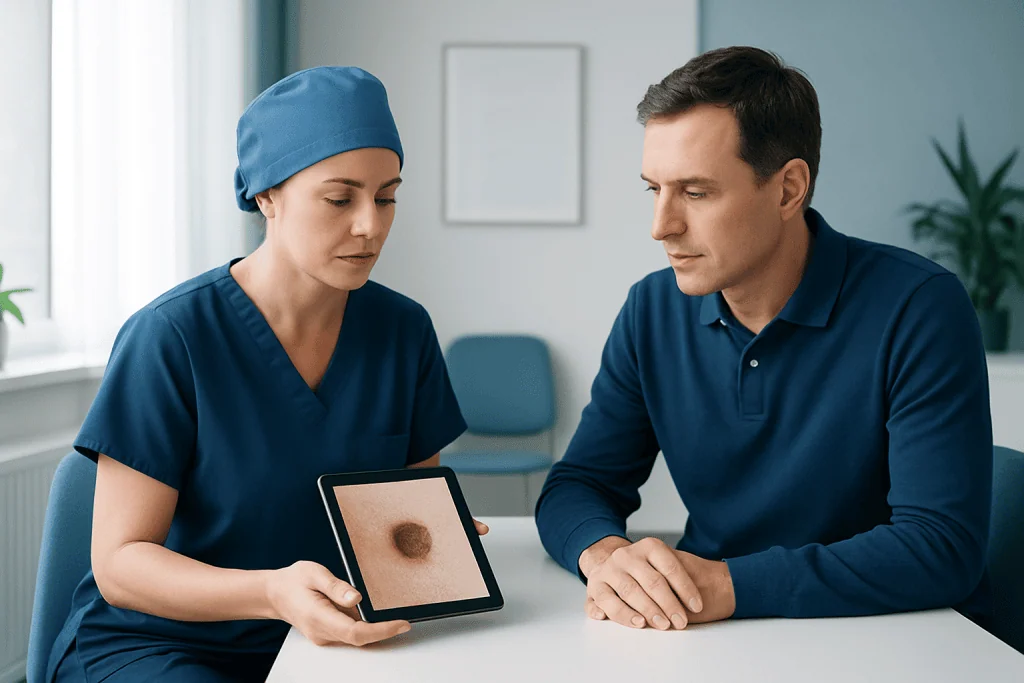
Most people diagnosed with primary cutaneous melanoma are candidates for surgical removal. Surgery may also be considered for selected melanoma deposits in nearby skin, lymph nodes or distant sites, often as part of a wider treatment plan. The aim may be curative, to obtain important diagnostic information, or to control symptoms caused by a localized tumor.
Before recommending an operation, the clinical team considers the biopsy findings, including melanoma thickness, ulceration and whether the tumor involves the edge of the biopsy sample. They also assess the location of the melanoma, a person’s general health, medications that affect bleeding, previous operations and personal preferences.
A sentinel lymph node biopsy may be discussed when the melanoma has features associated with a meaningful risk of reaching nearby lymph nodes. This is a staging procedure rather than a treatment for every person with melanoma. It can provide more accurate information about prognosis and help determine whether additional treatment or closer surveillance should be considered.
In situ melanoma, meaning melanoma limited to the top layer of the skin, is generally treated with local excision alone. More advanced melanoma may require surgery alongside immunotherapy, targeted therapy, radiotherapy or other treatments recommended by an oncology team.
Melanoma surgery step by step
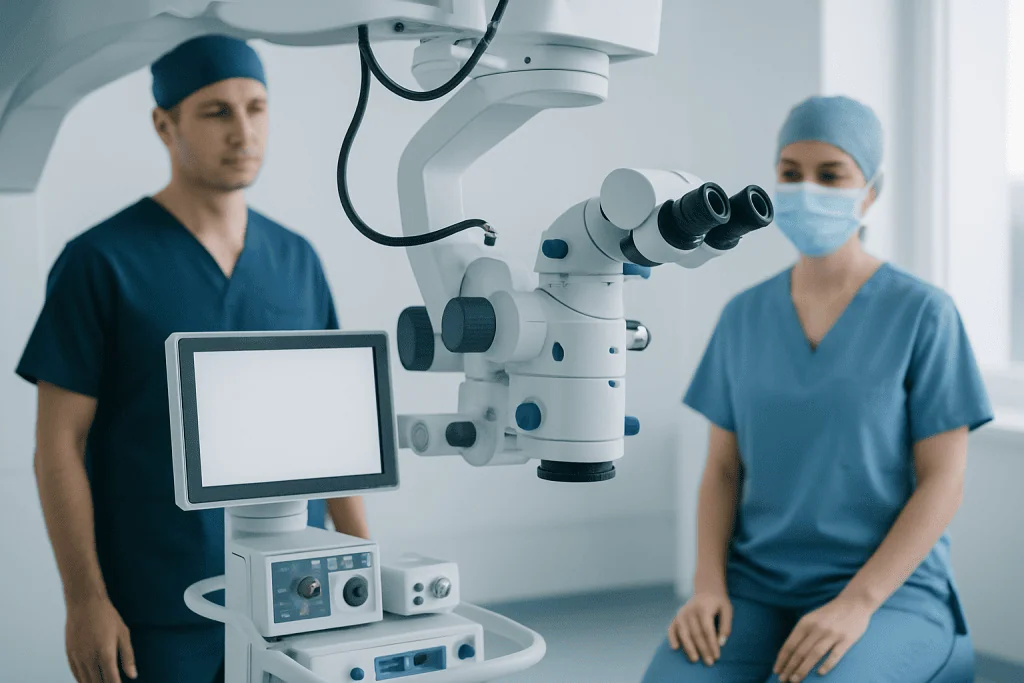
Planning begins with a review of the biopsy report and an examination of the skin and nearby lymph node areas. The surgeon marks the planned excision, explains the expected scar and closure options, and discusses anesthesia. Smaller excisions are often done with local anesthetic, while larger or more complex procedures may require sedation or general anesthesia.
During wide local excision, the surgeon removes the previous biopsy site or visible melanoma with the planned margin and a layer of tissue beneath it. The removed tissue is sent for laboratory examination. Depending on the wound size and site, it may be closed directly with stitches, allowed to heal naturally, or repaired with a skin graft or skin flap.
If a sentinel lymph node biopsy is planned, a small amount of radioactive tracer, blue dye or both is injected around the melanoma site before or during surgery. These materials help identify the first lymph node or nodes that drain the area. The surgeon removes these nodes through a separate small incision, and the pathologist checks them for melanoma cells.
Operations for melanoma affecting the face, hands, feet or other functionally important locations can require reconstructive planning. In such cases, a plastic or reconstructive surgeon may help preserve movement, contour and wound healing. Results are reviewed after final pathology is available, commonly within days to a few weeks depending on the tests required.
Recovery timeline, benefits and possible risks
After a simple excision, many people go home the same day and can return to light activities within a few days. Tenderness, mild swelling, bruising and tightness around the wound are common at first. Stitches may be removed after roughly one to two weeks, though the timing varies by body area and the type of closure.
Larger excisions, skin grafts, flap repairs and lymph node procedures usually need a longer recovery period. Activity restrictions may be advised to avoid pulling on the wound, bleeding or delayed healing. The scar often changes for many months, initially appearing red or firm before gradually softening and fading, although its final appearance varies.
The central benefit of surgery is that it can completely remove localized melanoma and provide precise pathology information. Possible risks include bleeding, infection, pain, numbness, wound separation, visible scarring and delayed healing. Sentinel lymph node biopsy can occasionally cause fluid collection, nerve irritation or lymphedema, although the latter is more likely after more extensive lymph node surgery.
Patients should follow their surgeon’s wound-care instructions closely and contact the care team if there is increasing redness, warmth, swelling, drainage, fever, worsening pain, persistent bleeding or a wound that opens. Acibadem International’s multidisciplinary specialists and JCI-accredited hospitals provide diagnosis and treatment planning for international patients with melanoma.
What should I do with my skin after melanoma removal surgery?
After melanoma removal surgery, the wound should be kept clean and cared for exactly as the surgical team advises. Patients should not remove dressings, apply creams or ointments, soak the wound, swim or resume strenuous activity until their clinician confirms it is appropriate. Once the wound has healed, protecting the scar from sunlight can help reduce discoloration; a clinician may recommend covering it and using broad-spectrum sunscreen on exposed skin.
Long-term skin care focuses on reducing ultraviolet exposure and finding suspicious changes early. This includes seeking shade when practical, wearing protective clothing and a wide-brimmed hat, using broad-spectrum sunscreen, and avoiding tanning beds. Sunscreen is helpful but should be used alongside, rather than instead of, shade and clothing.
Regular self-examination is also important. A person can check the entire skin surface, including the scalp, backs of the legs, soles of the feet, nails and between the toes, and ask for help with difficult-to-see areas. New or changing spots, a changing scar area or an enlarged lymph node should be discussed promptly with a clinician.
Can melanoma come back after removed?
Yes. Melanoma can recur after it has been removed, even when the original surgery had clear margins. A recurrence may develop in or near the surgical scar, in nearby lymph nodes or elsewhere in the body. A person who has had melanoma can also develop a separate new melanoma, which is one reason continued skin surveillance is important.
The chance of recurrence depends on factors such as the original melanoma stage, thickness, ulceration, lymph node findings and whether it had spread beyond the skin. Early-stage melanomas have a lower risk than thicker melanomas or those involving lymph nodes, but no individual outcome can be predicted from one feature alone.
Follow-up schedules are tailored to the melanoma stage and personal risk factors. Visits may include a full-skin examination, examination of regional lymph nodes and education on self-checks. Imaging and blood tests are not routine for every person, but may be used when clinically indicated in higher-risk disease or when symptoms raise concern.
Prompt assessment of a new pigmented lesion, unexplained lump, persistent cough, new neurologic symptom, unexplained weight loss or continuing bone pain is sensible, especially in someone with a history of melanoma. These symptoms often have causes other than melanoma, but they deserve medical review.
What are the different stages of melanoma skin cancer?
Melanoma staging describes how deeply the cancer has grown and whether it has spread. Doctors use the American Joint Committee on Cancer staging system, which incorporates the thickness of the primary melanoma, ulceration, lymph node involvement and spread to distant organs. Staging helps estimate prognosis, guide follow-up and determine whether additional treatment may be useful.
Stage 0, also called melanoma in situ, is limited to the outermost skin layer. Stages I and II are localized melanomas that have not spread to lymph nodes or distant organs; stage II generally includes thicker or ulcerated tumors and carries a higher risk than stage I. Surgery is usually the main treatment for localized disease.
Stage III means melanoma has spread to nearby lymph nodes or nearby skin or lymphatic channels. Stage IV means it has spread to distant parts of the body. Treatment at these stages may include surgery in selected circumstances, together with systemic therapies such as immunotherapy or targeted therapy when appropriate.
Stage labels are important but do not tell the whole story. The pathology report, imaging findings when needed, overall health and response to treatment all contribute to an individual care plan. Patients can ask their team to explain the stage in plain language and how it affects recommended treatment and follow-up.
What is the average life expectancy after being diagnosed with melanoma?
There is no single average life expectancy after a melanoma diagnosis because outcomes vary widely by stage at diagnosis, tumor features, age, overall health and response to treatment. Many people with melanoma diagnosed early and removed completely have an excellent outlook and may live a normal lifespan. Population survival estimates cannot predict what will happen for one individual.
For melanoma that has reached lymph nodes or distant organs, prognosis is more variable, but treatments have advanced substantially in recent years. Immunotherapy and targeted therapies have improved outcomes for many people with advanced melanoma. An oncology team can explain what the pathology and stage mean in the context of the individual patient.
It is often more useful to discuss stage-specific outlook, the purpose of treatment and the recommended follow-up plan than to focus on one life-expectancy number. Patients may wish to bring a family member to appointments, write down questions and request a clear explanation of any survival information that is discussed.
When to seek medical care
Medical assessment is important for a new or changing mole, a spot that differs from other marks on the skin, or a lesion that changes in asymmetry, border, color, diameter or evolution. A sore that does not heal, a dark streak beneath a nail, a new bump in or near a melanoma scar, or an enlarged lymph node also warrants review.
After surgery, patients should contact their surgical team for signs of infection, uncontrolled pain, active bleeding, rapidly increasing swelling, wound opening or a fever. Urgent care is appropriate for severe symptoms such as trouble breathing, chest pain or sudden weakness, regardless of whether they are thought to be related to melanoma.
People with a personal or strong family history of melanoma, many unusual moles, significant sun exposure or a weakened immune system may benefit from planned dermatology follow-up. A qualified clinician can recommend an examination schedule and advise whether genetic counseling or additional monitoring is appropriate.
Frequently asked questions
How long does melanoma surgery take?
A simple wide local excision may take less than an hour, although preparation and recovery time make the visit longer. Surgery can take longer when sentinel lymph node biopsy, a skin graft, a flap reconstruction or a complex location is involved. The surgical team can provide an estimate based on the planned procedure.
Is melanoma surgery painful?
The area is numbed for procedures performed under local anesthetic, and anesthesia is used for more extensive operations. Some soreness, pulling or tenderness is common after surgery and is usually managed with the pain-relief plan provided by the clinical team. Worsening or severe pain should be reported.
Will I need a skin graft after melanoma removal?
Not everyone needs a skin graft. Many excisions can be closed directly with stitches, but a graft or flap may be needed when a larger area is removed or when direct closure would cause too much tension or affect function. This is more common in certain body locations.
Do clear surgical margins mean melanoma cannot return?
Clear margins mean no melanoma cells were found at the outer edge of the removed specimen, which is an important sign that the local tumor was fully excised. However, clear margins do not eliminate the possibility of recurrence elsewhere or of a separate new melanoma. Regular follow-up remains necessary.
Can I exercise after melanoma surgery?
Light walking may often be possible soon after a simple procedure, but vigorous exercise, lifting, stretching and swimming may need to be delayed. The safe timing depends on the wound location, closure method and whether lymph node surgery was performed. Patients should follow the surgeon’s specific activity instructions.
How often should skin checks happen after melanoma?
Follow-up frequency depends on the melanoma stage, treatment history and personal risk factors. Early-stage melanoma generally requires periodic clinical skin examinations, while higher-risk melanoma may need more frequent visits and additional monitoring. The dermatologist or oncology team will create an individualized plan.
References
- American Academy of Dermatology
- American Cancer Society
- National Cancer Institute
- American Joint Committee on Cancer
- National Comprehensive Cancer Network
This article is for general information only and is not a substitute for professional medical advice. Please consult a qualified doctor about your individual situation.
Skin Cancer in Turkey — costs, top hospitals & a free quote
JCI-accredited · board-certified surgeons · reply within 24h