Spect: A Complete Medical Overview
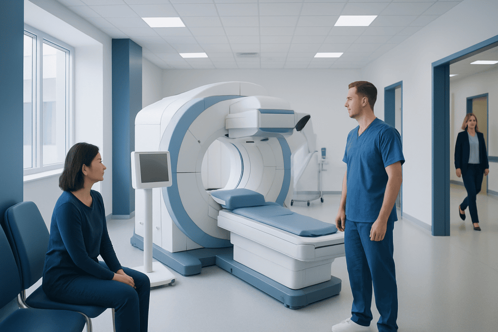
SPECT stands for single photon emission computed tomography and is a type of nuclear imaging. The scan uses a small amount of radioactive tracer to show organ function and blood flow.
Key Takeaways
- SPECT stands for single photon emission computed tomography and is a type of nuclear imaging.
- The scan uses a small amount of radioactive tracer to show organ function and blood flow.
- SPECT is often used for heart, brain, and bone evaluation, depending on the symptoms and clinical question.
- The test is generally safe, with low radiation exposure and usually only mild, short-lived side effects if any.
- Results are interpreted together with symptoms, medical history, and other tests for the most accurate diagnosis.
SPECT is a nuclear medicine imaging test that shows how blood flows through organs and how tissues are functioning, not just what they look like. It is commonly used to help evaluate the heart, brain, bones, and certain other conditions when doctors need functional information alongside standard imaging.
Overview: what SPECT is and what it shows
SPECT, short for single photon emission computed tomography, is an imaging test that helps doctors see how well certain organs and tissues are working. Unlike a regular X-ray, CT, or MRI that mainly shows anatomy, SPECT provides functional information. It can reveal patterns of blood flow, areas of reduced activity, or changes in tissue metabolism that may not be obvious on structural scans alone.
The test works by using a small amount of a radioactive tracer. After the tracer is given, a special camera detects the energy it emits and creates 3D images. These images help doctors assess how blood reaches the heart muscle, how different parts of the brain are functioning, or whether certain bones are unusually active because of injury, infection, or other problems.
SPECT is not a disease itself; it is a diagnostic tool. It is often requested when symptoms, exam findings, or other tests suggest that more detail is needed about organ function. In many cases, it helps confirm a diagnosis, estimate severity, or guide treatment planning.
Why doctors recommend a SPECT scan
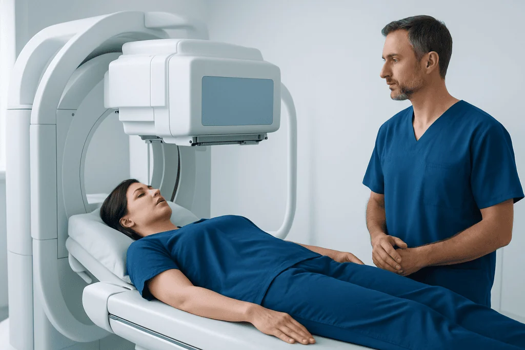
Doctors may recommend a SPECT scan when they need information that other imaging tests cannot fully provide. One of its best-known uses is in cardiology, where it helps evaluate blood flow to the heart muscle at rest and during stress. This can help identify areas that may not be getting enough oxygen because of narrowed or blocked arteries.
SPECT is also used in neurology. It may help assess blood flow patterns in the brain in people with seizures, memory problems, certain types of dementia, head injury, or movement-related symptoms. In some situations, it adds useful detail alongside MRI, CT, or neurological examination. For related brain conditions, doctors may also evaluate patients for Parkinson’s disease or other neurological disorders as part of a broader workup.
Another important use is bone imaging. A SPECT-based bone scan can help locate areas of increased bone activity, which may occur with stress fractures, arthritis, infection, inflammation, or spread of certain cancers to bone. Depending on the situation, SPECT may also be combined with CT as SPECT/CT, which allows doctors to compare function and anatomy more precisely.
- Heart blood flow and possible coronary artery disease
- Seizure focus evaluation in selected patients
- Certain memory and cognitive disorders
- Bone pain, stress injuries, or unexplained skeletal findings
- Some infections, inflammation, or tumor-related assessments
How to prepare and what happens during the test

Preparation depends on why the scan is being done. Patients are usually given specific instructions in advance. For some scans, fasting may be recommended for a few hours. For cardiac SPECT, people may need to avoid caffeine, smoking, or certain medications before the test because these can affect heart blood flow or stress testing results.
At the appointment, a small amount of radioactive tracer is usually injected into a vein. In some cases, there is a waiting period before images are taken so the tracer can travel to the organ or tissue being studied. The patient then lies still on a scanning table while a gamma camera rotates around the body or around a targeted body region.
The test itself is generally painless. The most common discomfort is from the intravenous line or from lying still for a period of time. A heart SPECT study may involve exercise on a treadmill or medicine that mimics the effect of exercise if the person cannot exercise safely. If stress-related heart symptoms are being investigated, a doctor may also consider cardiology evaluation and treatment as part of the broader care pathway.
After the scan, most people can return to normal activities unless they are told otherwise. Drinking fluids may be advised to help the body clear the tracer. Instructions may differ slightly for breastfeeding patients or for those who are pregnant or think they may be pregnant.
Common uses in heart, brain, and bone conditions
In heart care, SPECT is commonly used for myocardial perfusion imaging. This means the scan shows how blood reaches different parts of the heart muscle. If one area receives less blood during exercise or medical stress than it does at rest, this can suggest reduced blood flow from coronary artery narrowing. If an area shows persistently low uptake, it may indicate previous damage such as scarring after a heart event.
In brain imaging, SPECT can help show regional blood flow patterns. Depending on the clinical question, it may assist in evaluating seizure disorders, some cognitive changes, and selected movement disorders. It is usually not used alone; results are interpreted with neurological history, examination, and other imaging such as MRI. If doctors are investigating complex neurological symptoms, they may also use neurology assessment and treatment for coordinated care.
For bone problems, SPECT can be helpful when pain is difficult to localize or when standard imaging does not fully explain symptoms. It may detect areas of increased bone turnover caused by tiny fractures, degenerative joint changes, inflammation, or infection. In oncology settings, it may support the evaluation of suspicious bone findings, though the exact role depends on the type of cancer and other test results.
Safety, risks, and radiation exposure
SPECT is generally considered a safe test. The tracer contains a low dose of radioactive material, and the amount used is carefully selected for medical imaging. For most people, the exposure is low and the benefits of accurate diagnosis outweigh the small risk associated with radiation.
Side effects are uncommon. Some people may notice mild soreness or bruising where the injection was given. Rarely, a person may have a reaction to the tracer, but serious allergic reactions are unusual. In stress SPECT for the heart, side effects may be related to exercise or to stress medicines, such as flushing, headache, shortness of breath, or temporary chest discomfort, which are monitored by medical staff.
Patients should always tell their healthcare team if they are pregnant, may be pregnant, or are breastfeeding. This does not automatically mean the scan cannot be done, but it helps the team decide whether the test should be postponed, modified, or replaced with another option. People with kidney problems, severe mobility limitations, or difficulty lying flat should also mention this before the study.
Understanding the results and what they mean
SPECT results are read by a radiologist, nuclear medicine physician, or another trained specialist. The report describes how the tracer was distributed and whether any areas show reduced uptake, increased activity, or unusual patterns. These findings are then compared with the reason for the test, the patient’s symptoms, and any other imaging or lab results.
An abnormal SPECT result does not always mean there is a serious disease. For example, a heart SPECT may show reduced blood flow that needs follow-up but not emergency treatment. A brain SPECT may suggest a functional pattern that supports a diagnosis, but it usually does not provide a final answer on its own. A bone SPECT may detect increased activity, yet that pattern can occur in both minor injury and more significant conditions.
Because interpretation depends on context, patients should review the results with the doctor who ordered the scan. That conversation usually covers whether additional testing is needed, whether treatment should begin, and how the scan findings fit into the larger clinical picture. In some cases, doctors may recommend follow-up imaging or referral to another specialist for further evaluation.
Limits of SPECT and how it compares with other imaging
SPECT can provide very useful functional information, but it has limitations. It does not always show fine anatomical detail as clearly as CT or MRI. For this reason, SPECT is often used together with other tests rather than as a standalone answer. Hybrid imaging such as SPECT/CT can improve accuracy by matching function with anatomy.
Compared with PET scans, SPECT is generally more widely available and often used for established clinical questions such as myocardial perfusion and certain bone scans. PET may offer higher image resolution or different tracer options in some settings, but the best test depends on the organ being studied, the suspected condition, and local expertise.
Ultrasound, CT, MRI, and standard nuclear medicine studies may all play complementary roles. For example, MRI may better define soft tissue structure, while SPECT may better demonstrate blood flow or tissue activity. The choice of imaging is guided by the medical question rather than by one test being universally better than another.
When to seek medical care
A person should seek medical care if they have symptoms that may require imaging-based evaluation, such as ongoing chest pain, unexplained shortness of breath, episodes of fainting, new seizures, persistent memory changes, or bone pain that does not improve. A doctor can decide whether SPECT or another test is the most appropriate next step.
Urgent medical attention is important for symptoms such as severe chest pain, signs of stroke, sudden confusion, severe shortness of breath, or a seizure emergency. In these situations, immediate care matters more than arranging outpatient imaging.
After a SPECT scan, patients should contact their healthcare team if they have unexpected symptoms such as worsening shortness of breath, persistent chest discomfort after stress testing, or signs of infection at the injection site. While complications are uncommon, follow-up is important if anything feels unusual.
For international patients who need coordinated evaluation, Acibadem International offers diagnosis and treatment through multidisciplinary specialists at JCI-accredited hospitals, including services related to brain and nerve surgery or advanced imaging-guided care when clinically appropriate.
Frequently asked questions
What does SPECT stand for?
SPECT stands for single photon emission computed tomography. It is a nuclear imaging test that uses a small amount of radioactive tracer to create images showing how organs and tissues are functioning.
Is a SPECT scan the same as a CT scan or MRI?
No. CT and MRI mainly show body structure, while SPECT shows function, such as blood flow or tissue activity. In some cases, SPECT may be combined with CT to improve anatomical detail.
How long does a SPECT scan take?
The total appointment time varies depending on the type of study and whether stress testing is involved. Many scans take a few hours from start to finish because there may be waiting time between the tracer injection and image acquisition.
Is SPECT painful?
The scan itself is usually not painful. Most people only feel a brief needle stick for the tracer injection and may find it mildly uncomfortable to lie still during the imaging.
How safe is the radioactive tracer used in SPECT?
The tracer is generally considered safe and is used in very small amounts. Radiation exposure is low, and serious reactions are rare, but patients should tell their doctor if they are pregnant, breastfeeding, or have special medical concerns.
What can a SPECT scan detect?
A SPECT scan can help detect areas of reduced or increased function in the heart, brain, bones, and some other tissues. It may help identify reduced blood flow, seizure-related brain changes, stress injuries, inflammation, or other abnormalities depending on the type of scan.
Do abnormal SPECT results always mean a serious problem?
Not always. An abnormal result means the scan found a pattern that needs medical interpretation, but the cause can range from mild or temporary changes to more significant disease. Doctors interpret SPECT findings together with symptoms, examination, and other tests.
References
- Radiological Society of North America
- American College of Radiology
- Society of Nuclear Medicine and Molecular Imaging
- National Heart, Lung, and Blood Institute
- National Institute of Neurological Disorders and Stroke
This article is for general information only and is not a substitute for professional medical advice. Please consult a qualified doctor about your individual situation.
Medical weight-loss options in Turkey — costs & eligibility
JCI-accredited · board-certified surgeons · reply within 24h