Thrombosis vs Embolism: Key Differences and How Doctors Tell Them Apart

Thrombosis means a clot forms in place inside a blood vessel. Embolism means material travels through the bloodstream and blocks a vessel at a different site.
Key Takeaways
- Thrombosis means a clot forms in place inside a blood vessel.
- Embolism means material travels through the bloodstream and blocks a vessel at a different site.
- Doctors use the pattern of symptoms, physical examination, blood tests, and imaging to tell them apart.
- Deep vein thrombosis can lead to pulmonary embolism, so early assessment matters.
- Treatment often includes blood thinners, but urgency and additional procedures depend on where the blockage is and how severe it is.
Thrombosis and embolism both involve blocked blood flow, but they are not the same. In thrombosis, a clot forms and stays in a blood vessel; in embolism, a clot or other material travels through the bloodstream and blocks a vessel elsewhere.
Overview: thrombosis vs embolism at a glance
Thrombosis vs embolism can be understood by focusing on where the blockage starts and whether it moves. A thrombosis happens when a blood clot forms inside a blood vessel and remains attached there. An embolism happens when a clot or another material breaks loose, travels through the bloodstream, and lodges in a narrower vessel somewhere else.
These terms are related, and one can lead to the other. For example, a clot in a deep vein of the leg may begin as deep vein thrombosis, then part of it can travel to the lungs and cause a pulmonary embolism. Because the words are often used together, many people assume they mean the same thing, but the distinction helps explain symptoms, tests, and treatment decisions.
The table below summarizes the main differences doctors look for first.
- Thrombosis: a clot forms in place inside a vein or artery.
- Embolism: a blockage travels from another part of the body and suddenly lodges in a vessel.
- Common sites of thrombosis: deep veins of the legs, coronary arteries, brain arteries, abdominal or arm veins.
- Common sites of embolism: lungs, brain, limbs, intestines, or other organs.
- Typical pattern: thrombosis may build up gradually or suddenly; embolism often causes abrupt symptoms.
- Why it matters: the cause, urgency, and treatment plan can differ depending on the location and whether the blockage is stationary or has traveled.
In everyday practice, doctors do not rely on terminology alone. They focus on where the blockage is, how quickly symptoms started, what risk factors are present, and whether there are signs of tissue damage from reduced blood flow.
How a clinician tells them apart
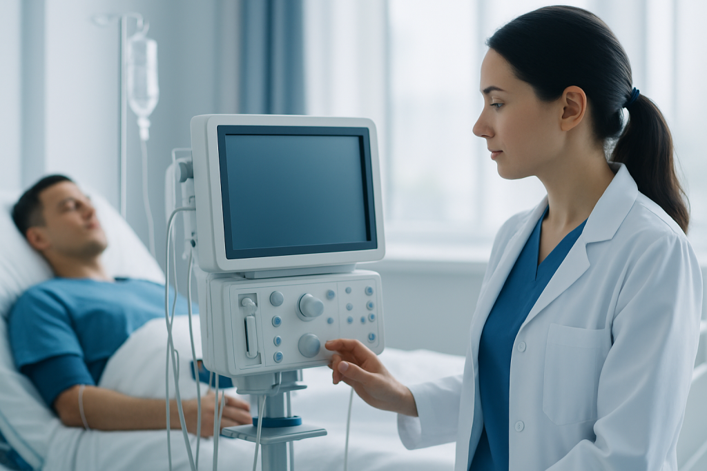
Doctors begin with the clinical story. Symptoms that build in one leg over hours or days, such as swelling, warmth, and pain, may suggest a clot forming in place. Sudden shortness of breath, sharp chest pain, fainting, or coughing up blood can raise concern that material has traveled to the lungs. Sudden weakness on one side of the body or trouble speaking may suggest a blockage affecting blood flow to the brain.
The next step is a physical examination and risk assessment. A clinician looks at the pattern of swelling, skin color, breathing rate, heart rate, oxygen levels, and circulation to the limbs. They also ask about recent surgery, prolonged travel, cancer, smoking, pregnancy, hormone therapy, atrial fibrillation, previous clots, trauma, and inherited clotting tendencies.
Tests help confirm the diagnosis and locate the blockage. Depending on symptoms, a doctor may order blood work such as D-dimer, but blood tests alone do not make the diagnosis. Imaging is usually the key step: ultrasound is often used for leg-vein clots, while CT pulmonary angiography may be used for suspected clots in the lungs. Echocardiography, electrocardiography, MRI, or other vascular imaging may be used when the heart, brain, or arteries are involved.
Clinicians also think about the source. An embolism is often suspected when symptoms are sudden and severe or when the patient has a condition that can send material into the bloodstream, such as atrial fibrillation or a known clot elsewhere. Finding both the blockage and its likely origin helps guide treatment and reduce the risk of another event.
Symptoms and warning signs

The symptoms of thrombosis or embolism depend mainly on which blood vessel is blocked. Vein-related clots often cause swelling, heaviness, tenderness, warmth, or a change in skin color. Artery-related blockages may reduce oxygen delivery to tissues and can cause pain, numbness, weakness, cool skin, or loss of function.
When a clot reaches the lungs, symptoms may appear suddenly. These can include shortness of breath, chest pain that may feel worse with deep breathing, a fast heartbeat, dizziness, or fainting. A clot affecting the brain may cause facial drooping, arm weakness, trouble speaking, confusion, vision changes, or severe imbalance.
Not everyone has classic symptoms. Some clots cause mild signs, and some embolic events are discovered because symptoms suddenly worsen after a period of seemingly minor leg discomfort. This is one reason doctors take clot symptoms seriously even when they seem vague.
- Possible leg-vein thrombosis: one-sided leg swelling, calf pain, warmth, redness, tenderness.
- Possible pulmonary embolism: sudden breathlessness, chest pain, rapid pulse, lightheadedness.
- Possible arterial blockage: sudden severe pain, pale or cool limb, weakness, numbness.
- Possible stroke-related embolism: facial droop, speech difficulty, one-sided weakness, sudden confusion.
Because symptoms overlap with many other conditions, a medical evaluation is needed to determine the exact cause. What matters most is the speed of onset, the body area affected, and whether blood flow appears to be at risk.
Causes and risk factors
Thrombosis often develops when blood flow slows down, the blood vessel lining is injured, or the blood is more likely to clot. This broad framework is sometimes described by clinicians when assessing clot risk. Common contributors include prolonged immobility, hospitalization, recent surgery, injury, smoking, obesity, cancer, inflammatory conditions, pregnancy, and certain medications such as estrogen-containing therapies.
Embolism is not a single disease but a mechanism of blockage. In many cases, the embolus is a clot that formed elsewhere and then traveled. A clot from a leg vein can move to the lungs. A clot can also form in the heart, especially in people with atrial fibrillation, and then travel to the brain or other organs. Less commonly, emboli may consist of fat, air, infected material, or fragments of plaque.
Age, personal or family history of clots, inherited clotting disorders, and long-distance travel can add to risk. The presence of central venous catheters, severe infection, or heart disease may also increase the chance of a thrombosis or embolic event. In some people, more than one risk factor is present at the same time.
Doctors pay close attention to whether the event is provoked or unprovoked. A provoked clot happens in the setting of a clear trigger, such as surgery or immobilization. An unprovoked event has no obvious short-term trigger and may lead to a broader medical review to understand ongoing risk and plan long-term prevention.
Diagnosis and tests
There is no single test that answers every clotting question. The diagnostic plan depends on where the blockage may be and how stable the patient is. Doctors first assess urgency by checking blood pressure, breathing, oxygen levels, mental status, and signs of poor circulation.
For suspected deep vein thrombosis, compression ultrasound is commonly used because it can show whether a vein is blocked by clot. For suspected pulmonary embolism, clinicians may use CT pulmonary angiography or, in some situations, a ventilation-perfusion scan. If a clot affecting the brain is suspected, urgent brain imaging is needed. If the source may be the heart, echocardiography and heart rhythm testing may be helpful.
D-dimer blood testing can support decision-making in selected patients when the clinical probability is low or intermediate. However, a positive result does not prove thrombosis or embolism, because many other conditions can raise D-dimer levels. Likewise, a normal result is most useful only in the right clinical setting.
Doctors may also look for underlying causes after the immediate problem is identified. This can include blood tests, cancer screening when appropriate, evaluation for inherited clotting conditions in selected patients, or rhythm assessment for atrial fibrillation. The goal is not only to treat the current blockage but also to reduce the chance of it happening again.
What doctors do for each case
Treatment depends on the location of the blockage, its severity, the risk of tissue injury, and the patient’s overall health. In many cases of thrombosis or embolism caused by blood clots, anticoagulant medicines, often called blood thinners, are the main treatment. These medicines do not instantly dissolve existing clots, but they help prevent clot extension and reduce the risk of new clots forming.
When the blockage is larger, causes severe symptoms, or threatens an organ, doctors may consider more urgent interventions. These can include clot-dissolving medicine in carefully selected patients, catheter-based procedures, or surgery. For example, vascular or endovascular procedures may be used for major arterial blockages or some high-risk venous or pulmonary cases. In centers with appropriate expertise, treatment plans may involve interventional radiology procedures or cardiovascular surgery when needed.
Doctors also treat the cause and protect the affected organ. This may include oxygen support, pain relief, compression measures in some venous conditions, heart rhythm control, or treatment of underlying illness. Some patients need short-term anticoagulation after a temporary risk factor, while others need longer treatment because the risk of recurrence remains higher.
Follow-up is important. Patients may need repeat imaging, blood test review, assessment for bleeding risk, and advice about activity, travel, hydration, and future procedures. If a clotting event is linked to the brain or other organs, specialists may coordinate care, including stroke rehabilitation after recovery from a stroke-related embolic event when appropriate.
Prevention, self-care, and recovery
Prevention focuses on lowering clot risk and recognizing symptoms early. Moving regularly during travel or after surgery, staying hydrated, stopping smoking, managing chronic conditions, and following medical advice on preventive medications can all help. People at increased risk may be advised to use compression stockings or anticoagulation in specific situations, but this should be based on a clinician’s recommendation.
During recovery, patients should take prescribed medication exactly as directed and attend follow-up visits. They should tell all healthcare professionals about any blood thinner use, because it can affect procedures, dental work, and the use of over-the-counter medicines or supplements. Sudden stopping of anticoagulation without medical advice can be unsafe.
Self-care also includes watching for warning signs of recurrence or bleeding. New swelling, sudden breathlessness, chest pain, severe headache, fainting, or unusual bleeding should be discussed promptly with a doctor. Moderate movement is often encouraged after a clot, but the timing and intensity should be individualized.
Near the end of treatment, doctors may review whether further testing or longer prevention is needed. At Acibadem International, multidisciplinary specialists in JCI-accredited hospitals evaluate and treat thrombosis and embolic conditions for international patients, with care plans based on the clot location, underlying cause, and recovery needs.
When to seek medical care
Urgent medical attention is needed for sudden shortness of breath, chest pain, fainting, coughing up blood, or signs of stroke such as facial drooping, one-sided weakness, or trouble speaking. These symptoms can indicate a serious embolic event or another emergency that requires immediate evaluation.
Prompt medical assessment is also important for one-sided leg swelling, calf pain, warmth, or unexplained redness, especially after travel, surgery, or a period of limited movement. Although these symptoms do not always mean a clot is present, they should not be ignored.
People already taking blood thinners should seek advice if they have a fall, a head injury, black stools, vomiting blood, severe bruising, or bleeding that does not stop. A doctor can determine whether symptoms are due to treatment effects, a new clot, or another condition entirely.
When in doubt, it is safer to ask for medical guidance than to monitor concerning symptoms alone. Early diagnosis can help prevent complications and support the most effective treatment plan.
Frequently asked questions
Is thrombosis the same as embolism?
No. Thrombosis means a clot forms and stays attached within a blood vessel, while embolism means a clot or other material travels and blocks a vessel elsewhere. The two are closely related because a thrombosis can become the source of an embolism.
Can a deep vein thrombosis turn into a pulmonary embolism?
Yes. A clot in a deep vein, usually in the leg, can break loose and travel to the lungs, where it becomes a pulmonary embolism. This is why leg swelling or pain should be assessed promptly when a clot is possible.
How do doctors confirm whether it is thrombosis or embolism?
Doctors combine the symptom pattern, physical examination, risk factors, and imaging tests. Ultrasound is commonly used for leg-vein clots, while CT scans or other vascular imaging may be used when the lungs, brain, or arteries are involved.
Are all embolisms caused by blood clots?
No. Many embolisms are caused by clots, but some are caused by other material such as fat, air, infected material, or cholesterol debris. The treatment depends on what caused the blockage and where it traveled.
What is the usual treatment for thrombosis or embolism?
Many patients are treated with anticoagulant medicines to prevent the clot from growing and to lower the risk of new clots. Some people need more urgent treatment, such as clot-dissolving therapy or a procedure, especially if a major organ or limb is at risk.
Can thrombosis or embolism be prevented?
Risk can often be reduced, though prevention is not always possible. Regular movement, hydration, smoking cessation, careful management after surgery or hospitalization, and following medical advice about preventive treatment can all help.
References
- Centers for Disease Control and Prevention
- National Heart, Lung, and Blood Institute
- American Heart Association
- National Blood Clot Alliance
- World Health Organization
This article is for general information only and is not a substitute for professional medical advice. Please consult a qualified doctor about your individual situation.
Explore treatments in Turkey — costs, top hospitals & a free quote
JCI-accredited · board-certified surgeons · reply within 24h
Check your numbers in seconds
BMI, calories, due date, blood pressure and 30+ more clinical calculators — free, instant, doctor-reviewed ranges.