Upj Obstruction: What Patients Need to Know

UPJ obstruction affects urine drainage from the kidney into the ureter. Some people have no symptoms, while others develop flank pain, infections, stones, or kidney swelling.
Key Takeaways
- UPJ obstruction affects urine drainage from the kidney into the ureter.
- Some people have no symptoms, while others develop flank pain, infections, stones, or kidney swelling.
- Diagnosis usually involves imaging tests that show kidney anatomy and how urine drains.
- Treatment may range from monitoring to surgery, often pyeloplasty, depending on symptoms and kidney function.
- Prompt medical review is important if there is fever, severe pain, repeated urinary infections, or worsening kidney swelling.
UPJ obstruction is a blockage or narrowing at the point where the kidney connects to the ureter, the tube that carries urine to the bladder. It can be present from birth or develop later, and treatment depends on symptoms, kidney drainage, and how well the kidney is working.
Overview
UPJ obstruction, also called ureteropelvic junction obstruction, is a narrowing or blockage where the kidney meets the ureter. This junction is the point where urine leaves the kidney and starts its journey to the bladder. When the opening is too narrow or becomes blocked, urine cannot drain normally, and pressure may build up inside the kidney.
This pressure can cause swelling of the kidney, known as hydronephrosis. Some people learn about UPJ obstruction before birth during a routine pregnancy ultrasound, while others are diagnosed later in childhood or adulthood because of pain, urinary tract infections, or kidney stones. In some cases, the condition is found by chance on imaging done for another reason.
Not every UPJ obstruction needs immediate treatment. The most important questions are whether the kidney is draining well, whether symptoms are present, and whether kidney function is being affected. Care is usually guided by a urologist, and the plan may include monitoring, temporary drainage, or surgery when needed.
How UPJ Obstruction Affects the Kidneys
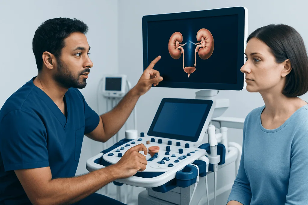
The kidneys constantly filter blood and make urine. For this process to work well, urine must flow freely from the collecting area inside the kidney into the ureter. In UPJ obstruction, that flow slows down or stops partly, which can stretch the kidney’s drainage system over time.
If the obstruction is mild, the kidney may continue to function well for years and the person may have few or no symptoms. If the obstruction is more significant, the trapped urine can increase pressure within the kidney. Over time, this may reduce kidney function, raise the chance of infection, or contribute to stone formation.
UPJ obstruction can affect one kidney or, less commonly, both. When only one kidney is involved, the other kidney often continues to work normally. Even so, preserving the affected kidney is important, especially in children and in adults who already have other kidney risks.
Hydronephrosis does not always mean severe obstruction, and obstruction does not always cause severe symptoms. That is why doctors often combine imaging with functional tests to understand how serious the problem is and whether intervention is likely to help.
Symptoms and Possible Complications
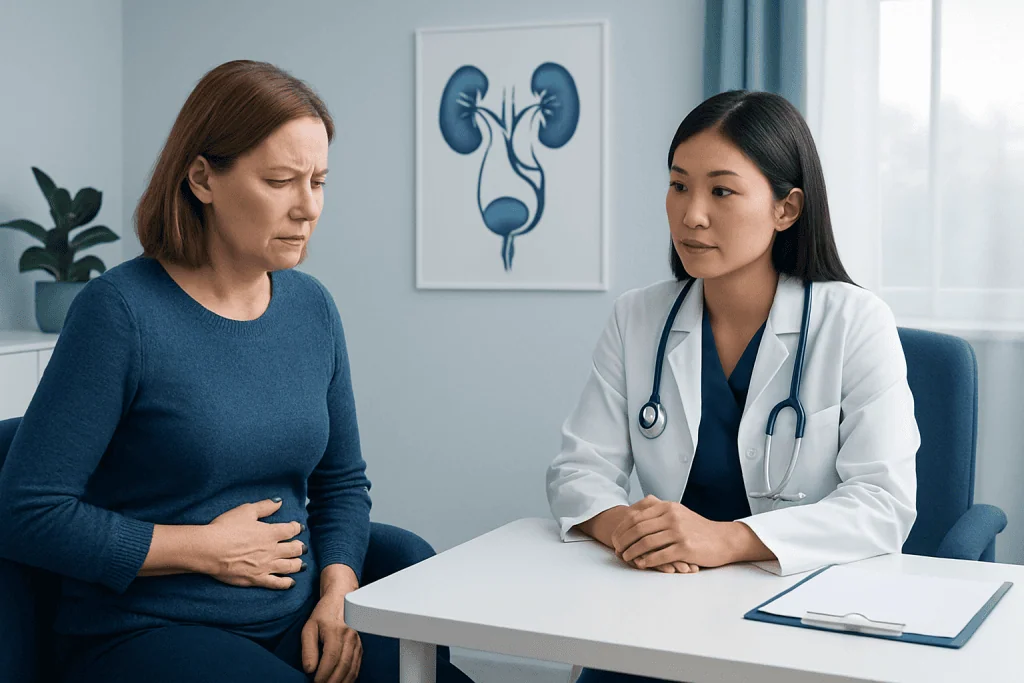
Symptoms of UPJ obstruction vary by age and by how much urine flow is blocked. In infants and young children, there may be no obvious symptoms at all, and the condition may only be seen on ultrasound. In older children and adults, symptoms often include pain in the side or back, especially after drinking a lot of fluids, nausea, vomiting, or repeated urinary tract infections.
Some people notice intermittent pain rather than constant discomfort. This can happen because urine flow may be partly blocked at some times and more limited at others. Blood in the urine may also occur, and some patients develop kidney stones because urine is not draining efficiently.
Potential complications include:
- Hydronephrosis, or swelling of the kidney
- Urinary tract infections
- Kidney stones
- Reduced function in the affected kidney
- Persistent flank or abdominal pain
Although these complications are important, many cases are manageable with appropriate follow-up and treatment. People who have severe pain, fever, vomiting, or symptoms of infection should seek prompt medical care because an infected obstructed kidney needs urgent attention.
Causes and Risk Factors
UPJ obstruction may be congenital, meaning a person is born with it, or acquired, meaning it develops later. Congenital cases are often due to the way the junction formed before birth. The muscle in that area may not work well, the channel may be too narrow, or the urine drainage pathway may be shaped in a way that slows flow.
In some people, a blood vessel crossing near the junction presses on the ureter from the outside and contributes to blockage. This is one reason why symptoms can appear later, even in adolescence or adulthood. Acquired UPJ obstruction is less common but may occur after scarring, inflammation, previous surgery, stones, or trauma affecting the urinary tract.
Risk factors are not always clear because many cases are developmental rather than caused by lifestyle. However, a history of kidney stones, urinary tract procedures, or recurrent infections can sometimes be part of the picture in acquired obstruction. Doctors may also consider other urinary tract conditions that can affect drainage, such as kidney stones.
Because UPJ obstruction can overlap with other causes of flank pain or hydronephrosis, careful evaluation is important. The goal is not only to identify the narrowing itself, but also to understand whether the problem is fixed, intermittent, or related to another condition.
Diagnosis and Tests
Diagnosis begins with a medical history, symptom review, and physical examination. The doctor will ask about pain, urinary infections, fever, kidney stones, and any imaging tests already done. In children, prenatal ultrasound findings and growth history may also be discussed.
Imaging is central to diagnosis. Ultrasound is often the first test because it can show hydronephrosis without radiation. Other imaging studies, such as CT or MRI, may help define the anatomy, look for crossing blood vessels, or check for stones. Functional nuclear medicine scans are especially useful because they show not only the shape of the kidney but also how well it drains and how much function each kidney contributes.
Urine and blood tests may also be used to look for infection and assess kidney function. Depending on the situation, a doctor may compare current and previous scans to see whether swelling is stable, improving, or worsening.
Some patients are also evaluated for related urinary tract issues. If a broader assessment is needed, care may involve specialists experienced in urology and kidney drainage disorders. The choice of tests depends on age, symptoms, pregnancy status, and whether urgent treatment is being considered.
Treatment Options
Treatment depends on symptoms, the degree of obstruction, and whether kidney function is at risk. If the person has mild hydronephrosis, no pain, and stable kidney function, the doctor may recommend monitoring with repeat imaging rather than immediate surgery. This is common in some children and in selected adults.
When symptoms are significant, drainage is poor, infections recur, or the kidney shows declining function, intervention is usually considered. The standard surgical treatment is pyeloplasty, which removes or bypasses the narrowed segment and reconnects the healthy parts to create a wider passage for urine. This can often be performed using minimally invasive techniques, including robotic surgery when appropriate.
In certain situations, a temporary ureteral stent or a nephrostomy tube may be used to relieve pressure and help the kidney drain before definitive treatment. Less commonly, an endoscopic incision of the narrowing may be an option, though long-term success depends on the exact cause and anatomy.
For patients with complicated anatomy, recurrent obstruction, or related stone disease, treatment planning may include advanced imaging and specialized kidney care. If symptoms are linked with obstruction and stone formation, doctors may also evaluate for kidney stone treatment. The best approach is individualized, and preserving kidney function is a key goal.
Living With UPJ Obstruction: Monitoring, Recovery, and Self-care
People with UPJ obstruction often do well with regular follow-up, whether they are being monitored or have already had treatment. Follow-up may include ultrasound, functional scans, urine tests, and office visits to check for symptoms. The schedule depends on age, severity, and whether kidney drainage has improved or remained stable.
After surgery such as pyeloplasty, recovery plans vary, but patients are usually advised to follow their surgeon’s instructions about activity, fluids, wound care, and follow-up imaging. A temporary stent may be placed during healing and removed later. Mild discomfort during recovery is common, but persistent fever, worsening pain, or trouble urinating should be reported promptly.
Self-care does not remove the obstruction, but it can support overall urinary health. Helpful steps may include staying well hydrated unless a doctor recommends otherwise, taking prescribed medicines as directed, and seeking care promptly for possible infection. Repeated antibiotics or pain medicines should not replace medical reassessment if symptoms continue.
For international patients who need diagnosis or treatment, Acibadem International’s multidisciplinary specialists at JCI-accredited hospitals evaluate conditions affecting the urinary tract, including cases related to hydronephrosis and UPJ obstruction.
When to Seek Medical Care
Medical care should be sought if there is ongoing flank pain, repeated urinary tract infections, blood in the urine, nausea with suspected kidney pain, or imaging that shows kidney swelling. Even mild symptoms can be worth discussing if they are recurring, because some cases worsen gradually rather than suddenly.
Urgent care is especially important if UPJ obstruction is suspected along with fever, chills, vomiting, severe pain, or difficulty passing urine. These symptoms may suggest infection or significant blockage and should not be ignored.
Parents of infants or children should also follow up if prenatal or childhood ultrasound has shown hydronephrosis, even if the child seems well. Early assessment helps determine whether the finding is temporary, mild, or part of a true obstruction that needs monitoring.
A qualified doctor can explain what the imaging means, whether kidney function is affected, and whether watchful waiting or treatment is the safest next step. Timely review is often the best way to protect kidney health and avoid complications.
Frequently asked questions
Is UPJ obstruction serious?
UPJ obstruction can be mild or more significant depending on how much it affects urine drainage and kidney function. Some people only need monitoring, while others need treatment to prevent pain, infection, or loss of kidney function. A doctor can determine the level of concern based on imaging and functional testing.
Can UPJ obstruction go away on its own?
In some infants and children, mild dilation seen on imaging may improve over time, which is why careful monitoring is sometimes recommended. However, a true fixed UPJ obstruction does not usually resolve on its own in adults. Decisions about observation versus treatment depend on symptoms and kidney drainage.
What does UPJ obstruction pain feel like?
Pain is often felt in the side, flank, or upper abdomen on the affected side. It may come and go, and some people notice it after drinking a large amount of fluid. Nausea or vomiting can occur if the blockage causes pressure to build up in the kidney.
How is UPJ obstruction diagnosed?
Doctors usually diagnose it with imaging tests that show both kidney swelling and how well urine drains. Ultrasound is often the first step, and a nuclear medicine renal scan may be used to assess function and obstruction more precisely. Blood and urine tests can help check kidney health and look for infection.
Is surgery always needed for UPJ obstruction?
No, surgery is not always necessary. If symptoms are absent and kidney function remains stable, a doctor may recommend regular follow-up instead. Surgery is more likely when pain, infections, poor drainage, or declining kidney function are present.
What is pyeloplasty?
Pyeloplasty is the main surgical treatment for UPJ obstruction. It repairs the narrowed area where the kidney meets the ureter so urine can flow more freely. It may be done through open, laparoscopic, or robotic techniques depending on the patient and the surgeon's recommendation.
This article is for general information only and is not a substitute for professional medical advice. Please consult a qualified doctor about your individual situation.
Explore treatments in Turkey — costs, top hospitals & a free quote
JCI-accredited · board-certified surgeons · reply within 24h
Add us as a Preferred Source to see more of our trusted health content across Google Search, AI Overviews and Discover.

More from the Health Library

Bipolar: A Complete Medical Overview

Blood Oxygen Level: What Patients Need to Know

Myrrh — Explained by Medical Evidence, Not Myths

Stinky Bowel: An Evidence-Based Guide for Patients

Best Tea for a Cold — Explained by Medical Evidence, Not Myths