Hemianopsia: A Complete Medical Overview

Hemianopsia means loss of half of the visual field, not total blindness. It most often happens after stroke, brain injury, tumor, or other conditions affecting the visual pathways.
Key Takeaways
- Hemianopsia means loss of half of the visual field, not total blindness.
- It most often happens after stroke, brain injury, tumor, or other conditions affecting the visual pathways.
- Diagnosis usually involves formal visual field testing and brain imaging.
- Treatment focuses on the underlying cause and may include rehabilitation strategies to improve daily function.
- New or sudden visual field loss needs urgent medical attention.
Hemianopsia is a type of visual field loss in which a person cannot see one half of the world to the left or right of where they are looking. It is usually a sign of a problem affecting the brain or visual pathways rather than the eye itself, so prompt medical assessment is important.
Overview: what hemianopsia means
Hemianopsia, also called hemianopia, is a condition in which one half of the visual field is missing. A person may still have normal sharpness of vision in the part they can see, but they do not detect objects, people, or movement on one side. This can affect reading, walking, driving, balance, and everyday safety.
Although many people think of vision loss as an eye problem, hemianopsia often develops because of damage along the brain’s visual pathways. These pathways carry information from the eyes to the brain, where images are processed. When part of this system is injured, the brain can no longer interpret information from one side of the visual field.
Hemianopsia can affect the right or left side and may involve both eyes in a matching pattern. Because it is commonly linked to neurological disease, it is not simply a vision complaint. It is a medical finding that can point to conditions such as stroke, traumatic brain injury, brain tumors, or less commonly inflammation or infection affecting the brain.
How hemianopsia affects vision and daily life
People with hemianopsia do not usually describe the problem as seeing darkness over one eye. Instead, they may notice that food is left on one side of the plate, they bump into doorframes, miss words at the beginning or end of a line, or fail to notice people approaching from one side. Some also feel disoriented in crowded places because the brain is receiving incomplete visual information.
The pattern of vision loss depends on which part of the visual pathway is affected. In homonymous hemianopsia, the same half of the visual field is lost in both eyes, such as the right half or the left half. This is the most common pattern after brain injury or stroke. In bitemporal hemianopsia, the outer halves of vision are lost, which can happen when the optic chiasm is affected, sometimes by a pituitary or nearby tumor.
Daily activities may become more tiring because the person has to make extra eye and head movements to scan the side they cannot see. Reading may slow down, mobility may feel less secure, and work tasks may take longer. With rehabilitation and practical adjustments, many people learn ways to compensate and function more confidently.
Symptoms and signs to recognize
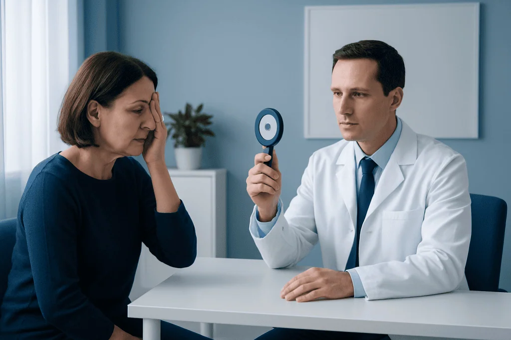
The main symptom of hemianopsia is missing part of the visual field, but the person may not realize at first that a specific visual half is absent. Instead, they may notice practical problems. Common experiences include stumbling over objects on one side, clipping furniture with the shoulder, becoming startled by people appearing from the affected side, or difficulty following lines of text while reading.
Some people also develop poor spatial awareness, headaches, dizziness, or trouble concentrating, especially if the cause is a recent neurological event. Others may have no eye pain and no obvious change in central sharpness, which can delay recognition. Family members sometimes notice the problem first when the person ignores one side of the room or turns the head repeatedly to compensate.
- Missing the left or right side of vision
- Reading difficulties or skipping words
- Bumping into objects or people
- Problems driving or crossing streets safely
- Reduced awareness of one side of the environment
- Symptoms of the underlying cause, such as weakness, speech changes, or severe headache
Sudden onset hemianopsia is especially important because it can be a warning sign of an acute neurological problem. If visual field loss appears together with weakness, numbness, trouble speaking, confusion, or facial drooping, emergency evaluation is needed.
Causes and risk factors
Hemianopsia happens when the optic tract, optic radiations, occipital lobe, or other parts of the visual pathway are damaged. The most common cause in adults is stroke, especially a stroke affecting the back part of the brain where visual information is processed. This is why visual field loss may be part of a broader neurological condition and should not be dismissed as simple eye strain.
Other possible causes include traumatic brain injury, brain tumors, bleeding in the brain, inflammation such as multiple sclerosis, brain infections, and complications after surgery. In some cases, pressure on the optic chiasm from a pituitary region tumor can lead to a different pattern of field loss. Children may develop hemianopsia after congenital brain injury, tumors, or severe neurological illness.
Risk factors depend on the underlying cause. They may include high blood pressure, diabetes, smoking, high cholesterol, heart disease, blood clotting disorders, prior stroke, or head trauma. A person already diagnosed with a neurological condition may also be at increased risk of visual field changes and may need ongoing monitoring.
Because the cause can range from temporary inflammation to urgent vascular disease, accurate diagnosis matters. Treating the underlying problem quickly may help limit further injury and improve overall outcomes.
How doctors diagnose hemianopsia
Diagnosis begins with a careful history and examination. A doctor will ask when the vision change started, whether it came on suddenly or gradually, and whether there are associated symptoms such as headache, weakness, speech difficulty, balance problems, or recent trauma. The examination may include visual acuity, pupil responses, eye movements, and a bedside check of the visual fields.
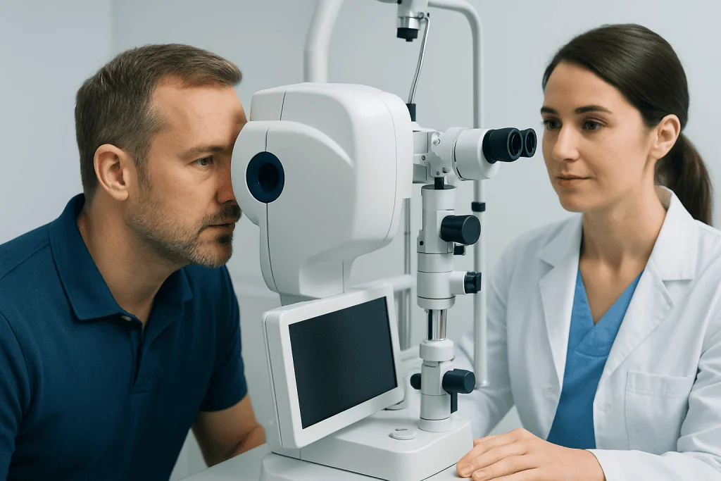
Formal visual field testing is usually needed to confirm the pattern and severity of loss. This may be done with computerized perimetry, which maps which parts of the visual field are seen and which are missing. If hemianopsia is present, brain imaging is commonly recommended to identify the cause, especially if the visual loss is new.
Depending on the situation, the workup may include MRI or CT imaging, blood tests, and referral to neurology, ophthalmology, or neuro-ophthalmology. If a vascular cause is suspected, doctors may also evaluate for brain and nerve disorders or evidence of an acute neurological event. The goal is not only to name the visual problem but also to find and treat the reason it happened.
Treatment options and rehabilitation
There is no single treatment that fits every case of hemianopsia because therapy depends on the cause. If the visual field loss follows an acute stroke, treatment focuses on urgent stroke care and long-term prevention. If a tumor, inflammation, or another brain condition is responsible, treatment is directed at that problem. In selected cases, management may involve brain tumor treatment or other neurological therapies recommended by specialists.
When the underlying condition is stabilized, rehabilitation becomes very important. Vision rehabilitation specialists may teach scanning techniques that train the person to turn the eyes and head more effectively toward the missing side. Reading strategies, mobility training, and occupational therapy can also help improve independence at home, school, and work.
Some patients may benefit from prism lenses or other visual aids that expand awareness of the affected side. Others may need support for associated problems such as balance changes, cognitive fatigue, or reduced confidence outdoors. If hemianopsia developed after a major neurological event, a broader rehabilitation plan may include stroke rehabilitation to address mobility, communication, and daily living skills.
Recovery varies. Some people experience partial improvement over time, especially in the first months after the injury, while others have lasting field loss and rely mainly on compensatory strategies. Regular follow-up helps monitor progress and adjust the rehabilitation plan safely.
Self-care, safety, and living with hemianopsia
Living with hemianopsia often requires practical changes rather than medical treatment alone. Good lighting, reducing clutter, and arranging frequently used items on the seeing side can make daily routines easier at first. Over time, many people learn to place reminders in their environment to encourage scanning toward the side they tend to miss.
Reading can become easier by using a straight edge or finger to guide the line, enlarging print, and taking regular breaks to reduce fatigue. At home, contrasting colors on stairs or doorways may improve safety. Family members can also help by approaching from the seeing side initially and allowing time for the person to orient.
Driving requires special caution. Laws and safety standards vary, and some people with hemianopsia may not meet legal or practical requirements for driving. A doctor or vision rehabilitation specialist can advise whether driving evaluation is appropriate. It is safest not to drive until the condition has been assessed and clear medical guidance has been given.
Near the end of the care journey, some patients may seek coordinated specialist assessment in an international center. Acibadem International’s multidisciplinary specialists and JCI-accredited hospitals diagnose and treat neurological and visual pathway conditions for international patients when a comprehensive evaluation is needed.
When to seek medical care
Sudden hemianopsia should always be treated as urgent. New loss of half of the visual field may be caused by stroke or another serious neurological condition, even if there is no eye pain. Immediate evaluation is especially important if the person also has weakness, numbness, trouble speaking, severe headache, confusion, or facial asymmetry.
Medical review is also needed if visual field loss develops gradually, seems to worsen, or interferes with reading, walking, work, or driving. Even when symptoms are mild, professional assessment can identify the cause and guide treatment or rehabilitation. Ongoing follow-up may be advised to monitor recovery, adjust therapy, and support daily function.
Frequently asked questions
Is hemianopsia the same as blindness in one eye?
No. Hemianopsia usually means loss of half of the visual field, often in a matching pattern across both eyes, rather than complete blindness in one eye. Many people still see clearly in the remaining half of their vision.
Can hemianopsia happen after a stroke?
Yes. Stroke is one of the most common causes of hemianopsia because it can damage the brain areas that process vision. Sudden visual field loss after or with stroke symptoms needs urgent medical care.
Can hemianopsia improve over time?
Some people have partial improvement, especially in the early months after the brain injury or illness. Others have more lasting vision loss but can still improve daily function through rehabilitation, scanning training, and safety strategies.
How is hemianopsia diagnosed?
Doctors usually confirm hemianopsia with visual field testing that maps which parts of vision are missing. Brain imaging such as MRI or CT is often used to find the underlying cause.
Is it safe to drive with hemianopsia?
Not always. Hemianopsia can reduce awareness of traffic, pedestrians, and hazards on one side, which may make driving unsafe. Whether a person can drive depends on local regulations, the severity of field loss, and specialist assessment.
What doctor treats hemianopsia?
Care may involve an ophthalmologist, neuro-ophthalmologist, neurologist, rehabilitation specialist, or neurosurgeon, depending on the cause. Because hemianopsia often reflects a brain or nerve pathway problem, coordinated evaluation is commonly needed.
References
- American Academy of Ophthalmology
- National Eye Institute
- National Institute of Neurological Disorders and Stroke
- Mayo Clinic
- American Stroke Association
This article is for general information only and is not a substitute for professional medical advice. Please consult a qualified doctor about your individual situation.
Explore treatments in Turkey — costs, top hospitals & a free quote
JCI-accredited · board-certified surgeons · reply within 24h
Check your numbers in seconds
BMI, calories, due date, blood pressure and 30+ more clinical calculators — free, instant, doctor-reviewed ranges.