Understanding Enchondroma Tumor: A Complete Patient Guide

An enchondroma is usually a noncancerous cartilage tumor located inside a bone. Many enchondromas are found incidentally on an X-ray performed for another reason.
Key Takeaways
- An enchondroma is usually a noncancerous cartilage tumor located inside a bone.
- Many enchondromas are found incidentally on an X-ray performed for another reason.
- Pain, a fracture after minor injury, or changes on imaging may lead to further evaluation.
- Most stable, symptom-free enchondromas can be monitored without surgery.
- Surgery may be considered when the lesion causes symptoms, weakens the bone, or cannot be clearly distinguished from a more serious tumor.
An enchondroma tumor is a growth of cartilage cells that develops within a bone, most often in the hands or feet. It is usually benign and causes no symptoms, but medical assessment is important to confirm the diagnosis and decide whether observation or treatment is appropriate.
What Is an Enchondroma Tumor?
An enchondroma tumor is a usually benign, or noncancerous, growth made of cartilage cells within a bone. Cartilage is the smooth, flexible tissue that helps shape bones during development and cushions joints. In an enchondroma, a small area of cartilage remains or grows inside the bone rather than at a joint surface.
These tumors are commonly identified in the small bones of the hands and feet. They can also occur in longer bones, such as the femur (thighbone), humerus (upper arm bone), or tibia (shinbone). An enchondroma may be discovered after an X-ray for an injury or another concern, even when it has never caused symptoms.
For most people, an isolated enchondroma is not cancer and does not require urgent treatment. However, cartilage tumors can sometimes have overlapping imaging features. Careful review by an orthopedic specialist, often with input from a musculoskeletal radiologist or orthopedic oncologist, helps ensure that the lesion is correctly characterized.
How Enchondromas May Affect the Bone
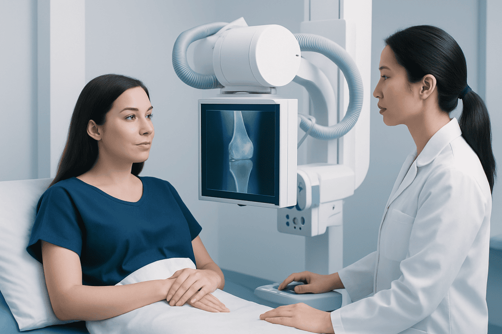
Enchondromas grow inside the central portion of a bone, known as the medullary cavity. They are often small and remain unchanged for years. A lesion may contain areas of calcification, which can create a characteristic pattern on an X-ray and help clinicians recognize cartilage tissue.
Although many lesions are harmless, an enchondroma can occasionally thin or expand the surrounding bone. In a small hand or foot bone, this may make the bone more vulnerable to a pathologic fracture, meaning a break that occurs through weakened bone after little or no force. In some cases, the fracture is the event that leads to diagnosis.
An isolated enchondroma is different from multiple enchondromas, which are uncommon and may occur in conditions such as Ollier disease or Maffucci syndrome. People with multiple lesions need individualized specialist follow-up because their risks and long-term monitoring needs differ from those of a person with one typical lesion.
Symptoms and Signs

Most enchondroma tumors do not cause symptoms. They may be found incidentally when imaging is performed for a sprain, arthritis, injury, or another unrelated reason. When an enchondroma is stable and painless, its presence alone does not necessarily affect daily activities.
Possible symptoms depend on the tumor’s size and location. A person may notice local discomfort, swelling, tenderness, or a visible change in the shape of a finger, toe, or other affected area. Pain may also occur if the bone has weakened or fractured. It is important to remember that pain near an enchondroma is not always caused by the tumor; tendon, joint, nerve, and injury-related problems can also be responsible.
Persistent pain at rest or at night, worsening discomfort, a growing lump, or a new fracture deserves clinical review. These features do not automatically mean cancer, but they can indicate the need for additional imaging or specialist assessment to clarify what is happening in and around the bone.
Causes and Risk Factors
The exact cause of a solitary enchondroma is not fully understood. It is generally considered to arise from cartilage cells left behind during normal bone growth or from changes in cartilage cells that occur after development. In most cases, it is not caused by an injury, exercise, diet, or something a person did or did not do.
Enchondromas can be diagnosed at many ages, though they are often recognized in adolescents and adults. They may be present for a long time before being seen on imaging. There is no established routine blood test or lifestyle factor that predicts who will develop one.
Rare inherited or developmental disorders can involve multiple enchondromas. A clinician may consider this possibility if several lesions are present, especially if there are bone shape differences, unequal limb length, or vascular skin changes. Genetic counseling or assessment may be appropriate in selected cases, but it is not routinely needed for a single typical enchondroma.
Diagnosis and Distinguishing Other Cartilage Tumors
Diagnosis begins with a medical history, symptom review, and examination of the affected area. Standard X-rays are usually the first and most useful test. They can show the lesion’s location, size, borders, internal calcification, and whether the outer bone cortex appears intact.
If the X-ray appearance is not fully typical, or if there is concerning pain or structural change, a doctor may request magnetic resonance imaging (MRI) or computed tomography (CT). MRI can provide more detail about cartilage, bone marrow, and nearby soft tissues. CT can be particularly useful for showing calcification and the bone’s structure. Repeat imaging over time may help determine whether a lesion is stable.
In selected cases, a biopsy may be advised. During this procedure, a small tissue sample is examined by a pathologist. Biopsy decisions are made carefully because cartilage tumors can be challenging to interpret from a small sample alone; imaging findings, symptoms, and pathology results are best considered together. The key aim is to distinguish a benign enchondroma from conditions that need different management, including chondrosarcoma, a malignant cartilage tumor.
Treatment Options and Follow-Up
For a small, painless enchondroma with reassuring imaging features, observation is often the preferred approach. This may include follow-up appointments and X-rays at intervals chosen by the treating clinician. Monitoring allows the team to confirm that the lesion remains stable while avoiding unnecessary procedures.
Surgery may be considered if the lesion causes persistent symptoms, contributes to a fracture, substantially weakens the bone, grows or changes on imaging, or has uncertain features. A common surgical approach is curettage, in which the surgeon removes the tumor tissue from inside the bone. The resulting space may be filled with bone graft, bone substitute, or another material when needed to support healing.
When a fracture is present, treatment may involve stabilizing the bone as well as addressing the lesion, depending on its location and severity. Recovery plans vary according to the bone involved, the procedure performed, and a person’s overall health. Follow-up imaging after surgery can help assess healing and identify the uncommon possibility of recurrence.
Care is ideally coordinated by clinicians experienced in bone tumors. Acibadem International’s multidisciplinary specialists and JCI-accredited hospitals diagnose and treat bone conditions for international patients, with treatment planning based on the individual’s imaging, symptoms, and overall needs.
Living Well and When to Seek Medical Care
There is no proven home treatment that makes an enchondroma disappear, and most people do not need to restrict normal activity solely because of a stable lesion. A clinician can advise whether any temporary changes are sensible, particularly when the tumor is in a weight-bearing bone or when the bone appears thin. Attending recommended imaging appointments is an important part of safe observation.
It is helpful to bring prior X-rays, MRI reports, and details about the onset and pattern of pain to appointments. Patients should tell their doctor if discomfort becomes more frequent, occurs at rest or at night, follows a new injury, or is associated with swelling or reduced function. This information helps clinicians decide whether the current follow-up plan remains appropriate.
Medical care should be sought promptly after a suspected fracture, such as sudden severe pain, marked swelling, deformity, or an inability to use the affected hand, foot, arm, or leg. A non-urgent appointment should be arranged for new or progressive bone pain, an enlarging area of swelling, or a known enchondroma that has not been reviewed as advised. In most cases, timely assessment provides clarity and supports straightforward management.
Frequently asked questions
Is an enchondroma tumor cancer?
An enchondroma is usually a benign cartilage tumor, meaning it is not cancer. Because some cartilage tumors can look similar on imaging, a doctor may recommend additional scans or follow-up to confirm that the lesion has reassuring features.
Can an enchondroma turn into cancer?
A solitary enchondroma is generally considered to have a very low risk of malignant change. The situation is different for people with rare disorders that cause multiple enchondromas, who may need more specialized long-term monitoring.
Does an enchondroma need to be removed?
Not always. Many painless enchondromas with typical, stable imaging findings are monitored rather than removed. Surgery may be considered for symptoms, a fracture risk, structural weakening, growth, or uncertainty about the diagnosis.
What does enchondroma pain feel like?
Many enchondromas do not cause pain at all. If pain is present, it may be local aching or tenderness, but pain can also come from a nearby joint, tendon, injury, or fracture. New, persistent, or night pain should be evaluated by a doctor.
How is an enchondroma diagnosed?
X-rays are typically the first test and often show the features of an enchondroma. MRI, CT, repeat imaging, or occasionally a biopsy may be used when the appearance is unclear or symptoms suggest that further assessment is needed.
Can people exercise with an enchondroma?
Many people can continue usual activities if the lesion is stable and the bone is not significantly weakened. The treating clinician may recommend modifying certain activities temporarily when there is pain, a high fracture risk, or recovery after surgery.
References
- American Academy of Orthopaedic Surgeons
- National Cancer Institute
- OrthoInfo
- Merck Manual Consumer Version
- World Health Organization Classification of Tumours
This article is for general information only and is not a substitute for professional medical advice. Please consult a qualified doctor about your individual situation.
Joint and spine care in Turkey — expert assessment & treatment
JCI-accredited · board-certified surgeons · reply within 24h
Add us as a Preferred Source to see more of our trusted health content across Google Search, AI Overviews and Discover.