Fenestrated: An Evidence-Based Guide for Patients

Fenestrated means containing an opening, window, or series of small holes. The term may describe medical devices, surgical techniques, anatomy, or imaging findings.
Key Takeaways
- Fenestrated means containing an opening, window, or series of small holes.
- The term may describe medical devices, surgical techniques, anatomy, or imaging findings.
- A fenestration is usually intentional when it refers to a medical device or procedure.
- The benefits and risks depend on the specific clinical context, such as airway care or blood vessel repair.
- A clinician can explain what fenestrated means in an individual medical report or treatment plan.
Fenestrated is a descriptive medical term meaning that something has one or more carefully placed openings, holes, or window-like sections. It is not a diagnosis on its own; its meaning depends on the body part, device, scan, or procedure being discussed.
What Does Fenestrated Mean?
Fenestrated means “having an opening” or “having windows.” The word comes from the Latin word fenestra, meaning window. In healthcare, clinicians use it to describe a structure, device, or treatment technique that includes one or more deliberate openings.
The term alone does not identify an illness or determine whether something is dangerous. For example, a fenestrated medical device may be designed to let air, blood, fluid, sound, or instruments pass through a controlled opening. In another setting, a report may use fenestrated simply to describe a natural anatomical pattern.
Understanding the surrounding words is important. “Fenestrated tracheostomy tube,” “fenestrated stent graft,” and “fenestrated membrane” describe very different situations. A patient who sees this term in a record can ask the treating clinician what structure is fenestrated, why it is relevant, and whether any follow-up is needed.
Where the Term Is Used in Medicine
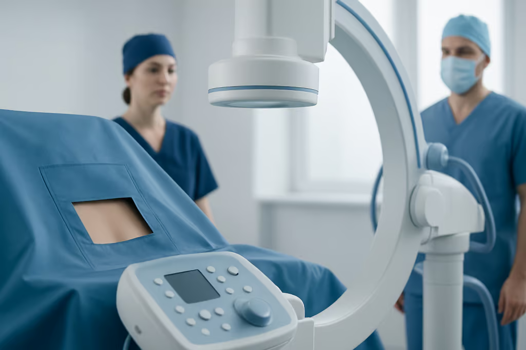
Fenestrated can be used in many areas of medicine. It most often refers to a device or procedure that has been adapted with precisely placed openings to preserve normal function while allowing treatment to take place. The opening may be round, oval, slotted, or shaped for a particular anatomical structure.
In airway care, a fenestrated tracheostomy tube has openings in part of the tube. In selected patients, these openings may help some air move upward through the voice box when the inner cannula is removed and the tube is managed appropriately. This may support speech in some circumstances, but suitability depends on the person’s airway, swallowing, breathing needs, secretions, and stage of recovery.
In vascular medicine, a fenestrated stent graft is a customized or specially designed graft with openings that align with branch arteries. It may be considered when treating certain complex aortic aneurysms, particularly where important arteries arise close to the area requiring repair. The openings are intended to help maintain blood flow to these branches while excluding the weakened part of the aorta.
Other uses include surgical materials, drainage devices, implants, and descriptions in anatomy or pathology. Therefore, the same word can have a routine, highly technical, or procedural meaning depending on the specialty involved.
Fenestrated Devices and Procedures: Why They Are Used
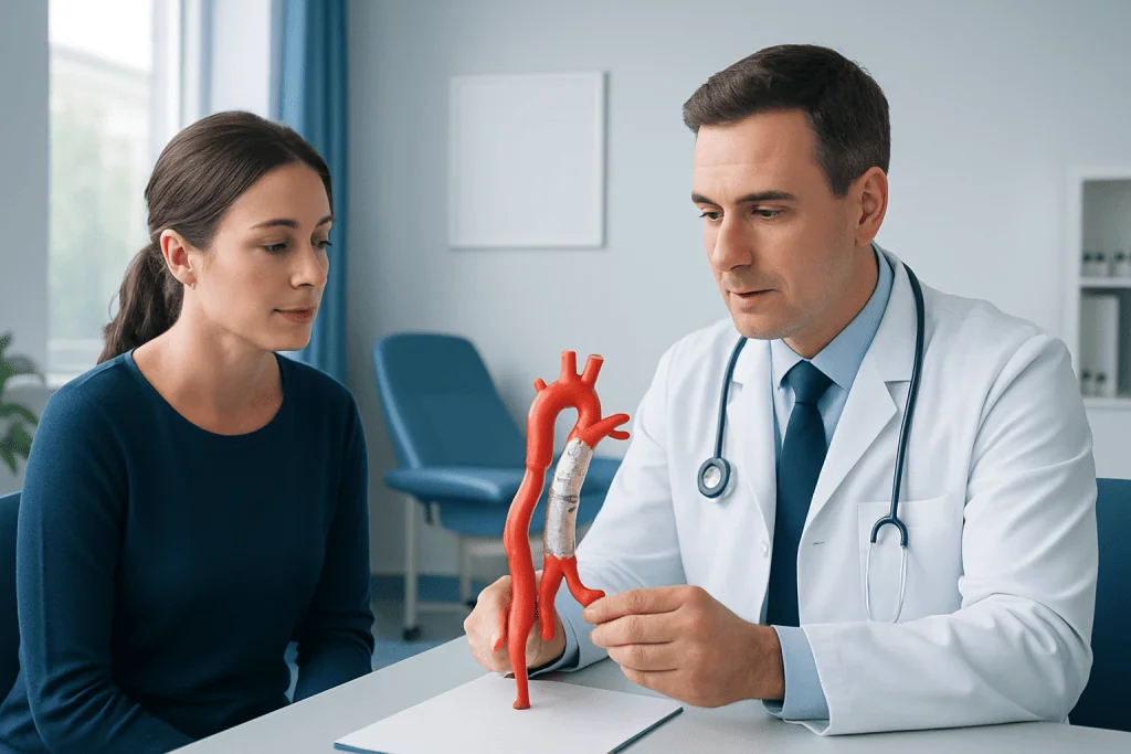
In clinical practice, fenestrations are created for a purpose. They may allow access to a target area, improve communication between spaces, preserve flow through a nearby vessel, or enable movement of air or fluid. The intended benefit is always specific to the device and the person’s medical needs.
A fenestrated approach can be useful when standard equipment would cover or obstruct an important structure. For example, a vascular graft without appropriately positioned openings could block an artery supplying the kidneys, intestines, or other organs. A fenestrated design may allow specialists to treat the diseased aortic segment while protecting blood flow through key branches.
Planning is especially important for procedures involving blood vessels. Clinicians use detailed imaging to assess the anatomy, the position of branch arteries, the size and shape of the vessel, and whether a fenestrated repair is technically appropriate. Treatment may include aneurysm treatment options selected according to the aneurysm’s location, size, symptoms, growth pattern, and the patient’s overall health.
Not every patient needs or benefits from a fenestrated device. A conventional device, open surgery, observation, or another approach may be more appropriate. The treating team balances possible benefits against procedure-related risks and the practical needs of long-term follow-up.
How Fenestrated Tracheostomy Tubes Work
A tracheostomy is an opening created through the neck into the windpipe to support breathing or help clear airway secretions. Some tracheostomy tubes are fenestrated, meaning they contain openings in the outer tube. When conditions are appropriate, this can allow a portion of exhaled air to pass upward through the upper airway and vocal cords.
Whether a fenestrated tube is used is an individualized decision. Healthcare professionals consider the reason for the tracheostomy, the need for mechanical ventilation, the amount and thickness of secretions, airway anatomy, cough strength, and swallowing safety. Speech and language therapists, respiratory therapists, nurses, and ear, nose, and throat specialists may all contribute to care.
Fenestrated tubes are not suitable for everyone. Openings can sometimes become blocked by dried secretions or tissue, and an unsuitable tube or incorrect use may affect ventilation or increase other complications. For this reason, tube selection, cleaning, suctioning, changes, and speaking-related trials should be directed by trained clinicians.
Anyone with a tracheostomy should follow their personalized airway-care plan. New breathing difficulty, increased work of breathing, inability to clear secretions, bleeding, a blocked tube, or accidental tube displacement requires urgent medical assessment.
What Fenestrated May Mean on a Scan or Medical Report
Medical reports may use fenestrated as a descriptive term rather than a treatment recommendation. In anatomy, it can refer to a structure that has tiny openings or a window-like pattern. In imaging or pathology, it may describe the appearance of tissue, bone, a membrane, or a blood vessel structure.
For example, some capillaries are naturally described as fenestrated because their lining has very small pores that support exchange of water and certain dissolved substances. These specialized capillaries are found in organs where rapid exchange is useful, including parts of the kidneys and endocrine system. This is a normal microscopic feature, not a disease.
In other cases, a clinician may describe an anatomical opening as a fenestration. Its importance depends on its location, size, cause, symptoms, and whether it affects normal function. A report should be interpreted alongside the patient’s history, examination, and any relevant test results.
Patients should avoid drawing conclusions from one unfamiliar word in a report. It is reasonable to ask for a plain-language explanation, including whether the finding is normal anatomy, an incidental observation, a sign of disease, or a feature that needs monitoring.
Benefits, Limitations, and Safety Considerations
A fenestrated design may offer practical advantages when it is carefully selected and correctly positioned. Depending on the context, it may preserve blood flow to vital branches, support communication in a person with a tracheostomy, or allow a surgeon to work through a controlled opening. These potential benefits are not automatic and vary between individuals.
All medical devices and procedures also have limitations. In vascular treatment, risks can include bleeding, infection, contrast-related concerns, injury to blood vessels, blood-flow problems involving branch arteries, and the need for continued imaging surveillance. In airway care, concerns may include blockage by secretions, difficulty with ventilation, irritation, and aspiration risk in some patients.
Specialists review the likely benefits, alternatives, and possible complications before recommending treatment. For complex aortic repair, planning commonly involves CT angiography or other detailed imaging and may involve surgeons, interventional specialists, anesthesiologists, and imaging teams. Follow-up remains important because implanted vascular devices require monitoring over time.
Acibadem International’s multidisciplinary specialists and JCI-accredited hospitals diagnose and treat complex conditions for international patients, including cases that may require advanced airway or vascular assessment.
When to Seek Medical Care
A person should contact their clinician if they have been told that a device, procedure, or scan finding is fenestrated and do not understand what it means. Clarifying the term can help patients follow instructions correctly and understand whether they need monitoring, additional imaging, or a specialist review.
Urgent medical care is needed for sudden or severe symptoms after a vascular procedure, such as chest, back, or abdominal pain; fainting; new weakness; a cold or painful limb; or symptoms of poor circulation. People with known or suspected aortic disease should seek urgent assessment for sudden severe chest, back, or abdominal pain, especially if it is associated with sweating, shortness of breath, collapse, or weakness. These symptoms can occur with serious conditions, including aortic aneurysm complications.
For a tracheostomy, emergency assessment is needed for severe breathing difficulty, suspected blockage, persistent bleeding, accidental decannulation, blue or grey lips, or reduced responsiveness. Caregivers should follow the emergency instructions provided by the airway team and call local emergency services when necessary.
Frequently asked questions
Is fenestrated a diagnosis?
No. Fenestrated is a descriptive word, not a diagnosis. It indicates that a structure, device, or technique has an opening or window-like feature, and its clinical meaning depends on the context.
What is a fenestrated tracheostomy tube?
A fenestrated tracheostomy tube contains openings in part of the tube. In selected patients, it may permit some airflow through the upper airway and vocal cords, potentially helping with speech. It should only be used and managed according to a clinician’s instructions.
What is a fenestrated stent graft?
A fenestrated stent graft is a vascular graft designed with openings that align with important arteries branching from the aorta. It may be used in selected complex aortic repairs to maintain blood flow to these arteries. Careful imaging and specialist planning are essential.
Does fenestrated mean there is a hole in an organ?
Not necessarily. The term can describe a designed opening in a medical device, a normal microscopic feature, or an anatomical pattern. The report or clinician should clarify exactly what is fenestrated and whether it has any health significance.
Are fenestrated medical devices safe?
They can be safe and effective when they are appropriate for the patient and used by trained healthcare professionals. Like all medical devices, they carry potential risks that vary by device type, procedure, and the person’s health. A treating team can explain the expected benefits, alternatives, and follow-up plan.
Should a patient worry if fenestrated appears in a test result?
The word by itself is not usually a reason for concern. It may simply describe normal anatomy or a technical feature of a device. Patients should discuss the full report with the clinician who ordered the test rather than interpreting one term in isolation.
References
- National Heart, Lung, and Blood Institute
- Society for Vascular Surgery
- American Thoracic Society
- National Institute of Diabetes and Digestive and Kidney Diseases
- Merck Manual Consumer Version
This article is for general information only and is not a substitute for professional medical advice. Please consult a qualified doctor about your individual situation.
Explore treatments in Turkey — costs, top hospitals & a free quote
JCI-accredited · board-certified surgeons · reply within 24h
Add us as a Preferred Source to see more of our trusted health content across Google Search, AI Overviews and Discover.

More from the Health Library


Related Specialists

Dr. Fikret Çolak
Radiology
Dr. Ayla Oktay
Pediatric Cardiology
Dr. Necati Çalı
Emergency Service
Assoc. Prof. Dr. Aslıhan Demirel
Infectious Diseases & Clinical Microbiology