Private mri scan Risks & Side Effects Explained by Surgeons
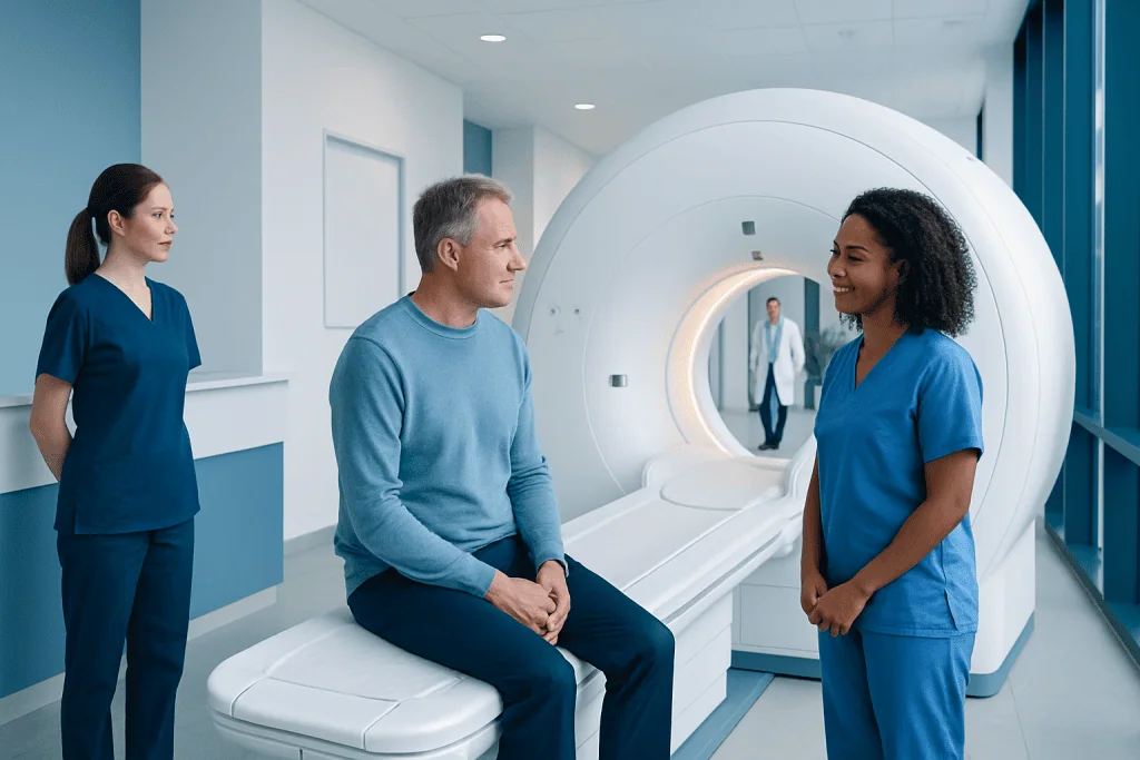
MRI uses powerful magnets and radio waves rather than ionising radiation. The most important safety step is declaring all implants, metal exposure, medicines and health conditions before the scan.
Key Takeaways
- MRI uses powerful magnets and radio waves rather than ionising radiation.
- The most important safety step is declaring all implants, metal exposure, medicines and health conditions before the scan.
- Gadolinium contrast is not needed for every MRI; serious allergic reactions are uncommon, but kidney function may need assessment.
- Noise, lying still and enclosed-space anxiety are common practical challenges and can usually be managed.
- A private MRI scan should be requested and interpreted in the context of symptoms, examination findings and appropriate follow-up.
Private MRI scan risks are generally low: MRI does not use X-rays or ionising radiation, and most people have no lasting side effects. Safe imaging depends on careful screening for implanted devices, metal fragments, kidney concerns, pregnancy considerations and possible reactions to contrast dye.
Overview: are private MRI scans safe?
A private MRI scan is usually a safe diagnostic test when it is appropriately requested and performed with standard safety checks. MRI, or magnetic resonance imaging, creates detailed pictures using a strong magnetic field and radio waves. Unlike CT scans and standard X-rays, it does not use ionising radiation, so there is no radiation exposure from the examination itself.
The main private MRI scan risks relate to the powerful magnet, not radiation. Some implanted devices or metal fragments can move, heat up or malfunction in the scanner. A screening questionnaire and discussion with the imaging team are therefore essential, even for people who have had an MRI before.
Some examinations use a gadolinium-based contrast agent to make blood vessels, inflammation or certain tissue changes easier to see. Most people tolerate contrast well, but the decision to use it should consider the clinical question, allergy history and kidney health. A private scan can offer timely access, but its value is greatest when a qualified clinician can explain whether imaging is needed and what the result means.
How MRI works and why safety screening matters
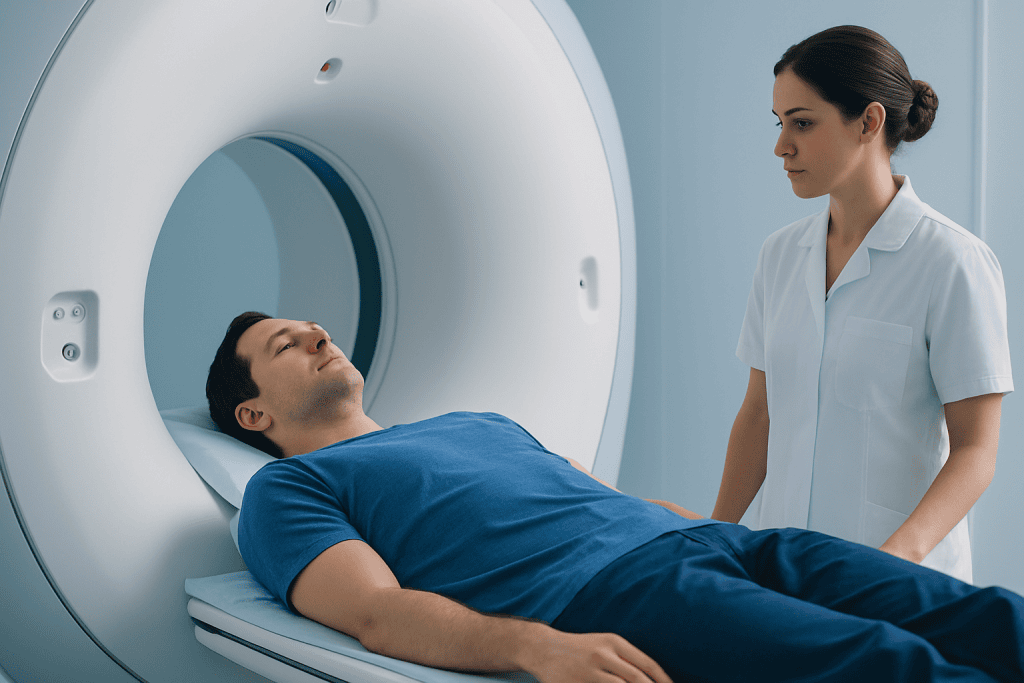
An MRI scanner is a large machine containing a very powerful magnet. During the scan, radio-wave pulses briefly alter the behaviour of hydrogen atoms in the body. As these atoms return to their usual state, they produce signals that a computer turns into detailed images of organs, joints, nerves, blood vessels and soft tissues.
The magnetic field is present around many MRI scanners continuously. For this reason, loose metal objects such as keys, coins, jewellery, hair clips, watches, hearing aids and bank cards must be removed before entering the scan room. Oxygen cylinders, wheelchairs and other equipment must also be MRI-compatible before they can be brought near the scanner.
The radiographer will ask about prior surgery, implants, pacemakers, neurostimulators, cochlear implants, aneurysm clips, medication pumps, dental work, tattoos and possible metal injury, especially to the eye. Many modern implants can be scanned under specific conditions, but this must be confirmed using the device information rather than assumed. More general information about arranging and preparing for the investigation is available through private MRI scan services.
Who may need additional assessment before an MRI

Most adults and children can have an MRI, but certain circumstances require extra planning or may make a particular scanner unsuitable. People with a pacemaker, implanted defibrillator, brain or spinal stimulator, cochlear implant, infusion pump, surgical clips or artificial joint should tell the referrer and imaging centre well in advance. The team can check whether the device is MRI-safe or MRI-conditional and identify any scanning limits.
Anyone who has worked with metal, had shrapnel injuries or may have a metal fragment in an eye needs to mention this. In some situations, an X-ray may be used to assess for metal before MRI. Pregnancy should also be discussed. MRI without contrast may be considered when clinically important, while gadolinium contrast is generally avoided during pregnancy unless the expected benefit clearly outweighs the potential risk.
Kidney disease, dialysis, previous reactions to contrast agents, asthma and severe allergies should be discussed before a contrast-enhanced scan. Claustrophobia, severe pain, difficulty lying flat, dementia or movement disorders may affect comfort and image quality. The team may suggest practical adjustments, a wider-bore scanner, a support person where permitted, or clinician-supervised medicine for anxiety when appropriate.
What happens during a private MRI scan
Before the appointment, the patient usually receives preparation instructions. In many cases, normal food, drink and regular medicines can continue, but fasting may be requested for selected abdominal scans or when sedation is planned. Comfortable clothing without metal is helpful, although a gown may be provided. The patient should bring details of implants, previous imaging and relevant medical records if requested.
On arrival, a radiographer reviews the safety questionnaire and explains the scan. If contrast is required, a small cannula may be placed in a vein. The patient lies on a padded table, and a receiving coil may be placed around the body part being examined. The table then moves into the scanner. The examination is painless, but the machine makes loud tapping and knocking sounds; ear protection is provided.
Each image sequence can last several minutes, and keeping still is important to avoid blurred images. The radiographer can see and speak to the patient throughout the scan, and an alarm device is usually available to call for help. Scan duration varies by body area and whether contrast is used, but patients should allow enough time for screening, positioning and any necessary post-contrast observation.
Afterward, images are reviewed and reported by an appropriately qualified radiologist. The report should be interpreted alongside symptoms, examination findings and any earlier tests. An abnormality on MRI does not always explain a person’s symptoms, while a normal MRI does not rule out every health condition.
Private MRI scan risks and possible side effects
The loud sounds, the need to remain still and the enclosed feel of the scanner are the effects most people notice. Some patients experience anxiety, claustrophobia or temporary discomfort from lying in one position. Ear protection helps reduce noise exposure, and the team can pause the examination if a patient feels unable to continue.
Magnet-related hazards are uncommon when screening is thorough, but they can be serious if a non-compatible implant or metal object enters the MRI environment. Potential concerns include movement or heating of metal, interference with an implanted device, and injury from an unsecured metal object being pulled toward the magnet. This is why patients should never enter the scan room with personal metal items unless staff have confirmed they are safe.
If gadolinium contrast is used, mild effects such as a cool sensation at the injection site, nausea, headache or itching can occur, though many people have no symptoms. Allergic-type reactions are possible and are usually mild; severe reactions are rare, and imaging teams are trained to respond promptly. Contrast can leak outside the vein, causing local swelling or discomfort, which should be reported to staff.
In people with severe kidney impairment, certain gadolinium agents have been associated with a rare condition called nephrogenic systemic fibrosis. Modern contrast selection and kidney assessment have substantially reduced this concern, but kidney disease must be declared before contrast is given. Tiny amounts of gadolinium may remain in the body after some contrast scans; the clinical importance of this in people with normal kidney function is still being studied, so contrast is used when it is expected to improve diagnostic information.
- MRI does not expose the patient to ionising radiation.
- Contrast is used only when it can add useful diagnostic detail.
- Patients should report burning, pain, rash, breathing difficulty, dizziness or swelling immediately.
Benefits, recovery and sensible next steps
MRI can provide highly detailed images without surgery and without radiation. It is particularly useful for many concerns involving the brain, spine, joints, muscles, pelvic organs, abdomen and blood vessels. Depending on the clinical situation, it may help identify inflammation, injury, infection, tumours or changes linked with conditions such as multiple sclerosis.
Recovery is usually immediate. Patients can generally eat, drink, work and drive after a routine MRI unless they have received sedation or have been given other specific instructions. After contrast, drinking fluids as normally tolerated may be advised. If sedating medicine was used, the patient needs a responsible adult to accompany them home and should avoid driving, alcohol and important decisions for the period advised by the clinical team.
A scan is most helpful when it answers a focused clinical question. Incidental findings are relatively common, particularly with imaging performed in people without symptoms. These are unexpected changes that may be harmless but can sometimes lead to further appointments or tests. Discussing the purpose, possible benefits, limitations and potential follow-up before booking supports an informed decision.
Acibadem International’s multidisciplinary specialists and JCI-accredited hospitals diagnose and treat patients from abroad, with imaging findings assessed as part of an individual clinical evaluation.
When to seek medical care
Before the scan, patients should contact the imaging provider or referring clinician if they discover they have an implant, may have metal in their body, are pregnant, have significant kidney disease, or have had a previous contrast reaction. It is better to clarify safety before arriving than to proceed with uncertainty.
During the scan, patients should alert staff immediately if they feel pain, heating, burning, severe distress, shortness of breath, itching, facial swelling or dizziness. The radiographer can stop the scan and assess the situation. A new or worsening rash, swelling, breathing problem or other concerning symptom after leaving the centre requires urgent medical assessment.
Patients should also arrange follow-up with the clinician who requested the MRI to discuss the report, especially if symptoms persist, worsen or include new weakness, loss of sensation, significant injury, severe headache or other urgent concerns. MRI results should guide care rather than replace a medical assessment.
Frequently asked questions
Does a private MRI scan have radiation risks?
No. MRI uses magnetic fields and radio waves rather than ionising radiation, so it does not have the radiation exposure associated with X-rays or CT scans. Its key safety issues are related to metal, implanted devices and, where used, contrast material.
What are the most common side effects after an MRI scan?
Most people have no side effects after a routine MRI. Some may feel temporarily uncomfortable from noise, remaining still or being in an enclosed space. If contrast is used, mild nausea, headache, itching or injection-site discomfort can occur.
Can someone with a pacemaker have an MRI?
Some people with pacemakers can have an MRI under carefully controlled conditions, particularly if the device is MRI-conditional. The exact device model, leads and implantation details must be checked by the relevant clinical and imaging teams before scheduling the scan.
Is MRI contrast safe for people with kidney disease?
Contrast may still be possible, but the decision needs individual assessment. The clinician and imaging team consider kidney function, the type of gadolinium agent and whether contrast is essential for the diagnostic question. People on dialysis or with severe kidney disease should always tell the team in advance.
How long does it take to recover after an MRI?
There is usually no recovery period after an MRI, and most people return to usual activities straight away. If sedation was used for anxiety or comfort, driving and certain activities should be avoided until the effects have fully worn off, following the care team’s instructions.
What should a patient do if they feel claustrophobic during an MRI?
They should tell the referring clinician and imaging centre before the appointment. Options may include detailed reassurance, music or ear protection, communication with the radiographer, a wider scanner where available, or appropriate clinician-prescribed medication. The scan can be paused if distress develops.
References
- American College of Radiology
- Radiological Society of North America
- U.S. Food and Drug Administration
- National Health Service
- World Health Organization
This article is for general information only and is not a substitute for professional medical advice. Please consult a qualified doctor about your individual situation.
Oncology care in Turkey — second opinion and treatment plan
JCI-accredited · board-certified surgeons · reply within 24h
Add us as a Preferred Source to see more of our trusted health content across Google Search, AI Overviews and Discover.