

Brain Neoplasm MRI: Preparation, Procedure and Results
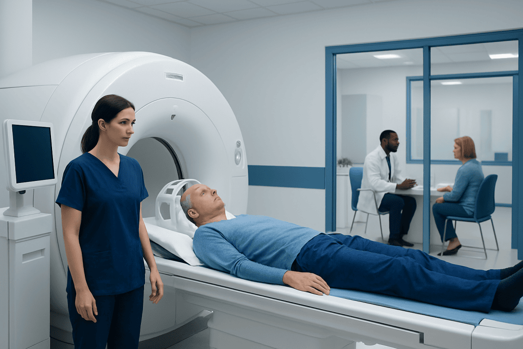
Brain MRI uses magnetic fields and radio waves rather than ionising radiation. Contrast material may be used to make certain tumours, inflammation and blood vessels easier to see.
Key Takeaways
- Brain MRI uses magnetic fields and radio waves rather than ionising radiation.
- Contrast material may be used to make certain tumours, inflammation and blood vessels easier to see.
- The scan is painless, but it requires lying still in a narrow scanner and can be noisy.
- MRI can strongly suggest the nature of a brain lesion, but it cannot always determine whether a tumour is cancerous.
- Results may be available within hours to several days, depending on the setting and whether specialist review is needed.
A brain neoplasm MRI is a magnetic resonance imaging scan used to examine a suspected or known brain tumour in detail. It can show the location, size and imaging features of an abnormal area, but tissue testing is often needed to confirm the exact tumour type and whether it is cancerous.
Overview: What Is a Brain Neoplasm MRI?
A brain neoplasm MRI is an MRI scan performed to investigate a possible or confirmed tumour in the brain. “Neoplasm” means an abnormal growth of cells; it may be benign (non-cancerous), malignant (cancerous), or uncertain until further tests are completed. The scan provides highly detailed pictures of brain structures and helps clinicians understand an abnormality’s size, location and relationship to nearby tissue.
MRI uses strong magnetic fields and radiofrequency waves to create images. Unlike CT scanning and standard X-rays, it does not use ionising radiation. Several image sequences are taken to highlight different features, such as fluid, swelling, blood products and tissue composition. In many cases, a contrast agent containing gadolinium is injected into a vein to show areas where the blood-brain barrier is disrupted or where blood supply is increased.
A brain neoplasm MRI may be requested after symptoms, an examination finding, or another imaging test raises concern. It is also used to plan treatment, guide biopsy or surgery, monitor a tumour over time, and assess the response to treatment. MRI findings are interpreted alongside symptoms, medical history and, when needed, pathology results.
How a Brain MRI Helps Assess a Suspected Tumour
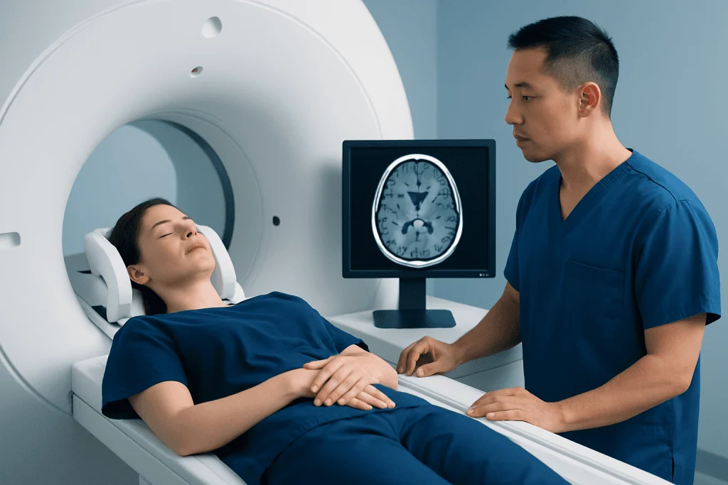
Brain MRI is one of the most useful imaging tools for evaluating a suspected brain tumour. It can identify whether an area appears to arise within brain tissue, the coverings of the brain, nerves, the pituitary region, or another structure. It can also show pressure effects, fluid build-up, bleeding, calcification patterns in some cases, and swelling around a lesion.
Specialised MRI techniques may add further information. Diffusion imaging can assess the movement of water within tissue, perfusion imaging can estimate blood flow, and spectroscopy can examine certain chemical signals. These techniques may help the radiologist and neurology or neurosurgery team narrow the possibilities, but they are not a substitute for a diagnosis based on tissue when this is clinically necessary.
Results can also help distinguish a tumour from conditions that may look similar on imaging, including infection, inflammation, stroke-related changes or vascular abnormalities. A multidisciplinary review may involve a radiologist, neurologist, neurosurgeon, oncologist, radiation oncologist and pathologist, depending on the findings.
When imaging suggests that a tissue sample is needed, the team may discuss brain tumour surgery or a targeted biopsy to establish the diagnosis and guide treatment planning.
Candidacy, Safety Checks and Preparation
Most people can undergo a brain MRI safely. Before the appointment, the imaging team will ask about implanted or retained metal and electronic devices. This includes pacemakers, cochlear implants, neurostimulators, aneurysm clips, insulin pumps, medication patches, shrapnel injuries and previous surgery. Many modern implants are MRI-compatible under specific conditions, but this must be checked in advance.
People should also tell the team about kidney disease, previous reactions to MRI contrast, pregnancy or the possibility of pregnancy, and severe claustrophobia. Gadolinium contrast is usually well tolerated, but it may not be appropriate for everyone, particularly those with severe kidney impairment. The imaging team may arrange blood testing or choose a non-contrast MRI protocol when appropriate.
What should you not do before a brain MRI? Patients should not enter the MRI room with metal objects or items that may contain metal. This includes jewellery, watches, hairpins, hearing aids, credit cards, phones, coins, belts, removable dental appliances and some cosmetics or clothing with metallic fibres. They should not stop prescribed medicines unless their doctor specifically advises this, and they should report all implants, past metal injuries and possible pregnancy honestly.
It is helpful to arrive early, bring relevant previous imaging if requested, and wear comfortable clothing without metal fasteners. If anxiety about enclosed spaces is likely, patients should mention it before the scan so that practical support or medication prescribed by a clinician can be considered.
Can I Eat or Drink Prior to a Brain MRI?
Can I eat or drink prior to a brain MRI? For most routine brain MRI examinations, people can eat and drink normally and continue their usual medicines. The imaging centre’s instructions should always take priority, as preparation can differ if sedation, anaesthesia, certain specialised studies or another procedure is planned on the same day.
If sedation is arranged because a person cannot remain still or has significant claustrophobia, fasting instructions may be required to reduce risks associated with sedation. People with diabetes, swallowing difficulties or other medical needs should ask for personalised guidance before changing meals, fluids or medication schedules.
Hydration is generally appropriate unless a doctor has advised fluid restriction. Patients who will receive contrast may be encouraged to drink normally after the scan as part of their usual daily intake, unless they have a medical condition that limits fluids.
Step by Step: What Happens During the Procedure?
On arrival, a radiology professional confirms identity, reviews the safety questionnaire and asks the person to remove metal items. If contrast is planned, a small intravenous cannula may be placed in an arm or hand. The person lies on a padded table, usually on their back, and a head coil is positioned around the head to obtain clear images.
The table then moves into the MRI scanner. The scan itself is painless, although the machine makes repetitive tapping, knocking or humming sounds. Earplugs or headphones are provided. It is important to remain as still as possible because movement can blur the images and may require sequences to be repeated. The radiographer can communicate with the patient throughout the examination.
Some sequences are completed before contrast is given. If contrast is used, it is injected through the cannula and additional images are taken. A person may notice a brief cool sensation in the arm, but many feel no different. A standard brain MRI often takes around 30 to 60 minutes, though advanced sequences or repeat imaging can take longer.
Young children and some adults who cannot stay still may need sedation or anaesthesia. This is arranged and monitored by appropriately trained clinical teams, with separate preparation and recovery requirements.
Risks, Benefits and Recovery Timeline
The main benefit of a brain neoplasm MRI is the detailed information it provides without radiation exposure. It can support timely decisions about observation, further testing, biopsy, surgery, radiotherapy, drug treatment or other care. It is also valuable for monitoring changes over time using comparable imaging sequences.
MRI is considered very safe when established screening procedures are followed. The most common challenges are noise, the need to remain still and discomfort in confined spaces. Rarely, a person may have an allergic-like reaction to contrast. Very rare risks related to gadolinium are more relevant in people with severe kidney problems, which is why the team reviews kidney health and contrast suitability beforehand.
Recovery after a standard MRI is immediate. Most people can return to work, travel, eat and resume normal activities straight away. If contrast was used, the cannula is removed before leaving. If sedation or anaesthesia was used, recovery takes longer; the person will need monitoring and should follow instructions about having an adult accompany them, avoiding driving and delaying important decisions.
For international patients, Acibadem International’s multidisciplinary specialists and JCI-accredited hospitals can coordinate MRI assessment, specialist review and further care when a brain lesion requires investigation.
Can You Tell From an MRI if a Brain Tumour Is Cancerous?
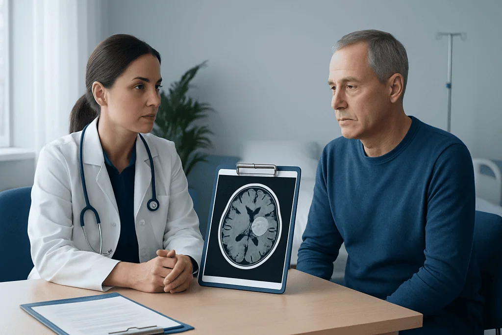
Can you tell from an MRI if a brain tumour is cancerous? MRI can show features that make a lesion more or less suspicious for a particular tumour type or grade, such as its margins, growth pattern, contrast enhancement, blood flow, surrounding swelling and effects on nearby structures. However, MRI alone cannot reliably confirm whether every tumour is cancerous or establish the exact tumour subtype.
Some brain tumours are benign but may still require treatment because of their location or effect on brain function. Conversely, a malignant tumour may not always have a single definitive imaging appearance. When the result will change treatment decisions, a biopsy or surgical removal of tissue may be recommended for examination by a pathologist and, increasingly, molecular testing.
A specialist team will explain what the MRI suggests, what remains uncertain and whether monitoring, additional imaging, blood tests, surgery or biopsy is appropriate. A finding on MRI is important, but it is only one part of a careful diagnostic process.
How Soon After a Brain MRI Do You Get Results?
How soon after a brain MRI do you get results? Timing varies by hospital, scan urgency and whether the images need review by a subspecialist neuroradiologist or multidisciplinary team. In urgent settings, preliminary findings may be communicated on the same day. For planned outpatient imaging, a formal report is commonly sent to the referring clinician within a few days.
The radiologist reviews all image sequences and compares them with prior scans when available. The referring doctor then discusses the results in the context of symptoms and medical history. If an unexpected finding requires urgent attention, the imaging team or referring clinician will generally arrange prompt contact and next steps.
Patients can ask at the appointment how and when results will be shared, and who to contact if they have not heard back within the expected timeframe. They should avoid interpreting technical report wording without clinical guidance, as many imaging terms require context.
When to Seek Medical Care
New or persistent neurological symptoms should be assessed by a qualified healthcare professional. These can include a new pattern of headaches, seizures, progressive weakness or numbness, difficulty speaking, changes in vision, worsening balance, persistent vomiting, unusual confusion or noticeable changes in thinking or personality. These symptoms have many possible causes and do not by themselves mean that a tumour is present.
Emergency medical care is appropriate for a first seizure, sudden severe headache unlike previous headaches, loss of consciousness, sudden weakness on one side of the body, new speech difficulty, severe confusion or rapidly worsening symptoms. These may indicate a time-sensitive neurological problem that needs immediate evaluation.
After a brain MRI, people should follow the plan given by their doctor and attend recommended follow-up appointments. If a suspected brain tumour is identified, timely review by relevant specialists helps ensure that further tests and treatment decisions are based on the complete clinical picture.
Frequently asked questions
Is a brain neoplasm MRI painful?
A brain neoplasm MRI is not painful. The person must lie still, and the scanner produces loud sounds, but ear protection is provided. If contrast is used, there may be brief discomfort from placing the intravenous line.
Do all brain tumour MRI scans need contrast?
Not all scans require contrast, but contrast is commonly used when evaluating a suspected brain tumour. It can make certain lesions and abnormal blood vessels easier to see. The radiologist and referring clinician decide whether it is needed based on the clinical question and safety considerations.
What if I am claustrophobic during a brain MRI?
Claustrophobia is common and should be mentioned when booking the scan. The imaging team may offer reassurance, communication during the examination, practical positioning measures, or discuss clinician-prescribed medication or sedation when suitable. Planning ahead is helpful.
Can metal dental fillings prevent a brain MRI?
Most routine dental fillings and many dental restorations do not prevent MRI, although they can sometimes create small image artefacts near the face or jaw. The MRI team should still be told about all dental work, implants and removable appliances. They will assess whether any special precautions are needed.
What happens if the MRI shows a brain lesion?
A brain lesion is a descriptive imaging term and does not automatically mean cancer. The clinician reviews its appearance, location, symptoms and any prior scans to decide whether observation, more imaging, referral, biopsy or treatment is appropriate. Further testing may be needed to determine the exact cause.
Can I drive after a brain MRI?
Most people can drive after a routine MRI, including one performed with contrast, if they feel well. Driving is not advised after sedation or anaesthesia, and patients should follow the discharge instructions given by the care team. Anyone who feels dizzy, unwell or anxious should arrange assistance instead.
References
- World Health Organization
- National Cancer Institute
- Radiological Society of North America
- American College of Radiology
- National Institute of Neurological Disorders and Stroke
This article is for general information only and is not a substitute for professional medical advice. Please consult a qualified doctor about your individual situation.
Oncology care in Turkey — second opinion and treatment plan
JCI-accredited · board-certified surgeons · reply within 24h

More from the Health Library



Related Specialists

Dr. Levent Karaman
Inpatient Clinic Physicians Clinical Service
Fzt. Tuğçe Kahraman
Physical Medicine and Rehabilitation
Prof. Dr. Ender Semiz
Cardiology
Dr. Mukhtar Shahgaldıyev
Physical Medicine and Rehabilitation