Hemangiomas Spine Treatment: How It Works, Results and What to Expect

Most spinal hemangiomas are benign blood-vessel growths found by chance on imaging and never cause symptoms. Treatment is tailored to symptoms, spinal stability, hemangioma location, and whether nerves or the spinal cord are affected.
Key Takeaways
- Most spinal hemangiomas are benign blood-vessel growths found by chance on imaging and never cause symptoms.
- Treatment is tailored to symptoms, spinal stability, hemangioma location, and whether nerves or the spinal cord are affected.
- Options may include observation, pain management, embolization, vertebroplasty, radiotherapy, decompression surgery, or spinal stabilization.
- New weakness, numbness, walking difficulty, or bladder or bowel changes require urgent medical assessment.
- Recovery depends on the treatment used, the level of nerve involvement, and whether spinal stabilization is required.
Hemangiomas spine treatment is usually not needed for an incidental, symptom-free vertebral hemangioma. If a hemangioma causes persistent pain, weakens the vertebra, or presses on spinal nerves, specialists may use targeted procedures, radiotherapy, or surgery to relieve pressure and stabilize the spine.
Hemangiomas Spine Treatment: How It Works
Hemangiomas spine treatment is designed around the individual finding rather than the scan result alone. A spinal, or vertebral, hemangioma is a benign cluster of blood vessels within a spinal bone. Many are small, stable, and discovered incidentally during imaging performed for another reason. In these cases, reassurance and observation are often the most appropriate approach.
Treatment is considered when the lesion is thought to be causing pain, expands beyond the vertebral bone, weakens the spine, or compresses the spinal cord or nerve roots. The main goals are to control symptoms, reduce blood flow to the lesion when necessary, relieve pressure on nerves, and maintain or restore spinal stability.
Care is typically planned by a multidisciplinary team that may include spine surgeons, neurosurgeons, interventional radiologists, radiation oncologists, neurologists, and rehabilitation specialists. The best option depends on the vertebra involved, imaging features, symptoms, overall health, and the urgency of any nerve-related changes.
How Serious Is a Hemangioma on the Spine?
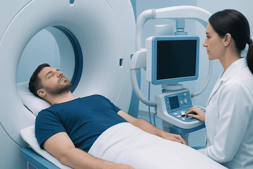
Most hemangiomas on the spine are not serious. They are benign, meaning they are not cancer and do not spread to other parts of the body. A typical hemangioma remains within the vertebral body and causes no symptoms, so it may only need periodic review if a clinician considers follow-up appropriate.
A smaller group are described as aggressive because they may extend into the spinal canal or nearby tissues, affect the structural strength of the vertebra, or put pressure on the spinal cord or nerve roots. This can lead to significant pain, numbness, weakness, or problems with walking. Although this situation needs timely specialist assessment, effective treatments are available.
Imaging helps distinguish a typical lesion from one needing closer attention. Clinicians also consider whether the symptoms match the affected spinal level, as back pain is common and may have causes unrelated to a hemangioma.
What Does Spinal Hemangioma Pain Feel Like?

When a spinal hemangioma causes symptoms, pain may be localized to the affected area of the neck, mid-back, or lower back. It can be persistent, aching, deep, or gradually worsening, and may become more noticeable with movement or when the vertebra is under mechanical stress. However, pain alone does not prove that a hemangioma is responsible.
If the lesion irritates or compresses a nerve root, pain may travel into an arm, chest wall, buttock, or leg depending on its location. Nerve pressure can also cause tingling, reduced sensation, weakness, or changes in coordination. Spinal cord compression may cause symptoms in both legs, trouble walking, or changes in bladder or bowel function.
A clinician should assess new or persistent spinal pain, particularly when it is associated with neurological symptoms. This helps identify the cause and determine whether the hemangioma is incidental or requires treatment.
Do Spinal Hemangiomas Stop Growing?
Many spinal hemangiomas remain stable over time and do not require active treatment. They are often longstanding findings rather than lesions that continue to enlarge steadily. For a typical, symptom-free hemangioma, doctors may recommend no routine intervention or may arrange follow-up imaging based on the individual imaging appearance and clinical context.
Occasionally, a hemangioma can become more active or extend in a way that affects the spinal canal or vertebral stability. Growth or progression is more clinically important when it causes symptoms or threatens nerves. Repeat magnetic resonance imaging (MRI) or computed tomography (CT) may be used to clarify changes when symptoms develop or when the original scan has features that need surveillance.
People should not assume that worsening back symptoms are due to a known hemangioma. A new assessment is important because disc problems, arthritis, fractures, infections, and other spinal conditions can produce similar symptoms.
Who May Need Treatment and How Is It Planned?
Candidates for treatment are usually people with pain that is clearly linked to the hemangioma and has not improved with appropriate conservative care, or people with neurological symptoms, spinal cord compression, vertebral collapse, or spinal instability. Urgent treatment may be required when there is progressive weakness, difficulty walking, or loss of bladder or bowel control.
Assessment often includes a neurological examination and detailed imaging. MRI is especially useful for evaluating the spinal cord, nerves, and soft tissues, while CT can show the bony structure of the vertebra. In selected situations, angiography may map the lesion’s blood supply before an endovascular procedure or surgery.
The team weighs expected benefit against the location and vascular nature of the lesion. Treatment planning may combine more than one method, such as embolization before surgery to reduce bleeding risk, followed by decompression or stabilization if the spinal cord is under pressure.
- Observation may suit a typical hemangioma without symptoms or instability.
- Targeted procedures may support a weakened vertebra or reduce blood flow to the lesion.
- Surgery is more often considered for significant nerve compression or spinal instability.
Procedure Options: Step by Step
For an asymptomatic hemangioma, the process may simply involve review of the scan, education about symptoms to report, and follow-up only when clinically indicated. For painful lesions without nerve compression, clinicians may first recommend non-surgical measures such as activity modification, physical therapy, and suitable pain relief under medical guidance.
Embolization is a minimally invasive procedure performed by an interventional specialist. Through a small catheter inserted into a blood vessel, materials are delivered to reduce blood flow to the hemangioma. It may be used alone in selected cases or before surgery to help reduce operative blood loss. Vertebroplasty or a similar bone-strengthening procedure may be considered when a vertebra needs additional internal support; bone cement is injected under image guidance.
Radiotherapy may be an option for selected symptomatic hemangiomas, particularly when surgery is not suitable or residual lesion remains after another treatment. It uses carefully planned radiation to control the lesion over time. When spinal cord or nerve roots are compressed, spine surgery may be performed to remove pressure from neural structures and, where needed, stabilize the spine with implants or bone grafting.
Before any intervention, the treating team explains the expected purpose, alternatives, anesthesia plan, and individual risks. The exact sequence varies: for example, embolization may be scheduled shortly before surgery, while radiotherapy works gradually and is not usually used to address rapidly progressive neurological compression on its own.
Benefits, Risks and Recovery Timeline
The potential benefits of treatment include improved pain control, protection or recovery of neurological function, reduced risk of further spinal instability, and improved ability to move safely. Outcomes depend largely on how much nerve compression was present before treatment, how long symptoms had been present, and whether there were other spinal conditions contributing to pain.
All procedures have risks. Embolization can rarely affect nearby blood vessels or nerves. Vertebroplasty may involve cement leakage, infection, bleeding, or injury to surrounding structures. Radiotherapy can cause temporary fatigue or skin effects and has carefully considered risks to nearby tissues. Surgery carries risks including bleeding, infection, blood clots, fluid leakage around the spinal cord, nerve injury, and complications related to anesthesia or implants. The vascular nature of hemangiomas makes careful pre-procedure planning particularly important.
Recovery after an image-guided procedure may take days to a few weeks, depending on symptoms and the procedure performed. Recovery after decompression and stabilization surgery is typically longer and may include a hospital stay, wound care, gradual activity progression, and physiotherapy over weeks to months. Follow-up visits and imaging help the team assess healing, spinal alignment, and symptom improvement.
Are Spinal Hemangiomas a Disability? When to Seek Medical Care
A spinal hemangioma is not automatically a disability. Most people with an incidental, symptom-free lesion can continue their usual daily activities. If the lesion causes ongoing pain, weakness, impaired mobility, or neurological deficits, it may affect work and function; any formal disability determination depends on local rules, medical evidence, and the person’s functional limitations rather than the diagnosis alone.
Medical review is appropriate for persistent or worsening neck or back pain, pain that radiates into the limbs, numbness, tingling, or reduced strength. Prompt assessment is especially important if there is new difficulty walking, worsening balance, rapidly increasing weakness, numbness in the groin or saddle area, or loss of bladder or bowel control. These symptoms can indicate nerve or spinal cord compression and should not be delayed.
People with a known spinal hemangioma should discuss safe activity and follow-up with their doctor rather than restricting all movement unnecessarily. When specialist treatment is needed, Acibadem International’s multidisciplinary specialists and JCI-accredited hospitals evaluate and treat spinal conditions for international patients.
Frequently asked questions
Can a spinal hemangioma turn into cancer?
Spinal hemangiomas are benign vascular lesions and are not considered cancer. They do not typically transform into cancer or spread elsewhere in the body. A doctor may order further imaging if scan features are unusual or if symptoms need another explanation.
Is surgery always necessary for a spinal hemangioma?
No. Most spinal hemangiomas do not require surgery because they cause no symptoms. Surgery is generally reserved for cases with substantial spinal cord or nerve compression, vertebral instability, or symptoms that cannot be adequately managed with less invasive options.
Can exercise make a spinal hemangioma worse?
Many people with an incidental spinal hemangioma can remain active. The right level and type of exercise depend on pain, spinal stability, and whether neurological symptoms are present. A clinician or physiotherapist can recommend appropriate activity modifications when needed.
How is a spinal hemangioma diagnosed?
A spinal hemangioma is commonly identified on CT or MRI scans, often performed for another reason. A clinician combines imaging findings with symptoms and a neurological examination to decide whether it is a typical incidental lesion or one that needs further evaluation.
Can spinal hemangioma symptoms improve after treatment?
Pain and neurological symptoms may improve when treatment successfully relieves compression, reduces lesion activity, or stabilizes the affected vertebra. The degree of recovery varies, particularly if nerves or the spinal cord were compressed for a prolonged period. Early evaluation of progressive neurological symptoms supports timely treatment planning.
What type of doctor treats spinal hemangiomas?
Care may involve a spine surgeon or neurosurgeon, often working with an interventional radiologist and radiation oncologist. The most appropriate specialist depends on whether the hemangioma is causing pain, structural weakness, or compression of nerves or the spinal cord.
References
- American Association of Neurological Surgeons
- National Institute of Neurological Disorders and Stroke
- Radiopaedia
- American College of Radiology
This article is for general information only and is not a substitute for professional medical advice. Please consult a qualified doctor about your individual situation.
Joint and spine care in Turkey — expert assessment & treatment
JCI-accredited · board-certified surgeons · reply within 24h