Broken Heel Treatment: How It Works, Results and What to Expect

A broken heel is usually a fracture of the calcaneus, the large bone beneath the ankle. Stable fractures may heal without surgery, while displaced or joint-involving fractures may need surgical fixation.
Key Takeaways
- A broken heel is usually a fracture of the calcaneus, the large bone beneath the ankle.
- Stable fractures may heal without surgery, while displaced or joint-involving fractures may need surgical fixation.
- Keeping weight off the injured foot during the early healing phase is essential to protect alignment and healing.
- Recovery commonly takes months, and some stiffness, swelling or discomfort may persist while function improves.
- Early medical assessment is important after a fall, jump, crush injury or severe heel pain.
Broken heel treatment depends on whether the heel bone fracture is stable, displaced, involves the joint, or has caused skin or soft-tissue injury. Care may include immobilization and protected weight-bearing, or surgery to restore alignment, followed by a carefully paced rehabilitation program.
Overview: how broken heel treatment works
Broken heel treatment aims to help the calcaneus, or heel bone, heal in the best possible position while protecting the surrounding joints, skin, nerves and tendons. The treatment plan is individualized. It is based on fracture pattern, whether the fragments have moved out of place, involvement of the subtalar joint below the ankle, the condition of the soft tissues, and the person’s overall health and activity needs.
Some calcaneus fractures are stable and can be treated without an operation using a splint, cast or removable boot. More complex fractures, particularly those with displaced pieces or joint surface disruption, may require surgery to realign the bone and hold it with plates, screws or other fixation devices. Treatment also includes pain management, swelling control, protection from weight-bearing and rehabilitation.
Even with appropriate care, recovery is gradual because the heel carries substantial load during standing and walking. The immediate priority is safe healing; the longer-term goal is to regain mobility, strength, balance and the ability to return to daily activities as comfortably as possible.
Who may need non-surgical care or surgery
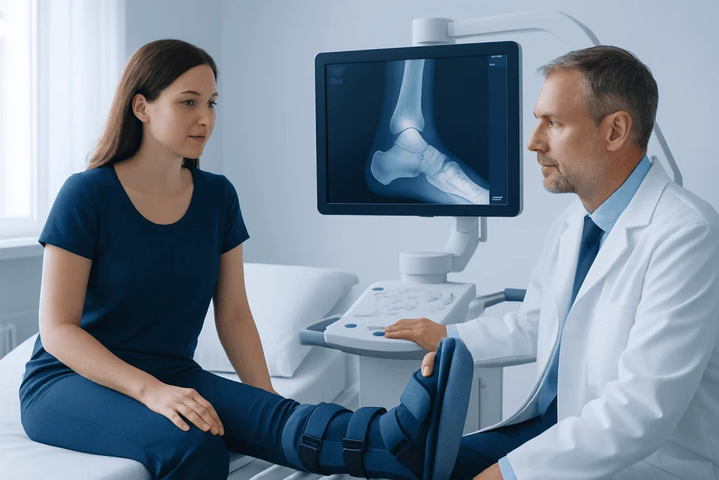
Non-surgical management may be appropriate when the fracture fragments remain well aligned, the joint surface is not significantly displaced, or an operation would carry greater risk than likely benefit. A clinician may use a splint initially to accommodate swelling, followed by a cast or walking boot. Crutches, a walker, knee scooter or wheelchair may be needed while weight-bearing is restricted.
Surgery may be considered when the heel has widened or lost height, bone fragments have shifted, the subtalar joint is affected, or the fracture has created an unstable pattern. Open fractures, in which bone communicates with a wound, require urgent specialist care. Surgical timing is often influenced by swelling and skin condition; operating too early through severely swollen tissue can increase wound-healing problems.
Suitability for an operation is discussed carefully with an orthopedic trauma or foot-and-ankle specialist. Factors such as smoking, diabetes, circulation problems, severe swelling, infection risk, age, other injuries and the ability to follow post-treatment restrictions can affect the recommended approach. The decision is not based on imaging alone; symptoms, function and personal priorities matter as well.
- Stable, non-displaced fractures may be treated with immobilization and monitoring.
- Displaced intra-articular fractures may benefit from surgical reconstruction in selected patients.
- Severe soft-tissue injury may require staged care before definitive fixation.
- Associated spine, ankle or leg injuries may need assessment after high-energy trauma.
Assessment and planning before treatment
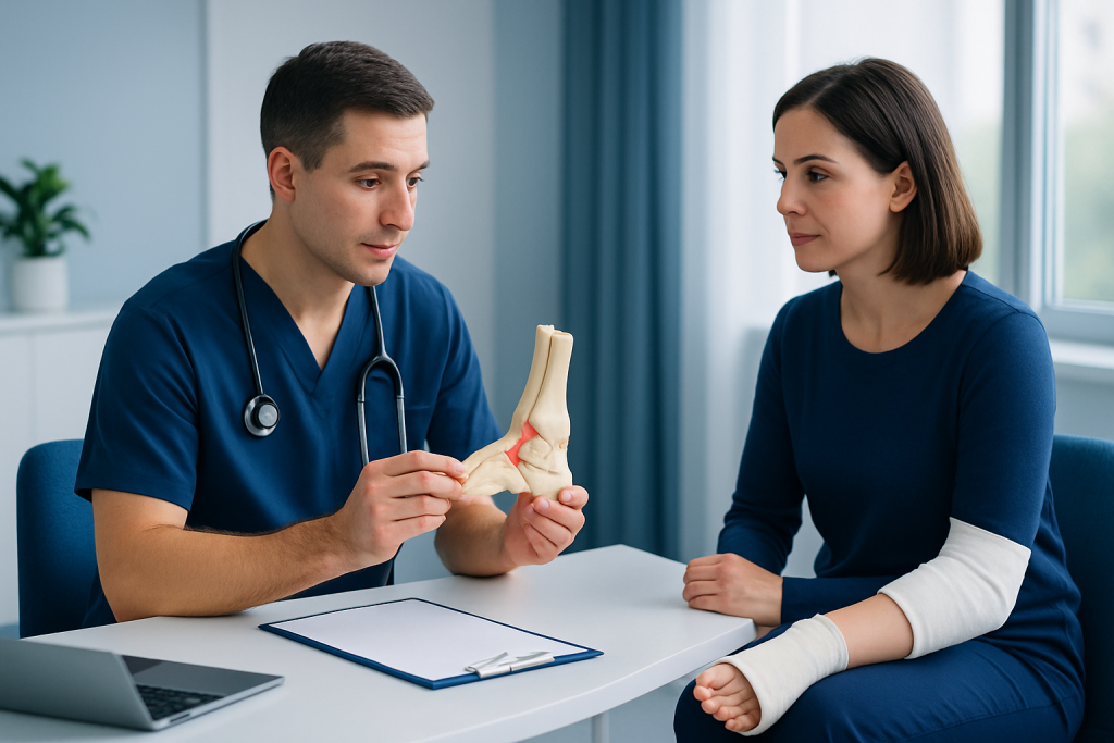
Clinicians begin by asking how the injury happened and examining the foot, ankle and lower leg. Bruising, swelling, tenderness, inability to bear weight, altered heel shape and numbness can be signs of a significant injury. The skin is examined closely, since pressure from displaced bone fragments can threaten the skin and require urgent attention.
X-rays usually confirm a heel fracture and show its overall alignment. A CT scan is often used when a fracture is complex or surgery is being considered, because it provides detailed views of the joint surfaces and individual fragments. Additional imaging may be needed if there is concern about injuries elsewhere, especially after a fall from height or major vehicle collision.
Before surgery, the clinical team reviews medical history, medications, allergies and anesthesia considerations. People who smoke are usually advised to stop, as tobacco exposure can impair bone and wound healing. The team may also discuss practical preparation, including mobility support at home, work arrangements and plans for keeping weight off the foot after treatment.
What happens during heel fracture surgery
Heel fracture surgery is commonly performed under regional anesthesia, general anesthesia, or a combination chosen by the anesthesia and surgical teams. The procedure is tailored to the fracture. For some patterns, the surgeon uses small incisions and imaging guidance to reposition fragments and place screws. More complex injuries may require a larger incision so the joint surface and heel shape can be restored as accurately as possible.
Once the bone fragments are aligned, they may be secured using screws, a plate and screws, or other fixation methods. Bone graft material may occasionally be used when there is a substantial gap after restoring the heel’s shape. The incision is then closed and the foot is protected in a splint, cast or boot.
The purpose of surgery is not simply to place metal implants. It is to improve alignment, preserve the height and width of the heel where possible, and support the joint surface so it can heal. Orthopedic evaluation and treatment can help determine whether surgical or non-surgical care is most appropriate for an individual fracture.
After the operation, the foot is elevated and monitored for swelling, circulation, sensation and wound concerns. Pain relief, instructions for safe mobility and follow-up appointments are arranged before discharge. The specific length of hospital stay varies with the injury, procedure, other health needs and home support.
Recovery timeline and rehabilitation
In the first days and weeks, protecting the injured heel is central to recovery. Elevation can help limit swelling, and the care team provides guidance on keeping the splint, cast or surgical dressing dry and intact. Patients should follow instructions on pain medicines and should not put weight through the injured foot until their clinician says it is safe.
Bone healing commonly requires several weeks, but a return to unrestricted walking and higher-impact activity often takes several months. Follow-up visits and repeat X-rays allow the clinician to check alignment and healing. Weight-bearing is usually reintroduced gradually, rather than all at once, because doing too much too soon can compromise recovery.
Physical therapy may begin with gentle movement of the toes, ankle and nearby joints when appropriate. As healing progresses, rehabilitation focuses on ankle and subtalar joint mobility, calf and foot strength, balance, walking pattern and tolerance for daily activity. Physical therapy and rehabilitation can support a structured, individualized return to movement.
Swelling and stiffness can last for many months, especially after more severe fractures. Comfortable supportive footwear, pacing activities and continued exercises may be useful. Recovery is not always linear; a temporary increase in soreness after therapy or increased activity can occur, but persistent or worsening symptoms should be discussed with the treating team.
Benefits, limitations and possible risks
The potential benefit of non-surgical treatment is avoiding an incision and the wound-related risks of surgery while allowing a stable fracture to heal. Surgical treatment may improve alignment in selected displaced fractures and can help restore the shape of the heel and the congruence of the subtalar joint. Neither approach can guarantee that the foot will feel exactly as it did before the injury.
Possible complications of a heel fracture include prolonged swelling, stiffness, reduced subtalar joint movement, changes in walking pattern, chronic pain and post-traumatic arthritis. More severe fractures and fractures involving the joint have a greater chance of lasting symptoms. Some people may later need treatment for arthritis or painful hardware, although this is not necessary for everyone.
Surgical risks include infection, delayed wound healing, bleeding, blood clots, nerve or tendon irritation, problems with bone healing, implant-related discomfort and the need for additional procedures. Risk can be reduced, though not eliminated, by careful surgical planning, following wound-care advice, avoiding tobacco and attending follow-up appointments.
It is helpful for patients to ask their specialist about the expected functional outcome for their particular fracture, the alternatives to surgery, the likely duration of protected weight-bearing and the signs of complications to watch for. Clear expectations and consistent rehabilitation are important parts of treatment.
Self-care and when to seek medical care
After a diagnosed heel fracture, patients should use mobility aids exactly as advised and avoid walking on the injured foot until cleared. Keeping follow-up appointments is important, since treatment can be adjusted according to swelling, wound condition and X-ray findings. A balanced diet with adequate protein and nutrients that support bone health may be helpful, while smoking and nicotine products should be avoided because they can delay healing.
Seek urgent medical care after a fall, jump, crush injury or collision if there is severe heel pain, inability to stand, marked swelling, deformity, an open wound, numbness, a cold or pale foot, or rapidly worsening pain. These features may indicate a significant fracture, circulation problem or soft-tissue emergency. A person with a heel injury after a high-energy accident should also be assessed for other injuries.
During recovery, medical advice should be sought promptly for fever, increasing redness or drainage around an incision, uncontrolled pain, new numbness, calf pain or swelling, shortness of breath, or a cast or boot that feels excessively tight. These symptoms do not always indicate a serious complication, but they need timely clinical assessment.
Acibadem International’s multidisciplinary orthopedic, imaging, anesthesia and rehabilitation specialists at JCI-accredited hospitals diagnose and treat heel fractures for international patients, with care plans based on the injury and recovery needs.
Frequently asked questions
Can a broken heel heal without surgery?
Yes. A stable heel fracture with little or no displacement may heal with immobilization, restricted weight-bearing and follow-up imaging. A specialist determines whether non-surgical care is suitable by reviewing the fracture pattern, joint involvement, swelling and the person’s overall health.
How long does recovery from a broken heel take?
Early bone healing often takes several weeks, but full recovery commonly takes months. The timeline depends on the severity of the fracture, whether surgery was needed, adherence to weight-bearing restrictions and participation in rehabilitation.
When can someone walk after heel fracture surgery?
The timing is individualized and guided by the surgeon based on healing seen during follow-up. Many people need a period of no weight-bearing first, followed by gradual progression in a boot or supportive shoe under clinical guidance.
Is heel fracture surgery always necessary?
No. Surgery is generally reserved for selected fractures, such as those that are displaced, unstable or involve an incongruent joint surface. Non-surgical care may be effective for fractures that are stable or for people whose surgical risks outweigh potential benefits.
Will a broken heel cause arthritis later?
A heel fracture that affects the subtalar joint can increase the risk of post-traumatic arthritis. The risk is related to the injury’s severity and joint damage, and symptoms may range from mild stiffness to more persistent pain or limited movement.
What should not be done with a suspected broken heel?
A person should not try to walk through severe heel pain or force the foot back into position. Avoid putting weight on the foot, and seek urgent assessment if there is deformity, an open wound, numbness, color change or severe swelling.
References
- American Academy of Orthopaedic Surgeons
- American Orthopaedic Foot & Ankle Society
- National Institute of Arthritis and Musculoskeletal and Skin Diseases
- Mayo Clinic
This article is for general information only and is not a substitute for professional medical advice. Please consult a qualified doctor about your individual situation.
Joint and spine care in Turkey — expert assessment & treatment
JCI-accredited · board-certified surgeons · reply within 24h
Add us as a Preferred Source to see more of our trusted health content across Google Search, AI Overviews and Discover.