Ureterocele Ultrasound: Preparation, Procedure and Results

Ultrasound uses sound waves rather than radiation and is commonly used in children and adults with suspected ureterocele. A ureterocele often appears as a rounded, thin-walled fluid-filled structure inside the bladder near a ureter opening.
Key Takeaways
- Ultrasound uses sound waves rather than radiation and is commonly used in children and adults with suspected ureterocele.
- A ureterocele often appears as a rounded, thin-walled fluid-filled structure inside the bladder near a ureter opening.
- The scan may show associated findings such as hydronephrosis, duplicated collecting systems or incomplete bladder emptying.
- Not every ureterocele needs immediate surgery; treatment depends on symptoms, infections, obstruction and kidney function.
- Seek timely medical assessment for fever with urinary symptoms, severe flank pain, vomiting, or difficulty passing urine.
A ureterocele ultrasound is a noninvasive imaging test used to assess a balloon-like widening at the lower end of a ureter, usually where it enters the bladder. It can help identify urinary blockage, kidney swelling and related urinary tract changes, although other tests may sometimes be needed to confirm the diagnosis and plan care.
Ureterocele ultrasound: what the test shows
A ureterocele ultrasound is a painless imaging examination that helps clinicians look at the kidneys, ureters and bladder. A ureterocele is a cyst-like enlargement of the lower end of a ureter, the tube that carries urine from a kidney to the bladder. It may narrow the flow of urine and, in some people, contribute to urinary tract infections, kidney swelling or stones.
Ultrasound is often the first test because it does not use ionizing radiation and can be performed in people of all ages, including babies and children. It can show the ureterocele itself, assess whether urine is backing up into a kidney, and identify features that may suggest another urinary tract difference. The results are interpreted alongside symptoms, urine tests and, when necessary, additional imaging.
A ureterocele may be found during assessment of repeated urinary infections or urinary symptoms. It can also be detected before birth or incidentally during imaging performed for another reason. A scan alone does not always establish how well the urinary system drains, so the care team may recommend further evaluation when the findings are unclear or clinically important.
Who may need a ureterocele ultrasound?
Clinicians may request this scan when a person has symptoms or test results that raise concern about a urinary tract blockage. In children, it may be arranged after recurrent urinary tract infections, prenatal kidney dilation, poor urine flow or unexplained fever. In adults, it may be considered for recurrent infections, blood in the urine, flank discomfort, stones, or new urinary symptoms.
People with a known ureterocele may also have follow-up ultrasound examinations. These help monitor the kidneys and bladder over time, particularly if treatment is being observed rather than performed immediately. The frequency of follow-up is individualized and depends on the ureterocele type, symptoms, kidney drainage and prior treatment.
Ultrasound is especially useful when clinicians need a radiation-free overview of the urinary tract. However, it cannot always define every detail of ureter anatomy or demonstrate urine reflux. A urologist may therefore combine it with urine analysis, a voiding cystourethrogram, nuclear medicine renal imaging, CT, MRI, or cystoscopy, depending on age and clinical circumstances.
Preparation and step-by-step procedure

Preparation for a ureterocele ultrasound is usually simple. The imaging center may ask the person to arrive with a comfortably full bladder, because bladder filling can make the ureterocele ultrasound bladder findings easier to see. Instructions vary by age and the type of scan, so patients should follow the directions provided by their radiology department.
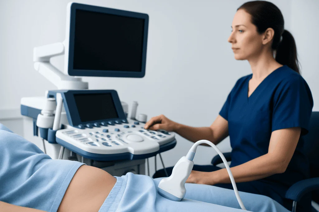
During the examination, the patient lies on an examination couch while a sonographer applies clear gel to the lower abdomen and sometimes the sides or back. A handheld transducer is moved gently over the skin to create live images. The sonographer may take pictures of both kidneys, measure any widening of the kidney drainage system, and obtain images of the bladder from several angles.
The procedure generally takes about 20 to 40 minutes, depending on the views required and whether images are also needed after the bladder is emptied. It should not be painful, though mild pressure from the probe or temporary discomfort from a full bladder may occur. Patients can return to normal activities immediately after the scan unless they have been given separate instructions for another test.
Ultrasound has important benefits: it is noninvasive, widely available and free of radiation. Its limitations are that image quality can be affected by bowel gas, body shape and bladder filling, and it may not fully show the cause or functional significance of obstruction. There are no known significant risks from standard diagnostic ultrasound.
What does a ureterocele look like on ultrasound?
The typical ureterocele ultrasound appearance is a round or oval, thin-walled, fluid-filled structure within the bladder. It is usually seen near the point where a ureter enters the bladder and may appear to change in size as the bladder fills or empties. Because it contains fluid, it is often described on ultrasound as an anechoic, or dark, cystic area.
A radiology report may include a ureterocele ultrasound description such as “cystic lesion at the ureterovesical junction” or “intravesical cystic dilatation of the distal ureter.” The report may describe its size, location, whether it is on the right or left, and whether it extends beyond the bladder neck. The exact appearance can vary according to anatomy and bladder fullness at the time of imaging.
The ultrasound also evaluates possible consequences. These include hydronephrosis, meaning dilation of the kidney collecting system; hydroureter, meaning widening of the ureter; bladder wall changes; stones; or signs of a duplicated collecting system. These findings help the urology team judge whether the ureterocele may be affecting urinary drainage.
Ultrasound findings are important, but they are not interpreted in isolation. A small ureterocele without kidney dilation may be managed differently from one associated with infections, reflux or marked obstruction. Further imaging may be advised before a treatment decision is made.
How serious is ureterocele?
How serious a ureterocele is depends on its size, position, effect on urine flow, and whether it is associated with other urinary tract differences. Some are small and cause no symptoms, while others can obstruct urine drainage, contribute to recurrent infections, or affect kidney function if not appropriately monitored and treated.
In children, ureteroceles may occur with a duplicated collecting system, in which a kidney has two drainage pathways. In adults, they may be identified after urinary symptoms, stones or infection. A urologist considers kidney ultrasound findings, urine infections, renal function and other imaging results when assessing the likely impact.
The reassuring point is that ureteroceles can usually be evaluated carefully and managed with observation, infection treatment, monitoring, or a procedure when needed. Early assessment is particularly important when there is kidney swelling, repeated febrile urinary infections, impaired drainage or concerns about kidney development.
For more general information on urinary tract health and evaluation, a specialist may discuss ureterocele and its management options where appropriate. The best plan is individualized rather than based on the ultrasound image alone.
What are the symptoms of a left side ureterocele?
A left side ureterocele can cause the same symptoms as a ureterocele on the right side. The location may influence where discomfort is felt, but many symptoms reflect urinary infection or impaired drainage rather than the side alone. Some people have no symptoms and learn about the condition only after an ultrasound.
Possible symptoms include recurrent urinary tract infections, burning or pain during urination, frequent urination, urgency, cloudy or bloody urine, fever, and pain in the lower abdomen or left flank. Infants and young children may have nonspecific symptoms such as fever, irritability, poor feeding, vomiting or poor growth, so medical review is important when urinary infection is suspected.
If a ureterocele significantly blocks drainage from the left kidney, it may lead to left-sided hydronephrosis and discomfort. However, pain is not a reliable measure of severity: some people with important obstruction have few symptoms. Imaging and urine testing are therefore central to assessment.
New symptoms should not be self-diagnosed as a ureterocele. Conditions such as urinary infection, kidney stones and other bladder or kidney problems can cause similar concerns. A qualified clinician can determine the cause and whether urgent treatment is needed.
Treatment options and ureterocele surgery recovery
Treatment is based on symptoms, age, urinary infections, kidney drainage, reflux and kidney function. A small ureterocele that is not causing obstruction or infection may be monitored with planned clinical reviews, urine testing and follow-up imaging. If an infection is present, it requires prompt medical treatment.
When drainage is obstructed or infections recur, a ureterocele procedure may be recommended. One commonly used approach is endoscopic puncture or incision, in which a urologist passes a small camera through the urethra and opens the ureterocele to improve urine drainage. Some people need reconstructive surgery, particularly when there is complex anatomy, reflux or a duplicated collecting system.
Ureterocele treatment may involve different approaches, and the treating urologist will explain the purpose, expected benefits, alternatives and possible complications for the individual situation. Potential considerations after an endoscopic procedure or surgery include temporary urinary discomfort, blood in the urine, infection, persistent obstruction or vesicoureteral reflux, which may require monitoring or additional care.
How long does it take to recover from ureterocele surgery? Recovery depends on the procedure and the person’s overall health. After a straightforward endoscopic procedure, many people resume light everyday activities within several days, although the clinical team may arrange follow-up testing over the following weeks or months. Open, laparoscopic or robotic reconstructive operations usually require a longer recovery period, often several weeks; the surgical team provides individualized activity, catheter and return-to-work guidance.
When to seek medical care
Medical care should be sought promptly for fever with urinary symptoms, chills, worsening flank or abdominal pain, vomiting, visible blood in the urine, or difficulty passing urine. These symptoms can indicate a urinary infection or obstruction and need timely clinical assessment, especially in infants, children, older adults and people with known kidney disease.
Emergency evaluation is appropriate for severe pain, high fever, confusion, inability to urinate, or signs of serious illness. Parents and caregivers should seek urgent advice for a baby or child with fever, poor feeding, vomiting, marked lethargy, or fewer wet diapers than usual.
For nonurgent concerns, patients can discuss recurring urinary infections, new bladder symptoms or an abnormal prenatal or ultrasound finding with a primary care clinician, pediatrician or urologist. Acibadem International’s multidisciplinary specialists and JCI-accredited hospitals diagnose and treat ureterocele and related urinary tract conditions for international patients.
Until assessment, adequate fluid intake may be appropriate for many people, but individuals with fluid restrictions or kidney, heart or liver conditions should follow their clinician’s advice. Antibiotics, pain medicines and surgical decisions should be guided by a qualified healthcare professional rather than started independently.
Frequently asked questions
Is a ureterocele ultrasound painful?
No. A ureterocele ultrasound is noninvasive and should not be painful. The person may feel mild pressure from the ultrasound probe or temporary discomfort if asked to have a full bladder.
Do I need to fast before a ureterocele ultrasound?
Fasting is usually not required for a standard kidney and bladder ultrasound. Some centers ask patients to drink water beforehand and avoid urinating so the bladder is comfortably full; the imaging department will provide the correct instructions.
Can ultrasound diagnose a ureterocele by itself?
Ultrasound can often strongly suggest or show a ureterocele, especially when a typical cystic structure is visible in the bladder. Additional tests may be needed to assess urine reflux, the degree of blockage, detailed anatomy and kidney function.
Can a ureterocele go away on its own?
A ureterocele is a structural change at the end of the ureter and does not usually disappear on its own. Some small, symptom-free ureteroceles can be monitored without immediate surgery, but follow-up should be guided by a urologist.
What happens after a ureterocele ultrasound?
A radiologist reviews the images and sends a report to the requesting clinician. Depending on the findings, the next step may be observation, urine testing, referral to a urologist, or further imaging to understand kidney drainage and urinary reflux.
Can a ureterocele cause kidney damage?
A ureterocele can affect kidney drainage and may contribute to kidney damage if there is significant, untreated obstruction or repeated serious infection. Regular assessment and appropriate treatment help protect kidney function when a clinically important ureterocele is present.
References
- National Institute of Diabetes and Digestive and Kidney Diseases
- American Urological Association
- European Association of Urology
- RadiologyInfo.org
- National Health Service
This article is for general information only and is not a substitute for professional medical advice. Please consult a qualified doctor about your individual situation.
Ultrasound in Turkey — costs, top hospitals & a free quote
JCI-accredited · board-certified surgeons · reply within 24h
Add us as a Preferred Source to see more of our trusted health content across Google Search, AI Overviews and Discover.

Check-up & Preventive Medicine
Comprehensive health screenings designed for early detection and prevention.
3 specialists in this unitMore from the Health Library



Related Specialists

Assoc. Prof. Dr. Zuhal Atan Uçar
Internal Medicine
Prof. Dr. Bora Peynircioğlu
Interventional Radiology
Assoc. Prof. Dr. Yetkin Zeki Yılmaz
Ear Nose & Throat
Ebru Uzun Saral
Physical Medicine & Rehabilitation