Radiation Therapy for Brain Tumour: How It Works, Results and What to Expect
Radiation can slow or control a brain tumour, but outcomes depend strongly on the exact tumour diagnosis and treatment goal. External-beam radiation is commonly delivered in daily sessions over several weeks, while stereotactic radiosurgery may be completed in one or a few sessions.
Key Takeaways
- Radiation can slow or control a brain tumour, but outcomes depend strongly on the exact tumour diagnosis and treatment goal.
- External-beam radiation is commonly delivered in daily sessions over several weeks, while stereotactic radiosurgery may be completed in one or a few sessions.
- A planning process using MRI or CT imaging helps the team target the tumour while protecting healthy brain tissue as much as possible.
- Tiredness, scalp or hair changes, and temporary swelling-related symptoms can occur; the care team monitors these closely.
- Tumour shrinkage is often gradual, and follow-up MRI scans are needed to assess response over time.
Radiation therapy for brain tumour uses carefully planned high-energy radiation to damage tumour-cell DNA and limit further growth. It may be used after surgery, instead of surgery in selected situations, or to control symptoms and spread, with the approach based on the tumour type, location, size and a person’s overall health.
Overview: how radiation therapy for brain tumour works
Radiation therapy for brain tumour is a local treatment that directs high-energy beams, usually X-rays, at a defined area of the brain. The radiation damages the DNA of tumour cells, making it harder for them to divide and survive. Healthy cells can often repair radiation damage more effectively than tumour cells, although surrounding brain tissue is still protected as carefully as possible.
It may be recommended after an operation to treat tumour cells that cannot be seen or safely removed, as the main treatment when surgery is not suitable, or when a tumour has returned or spread to the brain. Radiation may also help reduce pressure-related symptoms by controlling tumour growth. It does not work in the same way for every diagnosis; the plan for a slow-growing benign tumour is different from the plan for an aggressive primary brain cancer or brain metastases.
Modern treatment uses detailed imaging, immobilization masks and computer planning to shape the dose around the target. A multidisciplinary team, often including a radiation oncologist, neurosurgeon, neuroradiologist, medical oncologist and specialist nurses, reviews the diagnosis and recommends an individualized approach.
Who may be a candidate for brain tumour radiation?
Candidacy depends on pathology results when available, MRI findings, tumour size and location, whether there is more than one lesion, previous treatments, neurological symptoms and general health. Some tumours are best treated first with surgery, while others can be managed with radiation alone or with medicines such as chemotherapy, targeted therapy or immunotherapy. For some low-risk tumours, careful MRI monitoring may be appropriate before treatment begins.
External-beam radiotherapy is often used for larger areas at risk of microscopic disease or after removal of a malignant tumour. Highly focused treatment, often called stereotactic radiosurgery or fractionated stereotactic radiotherapy, can be useful for selected small, well-defined tumours and metastases. Despite its name, radiosurgery does not involve an incision; it is a precisely delivered form of radiation.
Before making a recommendation, clinicians consider important safety factors, including the tumour’s closeness to the optic nerves, brainstem, memory-related structures and other sensitive areas. They also review prior radiation exposure, because re-treatment requires especially careful dose planning. Related conditions may include brain tumours, which can vary widely in behaviour and treatment needs.
What happens before and during the procedure?

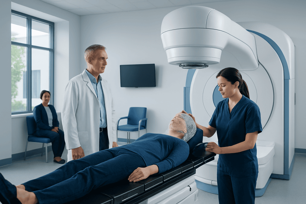
The process starts with a consultation and treatment-planning appointment. The radiation oncology team reviews scans and medical records, explains the purpose of treatment and discusses likely benefits and side effects. A thermoplastic mask is commonly made for head treatments. It is custom-fitted, open around the face, and helps the head remain in exactly the same position during each session.
A planning CT scan, usually combined with MRI information, maps the tumour and nearby healthy structures. Doctors and medical physicists then create a plan that selects beam angles, dose distribution and the number of sessions. This preparation can take several days because precision and safety checks are essential.
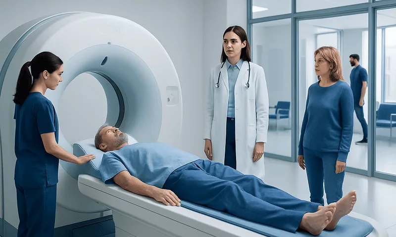
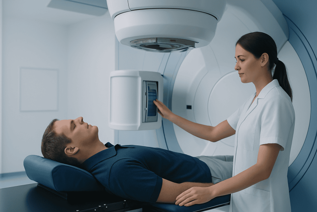
For most external-beam treatments, the person lies on a treatment table wearing the mask. The machine moves around the head but does not touch it, and radiation itself is painless and cannot be felt. Staff watch and communicate from outside the room. Each visit may take 15 to 45 minutes, although the actual beam delivery is often shorter. A focused option may be discussed as stereotactic radiosurgery when clinically appropriate.
Radiation does not make a person radioactive after external-beam treatment. They can generally be around family, friends and children as usual unless their clinical team gives different advice for another aspect of care.
How many rounds of radiation is normal for brain cancer?
There is no single normal number of rounds for brain cancer because the schedule is chosen for the diagnosis and treatment goal. Conventional fractionated radiotherapy is commonly given on weekdays over approximately two to six weeks. Dividing treatment into smaller daily doses, called fractions, allows normal tissue time to repair between sessions while building an effective dose in the target.
Shorter schedules may be appropriate for certain situations, including symptom-focused treatment or selected metastatic brain tumours. Stereotactic radiosurgery may be delivered in one session, while fractionated stereotactic treatment can involve several sessions. The team may use terms such as “fractions,” “sessions” or “treatments”; all refer to planned visits for radiation delivery.
Appointments should not be missed or changed without speaking with the radiation team. If fatigue, travel challenges or side effects make attendance difficult, the team can advise on practical support and whether any adjustment is medically appropriate. The schedule is designed to balance effectiveness, convenience and safety rather than to follow a fixed number for every patient.
Benefits, risks and recovery timeline
The main potential benefit of radiation is local tumour control: slowing growth, reducing the chance of recurrence after surgery, or relieving symptoms caused by a tumour. In some settings it can be curative; in others, it is used to extend control and preserve neurological function or comfort. The expected benefit should be discussed in relation to the specific tumour type, grade and molecular findings, rather than assumed from the term “brain tumour” alone.
Common short-term effects include tiredness, scalp tenderness or redness, temporary hair loss in the treated area, headache, nausea and a temporary increase in symptoms caused by treatment-related swelling. Doctors may prescribe corticosteroids when swelling needs treatment. Hair often regrows after radiation, but regrowth can be incomplete depending on the dose and area treated.
Recovery is gradual. Many people continue usual light activities during treatment but may need more rest, especially toward the end of a longer course and for several weeks afterward. Follow-up MRI scans are commonly scheduled after treatment, but timing varies because scans taken too early can be difficult to interpret. Radiation-related inflammation can sometimes resemble tumour growth on imaging.
Less common but important delayed effects may include cognitive changes, hormonal effects when areas near the pituitary are treated, tissue injury known as radiation necrosis, or effects on vision, hearing or movement depending on the treatment area. Advanced planning reduces these risks but cannot remove them completely. The team will explain the risks most relevant to the planned field.
How successful is radiation on brain tumors?
Radiation can be very successful at controlling many brain tumours, but “success” has different meanings depending on the diagnosis. For some tumours, the aim is to destroy or durably control a small target. For others, especially aggressive tumours, radiation is an important part of combined treatment that may slow progression, improve symptoms and help preserve quality of life, rather than guarantee a cure.
Outcome is influenced by whether the tumour began in the brain or spread from another cancer, its grade and molecular features, the amount safely removed by surgery, its size and location, and a person’s age and functional condition. It is also influenced by whether the tumour has received treatment before. The radiation oncologist can discuss what local control and symptom improvement are realistically expected in an individual case.
Response is not judged by symptoms alone. The care team compares serial MRI scans over time and considers neurological examination findings, steroid needs and daily functioning. A scan can show temporary changes from inflammation after radiation, so a single early image may not provide a final answer.
How long does it take radiation to shrink a brain tumor?
Brain tumours usually do not shrink immediately after radiation. Some respond over weeks to months, while others may remain stable rather than visibly smaller; stability can still mean that treatment is working. The timing depends on tumour biology, radiation technique, other treatments and whether swelling is present.
Symptoms may improve sooner if radiation reduces swelling or if supportive medicines control inflammation, seizures, nausea or headaches. In other cases, symptoms may briefly worsen during or shortly after treatment because of swelling. New or changing symptoms should be reported promptly so the clinical team can assess whether supportive treatment is needed.
Follow-up imaging is interpreted cautiously. After focused radiation, treated tissue may appear larger or different for a period because of inflammation and healing effects. The treating specialists use the scan pattern, timing, symptoms and sometimes additional imaging to distinguish treatment effects from active tumour when possible.
What I wish I knew before radiation and when to seek medical care
Before radiation, it helps to know that planning is a major part of treatment and that the first appointment is often not the first radiation session. Bringing a current medication list, arranging transport if fatigue is likely, asking how hair and scalp may be affected, and discussing work or caregiving responsibilities can make the course easier to manage. Patients may also wish to ask about the purpose of treatment, the planned number of fractions, expected side effects and the follow-up schedule.
It is useful to keep a simple record of symptoms, energy levels and questions between visits. Continue prescribed medicines exactly as directed, particularly anti-seizure medicines and steroids; steroids should not be stopped suddenly unless a clinician has advised a taper. Maintaining hydration, balanced meals, gentle activity as tolerated and regular rest can support recovery, but no supplement should be started without checking with the oncology team.
Urgent medical assessment is needed for a new seizure, sudden weakness or numbness, trouble speaking, severe or rapidly worsening headache, persistent vomiting, confusion, fainting, marked drowsiness, or a major change in vision or balance. These symptoms may have several causes, but they should not be managed at home. For non-urgent concerns, such as increasing fatigue, scalp discomfort or mild nausea, patients should contact their radiation team for advice.
Acibadem International’s multidisciplinary specialists at JCI-accredited hospitals evaluate and treat brain tumours for international patients, coordinating neurosurgery, oncology and radiation care where appropriate. Radiation oncology care should always be planned with a qualified team that can explain the expected benefits, alternatives and follow-up needs.
Frequently asked questions
Is radiation therapy painful for a brain tumour?
The radiation beam itself is painless, and most people do not feel it during treatment. The fitted mask may feel close-fitting, but the team can help with comfort and communication. Side effects, such as fatigue, scalp irritation or headache, may develop later and should be reported to the care team.
Will radiation therapy cause hair loss?
Hair loss can occur in the part of the scalp within the radiation field. It often begins a few weeks after treatment starts and may regrow over time, although the amount of regrowth varies with the dose and treated area. The radiation team can explain the likelihood for an individual plan.
Can a person drive during brain radiation treatment?
Some people can drive, but this depends on symptoms, seizure history, vision, medicines and local legal requirements. Fatigue or treatment-related swelling may affect safety. The treating clinician should give individualized advice before driving.
Can radiation therapy be given after brain tumour surgery?
Yes. Radiation is often used after surgery when there is a risk that remaining microscopic tumour cells could grow, or when complete removal was not safe. The need and timing depend on the pathology results, tumour location and recovery after surgery.
What is the difference between stereotactic radiosurgery and standard radiation therapy?
Stereotactic radiosurgery uses highly focused radiation to treat a small, defined target in one or a few sessions. Standard external-beam radiation usually treats a broader area with smaller daily doses over several weeks. The best option depends on tumour size, location, number of lesions and treatment history.
How often are MRI scans done after brain radiation?
The timing varies by diagnosis and treatment plan, but follow-up MRI is commonly arranged within months after treatment and then at regular intervals. Early scans can be affected by inflammation, so doctors interpret them alongside symptoms and previous images. The care team will provide a personalized surveillance schedule.
References
- National Cancer Institute
- American Cancer Society
- American Society for Radiation Oncology
- National Institute for Health and Care Excellence
- European Society for Medical Oncology
This article is for general information only and is not a substitute for professional medical advice. Please consult a qualified doctor about your individual situation.
Radiation Therapy in Turkey — costs, top hospitals & a free quote
JCI-accredited · board-certified surgeons · reply within 24h
Add us as a Preferred Source to see more of our trusted health content across Google Search, AI Overviews and Discover.

Radiation Oncology
Precision radiotherapy and radiosurgery using advanced linear accelerators and image-guided techniques.
26 specialists in this unit