Testicular Carcinoma on Ultrasound: Preparation, Procedure and Results

Scrotal ultrasound is painless, uses no radiation and is usually the first imaging test for a new testicular lump. Most testicular cancers arise inside the testicle and often appear as a solid, darker-than-normal lesion on ultrasound.
Key Takeaways
- Scrotal ultrasound is painless, uses no radiation and is usually the first imaging test for a new testicular lump.
- Most testicular cancers arise inside the testicle and often appear as a solid, darker-than-normal lesion on ultrasound.
- An ultrasound can strongly suggest a tumor but cannot reliably determine the exact cancer type on its own.
- Results may be available the same day, although formal reporting and specialist discussion can take longer.
- A new painless lump, swelling, heaviness or persistent change in a testicle should be assessed promptly.
Testicular carcinoma on ultrasound commonly appears as a solid area within the testicle that differs from the surrounding tissue, often with increased blood flow on Doppler imaging. Ultrasound is highly useful for identifying and locating a suspicious testicular mass, but diagnosis and treatment planning also require examination, blood tests and specialist review.
Testicular Carcinoma on Ultrasound: What the Scan Can Show
Testicular carcinoma on ultrasound may be seen as a solid mass within the testicle that looks different from normal testicular tissue. The scan is particularly helpful because it can distinguish a mass arising inside the testicle, which requires prompt urology assessment, from conditions affecting the skin, epididymis or surrounding tissues.
Ultrasound does not use radiation. It uses high-frequency sound waves to create real-time images of the testicles, epididymis and scrotum. Color Doppler ultrasound can also assess blood flow, which may add useful information when a lesion is present.
Although a suspicious intratesticular mass is treated seriously, an ultrasound result is not a final cancer diagnosis. Benign lesions, inflammation, injury and other conditions can sometimes produce overlapping appearances. A urologist considers the scan together with symptoms, physical examination, blood tumor markers and, when needed, further imaging and surgery.
How Testicular Ultrasound Works and Who May Need It
A testicular, or scrotal, ultrasound is commonly requested for a new lump, one-sided enlargement, a feeling of heaviness, persistent discomfort, an injury or an unexplained change in testicular shape. It may also be used when a clinician finds an abnormal area during an examination or when blood tests raise concern.
People of any age can have a scrotal ultrasound, including adolescents and adults. Testicular cancer is most often diagnosed in younger and middle-aged adults, but any persistent testicular change deserves medical attention. Ultrasound may also help assess noncancerous causes of symptoms, including cysts, varicocele, hydrocele, epididymal inflammation or testicular torsion.
The examination is generally safe for people with medical implants, kidney disease or medication allergies because it does not involve contrast dye or ionizing radiation. It is not usually a screening test for people without symptoms; instead, it is used to investigate a clinical concern.
Preparing for and Having a Testicular Ultrasound
Very little preparation is needed. A person can usually eat, drink and take regular medicines as normal unless their healthcare team gives different instructions. Wearing comfortable clothing may make the appointment easier, and the person may be asked to remove clothing from the waist down and wear a gown.
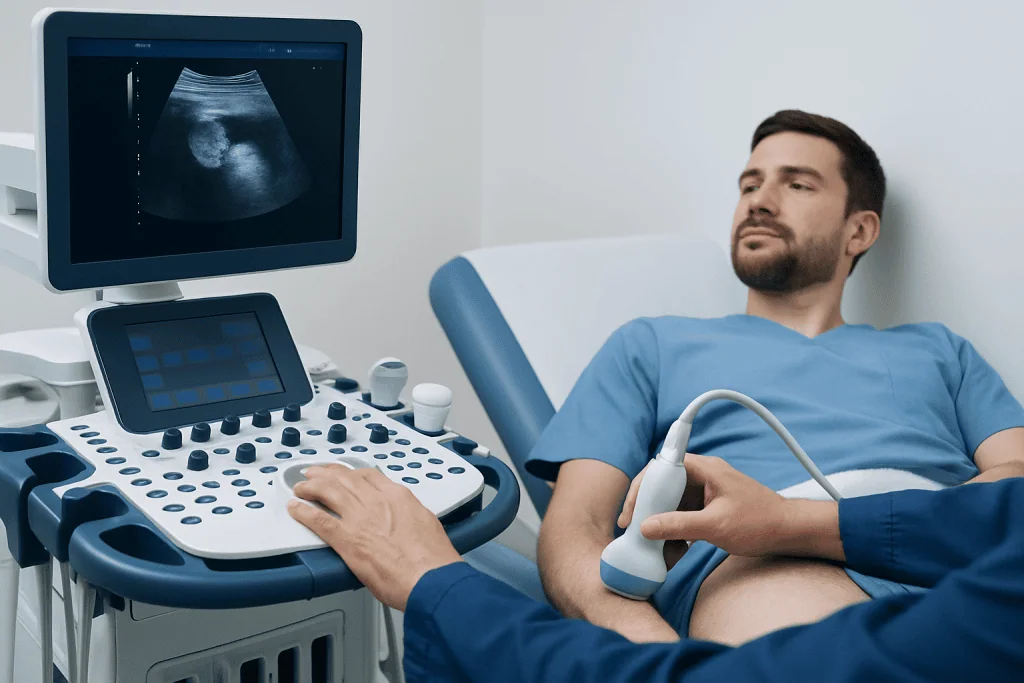
During the scan, the person lies on their back. A towel or support may be placed beneath the scrotum for comfort and stability. The sonographer applies warm or room-temperature gel to the skin and moves a small handheld probe gently over both sides of the scrotum.
Images are obtained of each testicle and the surrounding structures. Doppler images may be recorded to show blood flow. The examination commonly takes about 15 to 30 minutes. It should not be painful, although an already tender area may feel mildly uncomfortable when the probe passes over it.
Afterward, the gel is wiped away and normal activities can usually resume immediately. There is no recovery period, sedation or radiation exposure. If the scan was ordered urgently because of sudden severe pain, the clinician may discuss results immediately to rule out time-sensitive conditions such as torsion.
What Are the Typical Ultrasound Findings for Testicular Cancer?
The most typical ultrasound finding for testicular cancer is a solid lesion located within the testicle. It is often hypoechoic, meaning it appears darker than the normal surrounding testicular tissue. The lesion may be well defined or have less distinct borders, and it can be a single focus or, less commonly, involve more than one area.
Color Doppler imaging may show internal blood flow, particularly in many solid tumors. Other findings can include a change in the normal uniform texture of the testicle, small calcifications or associated fluid around the testicle. However, none of these features alone proves that a lesion is cancer.
Some testicular tumors have varied appearances, and a small number may be difficult to distinguish from benign abnormalities on imaging alone. A solid intratesticular lesion is therefore usually referred to a urologist without delay. The next steps may include blood tests for markers such as AFP, beta-hCG and LDH, along with staging scans if cancer is suspected.
When cancer is considered likely, the usual diagnostic and initial treatment approach is removal of the affected testicle through an incision in the groin, called testicular cancer treatment. A biopsy through the scrotal skin is generally avoided because it can affect local tissue planes and treatment planning.
How Long Does It Take to Get Testicular Ultrasound Results?
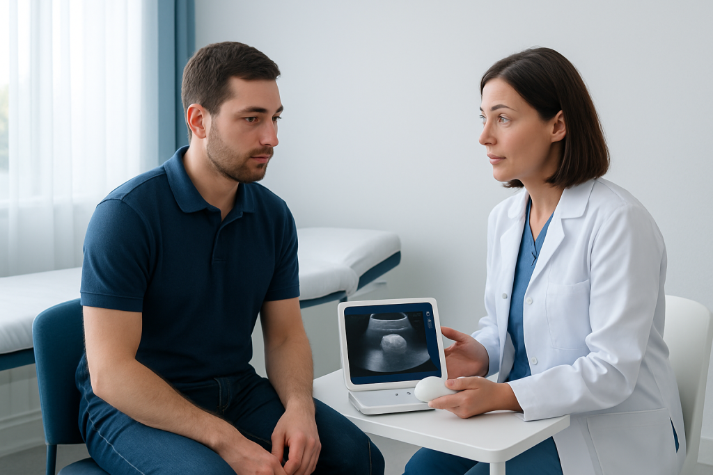
Images are created during the appointment, but the timing of the final result depends on the healthcare setting. In many centers, a radiologist reviews the images and issues a report on the same day or within a few days. The clinician who ordered the test then explains what the report means in the context of symptoms and examination findings.
If the ultrasound shows a suspicious intratesticular mass, the referring clinician may arrange an urgent urology appointment. In emergency situations, such as sudden pain where torsion is a concern, results are often communicated immediately because timely treatment matters.
A report may describe the size, location and imaging characteristics of a lesion, as well as Doppler blood flow and findings in the other testicle. It may also recommend follow-up imaging, blood tests or specialist assessment. Patients should ask when and how they will receive results before leaving the imaging department.
What Is the Typical Size of a Testicular Cancer Lump?
There is no single typical size for a testicular cancer lump. A tumor may be only a few millimeters across when found early, while others can grow to several centimeters before diagnosis. The lump may be felt as a firm, painless nodule, but some lesions are too small to be detected by touch and are found during ultrasound.
Size alone cannot determine whether a lump is cancerous, how fast it is growing or whether it has spread. Small intratesticular masses still need proper evaluation, while larger scrotal swellings can occur for noncancerous reasons such as fluid collection or infection.
Ultrasound provides accurate measurements in three dimensions and helps show whether the finding is inside the testicle or outside it. This distinction is important: masses outside the testicle are more often benign, whereas a solid mass inside the testicle is assessed more urgently.
How Accurate Is Ultrasound for Testicular Cancer?
Scrotal ultrasound is highly accurate for detecting and locating a testicular mass. It is especially effective at identifying whether an abnormality is intratesticular or extratesticular, and this information strongly guides the next clinical steps. It can also assess the opposite testicle and identify other explanations for symptoms.
However, ultrasound cannot confirm the precise cell type of a tumor or always distinguish every benign lesion from cancer. For this reason, clinicians combine the imaging findings with examination, blood tests and urology assessment. When a solid intratesticular mass is suspicious, surgery may be required both to establish the diagnosis and to treat the primary tumor.
The benefits of ultrasound include speed, accessibility, lack of radiation and detailed imaging of scrotal structures. Risks are minimal and mainly relate to brief discomfort from pressure on a tender area. There is no known harmful effect from diagnostic ultrasound when used appropriately.
Care may involve urologists, radiologists, medical oncologists, pathologists and fertility specialists. Before treatment, a person may be offered discussion of sperm banking because some treatments can affect fertility. Related evaluation may include information about testicular cancer and its staging and follow-up needs.
When to Seek Medical Care
A person should arrange medical assessment promptly for a new lump, firmness, swelling, enlargement, heaviness or persistent ache in either testicle. Other reasons to seek care include a change in shape, a dull lower abdominal or groin ache, or breast tenderness that is unexplained. These symptoms often have noncancerous causes, but it is important not to self-diagnose.
Sudden severe testicular pain, especially with nausea, vomiting or a high-riding testicle, needs emergency assessment. This pattern can be caused by testicular torsion, where reduced blood supply may threaten the testicle if not treated quickly.
After a suspicious ultrasound result, a person should attend all recommended urology appointments and ask about blood tests, fertility preservation, treatment choices and follow-up. Acibadem International’s multidisciplinary specialists and JCI-accredited hospitals support international patients with diagnostic assessment and treatment planning for testicular conditions.
Frequently asked questions
Can an ultrasound diagnose testicular cancer?
Ultrasound can identify a suspicious mass and show whether it is located inside the testicle, which is an important finding. However, it cannot always confirm cancer or identify the exact tumor type. A urologist uses the scan alongside examination, blood tests and, when appropriate, surgical pathology.
What does testicular cancer look like on an ultrasound?
Many testicular cancers appear as a solid, darker-than-normal area within the testicle. Doppler imaging may show increased blood flow within the lesion. Appearances vary, so the report must be interpreted by an experienced clinician.
Is a testicular ultrasound painful?
The scan is usually painless and does not require needles, anesthesia or radiation. The probe may cause temporary discomfort if the scrotum or testicle is already sore. Patients should tell the sonographer if pressure is uncomfortable.
Can a benign condition look like testicular cancer on ultrasound?
Yes. Inflammation, injury, infarction and some benign growths can sometimes resemble a tumor on ultrasound. A suspicious solid mass inside the testicle is still evaluated urgently because imaging alone may not provide a definite answer.
Can testicular cancer be too small to feel?
Yes. Some small testicular lesions are not easy to feel, particularly if they are deep within the testicle. Ultrasound can detect small abnormalities and measure them accurately.
What happens after an abnormal testicular ultrasound?
The next step is usually prompt review by a urologist. Further evaluation may include a physical examination, tumor marker blood tests and imaging to assess whether there is any spread. If findings are strongly suspicious, removal of the affected testicle through the groin is often recommended for diagnosis and treatment.
References
- American Cancer Society
- European Association of Urology
- National Cancer Institute
- Radiological Society of North America
- World Health Organization
This article is for general information only and is not a substitute for professional medical advice. Please consult a qualified doctor about your individual situation.
Ultrasound in Turkey — costs, top hospitals & a free quote
JCI-accredited · board-certified surgeons · reply within 24h
Add us as a Preferred Source to see more of our trusted health content across Google Search, AI Overviews and Discover.

Check-up & Preventive Medicine
Comprehensive health screenings designed for early detection and prevention.
3 specialists in this unit