Glomerulonephritis Ultrasound: Preparation, Procedure and Results

Ultrasound does not use ionising radiation and is generally safe, including during pregnancy. Glomerulonephritis may not produce a specific ultrasound appearance, particularly in early disease.
Key Takeaways
- Ultrasound does not use ionising radiation and is generally safe, including during pregnancy.
- Glomerulonephritis may not produce a specific ultrasound appearance, particularly in early disease.
- The scan helps clinicians assess kidney size, structure, blood flow when Doppler is used, and possible urinary blockage.
- Preparation often involves drinking water and arriving with a comfortably full bladder; instructions vary by facility.
- A radiologist interprets the images, and the referring clinician explains what the findings mean alongside laboratory results.
A glomerulonephritis ultrasound is a painless imaging test that uses sound waves to assess the kidneys and urinary tract. It can identify changes such as altered kidney size, swelling or obstruction, while blood tests, urine tests and sometimes a kidney biopsy are usually needed to diagnose glomerulonephritis.
Overview: what is a glomerulonephritis ultrasound?
A glomerulonephritis ultrasound is usually a kidney and bladder ultrasound performed when a clinician is investigating signs of kidney inflammation. Glomerulonephritis affects the glomeruli, the tiny filtering units inside the kidneys. It may be suspected after protein or blood is found in urine, kidney function blood tests are abnormal, blood pressure is raised, or swelling develops.
The examination uses a handheld probe and sound waves to create real-time images of the kidneys. It is non-invasive, does not involve radiation and commonly takes about 20 to 30 minutes. The scan is valuable for looking at kidney size and shape, signs of urinary obstruction, cysts, stones or other structural concerns that can contribute to reduced kidney function.
However, ultrasound is one part of a wider assessment. A clinician may combine it with urine testing, blood tests, blood pressure measurements and, in selected cases, a kidney biopsy to clarify the type and cause of suspected glomerulonephritis.
Does glomerulonephritis show on ultrasound?
Glomerulonephritis does not always show clearly on ultrasound. The inflammation occurs in microscopic kidney filters, so ultrasound cannot directly see the glomeruli or reliably identify the exact type of glomerulonephritis. In early or mild disease, the kidneys can appear normal on a scan.
In some forms of active inflammation, the kidneys may look enlarged or more echogenic, meaning they reflect more sound waves than expected. In long-standing kidney damage, they may become smaller with a thinner outer layer. These findings are not specific to glomerulonephritis and can also occur with other kidney conditions.
The major benefit of ultrasound is that it helps identify structural explanations or complications, such as an enlarged collecting system caused by blocked urine flow. It also helps the care team judge whether a biopsy may be appropriate and safe. Test results must be interpreted together rather than as a diagnosis on their own.
- Urinalysis can detect protein, blood and casts in the urine.
- Blood tests assess kidney filtration and may investigate immune or infectious causes.
- A kidney biopsy may be needed to identify the specific pattern of glomerular inflammation and guide treatment.
Who may need this scan and how does it work?

A clinician may request a kidney ultrasound for someone with persistent protein or blood in the urine, a falling estimated glomerular filtration rate, unexplained swelling, difficult-to-control blood pressure or symptoms suggesting a urinary tract blockage. It may also be used to establish a baseline before a biopsy or to monitor known chronic kidney disease.
Ultrasound works by sending high-frequency sound waves through the skin. The waves reflect differently from fluid, soft tissue and denser structures. A computer turns these returning echoes into images. If needed, Doppler ultrasound can assess the direction and pattern of blood flow in kidney vessels, although it is not required for every examination.
Most people are suitable for an ultrasound because it does not use contrast dye or radiation. Image quality can sometimes be reduced by bowel gas, body habitus, wounds or difficulty remaining still, but these factors do not usually prevent the test. The sonographer may adjust positioning or scanning angles to obtain useful views.
How should I prepare for an ultrasound kidney scan?
Preparation instructions differ according to the type of kidney ultrasound and local imaging protocol. Many centres ask patients to drink water before the appointment and avoid urinating for a period beforehand, so the bladder is comfortably full. A full bladder can improve views of the bladder and the lower urinary tract.
Patients should follow the instructions supplied by the imaging department, particularly if the scan includes the bladder, prostate, pelvic organs or abdominal blood vessels. It is sensible to wear loose, two-piece clothing, bring the referral information if provided, and tell the team about previous kidney imaging, operations or relevant medical conditions.
Regular prescribed medicines can usually be taken as normal unless a clinician has advised otherwise. People with diabetes, those who are pregnant, or anyone who has been instructed to fast for another test on the same day should ask the imaging team for individual guidance rather than changing medication or food intake independently.
What not to eat before a kidney ultrasound?
For a standard kidney and bladder ultrasound, people are often not required to avoid particular foods. Some departments may ask patients not to eat for several hours before an abdominal ultrasound because eating can increase bowel gas and make images less clear. This is more likely when the examination includes other abdominal organs.
If fasting is requested, the imaging centre will explain how long it should last and whether clear fluids are allowed. Patients should not assume that fasting is needed, as drinking water may be important to fill the bladder. Carbonated drinks and foods that cause substantial bloating may be discouraged in some protocols, but the centre’s specific instructions should take priority.
People should contact the imaging department if they have not received preparation details, have a medical condition that makes fasting difficult, or are scheduled for several tests at once. Clear preparation instructions help the team obtain the best possible images without unnecessary inconvenience.
What happens during and after the procedure?
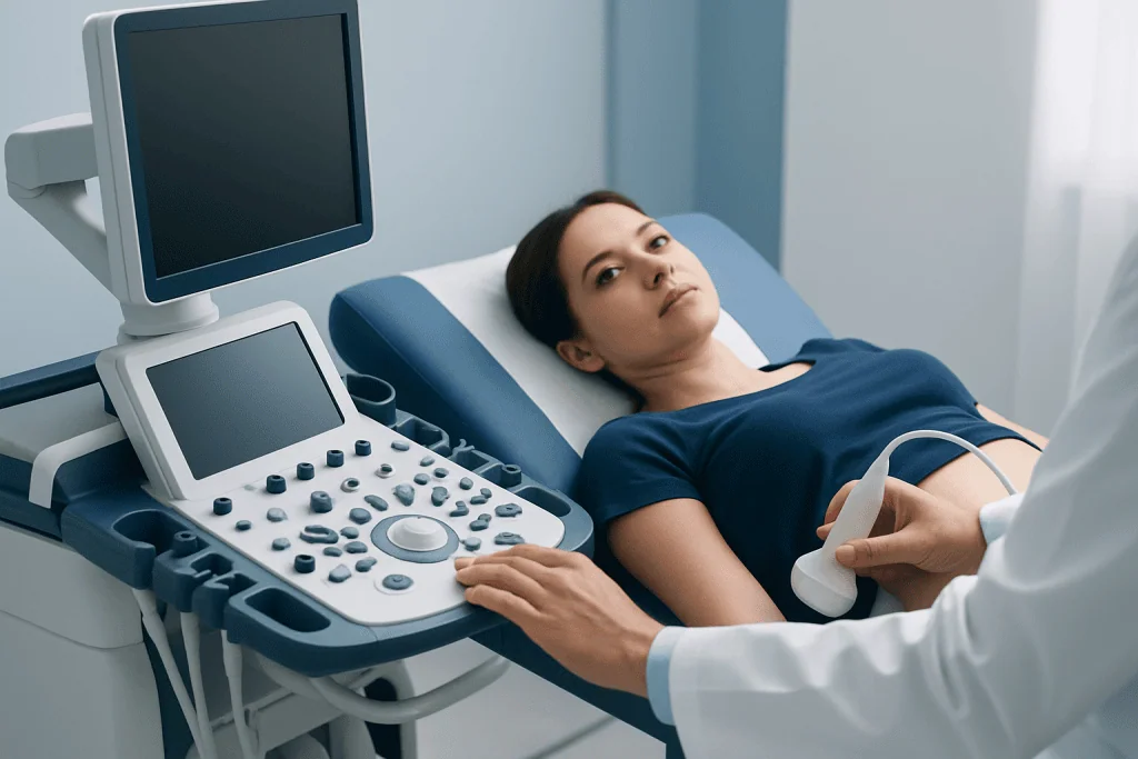
At the appointment, the patient is usually asked to lie on an examination couch and expose the side of the abdomen and back. A sonographer applies a clear gel to the skin, then moves the ultrasound probe over the area. The person may be asked to turn onto either side, take a deep breath or briefly hold their breath so the kidneys can be seen more clearly.
The probe may cause mild pressure, especially if the area is tender, but the scan should not be painful. No needles, sedatives or injections are normally needed for a routine renal ultrasound. The sonographer records images for review by a radiologist or another appropriately qualified clinician.
Once the scan is complete, the gel is wiped away and normal activities can usually resume immediately. There is no recovery period, driving restriction or special aftercare. Ultrasound is considered very low risk because it uses sound waves rather than radiation; its main limitation is that the results may not fully explain microscopic kidney disease.
How long do kidney ultrasound results take?
Image acquisition happens during the appointment, but the final result is usually available after a radiologist reviews the images and prepares a report. Timing varies between healthcare settings. In many non-urgent cases, the referring clinician receives the report within a few days, while urgent findings may be communicated more quickly.
The scan provider may not be able to give a full interpretation immediately, since the result should be considered with the reason for referral and other medical information. The clinician who ordered the test can explain whether the kidneys appear normal in size and structure, whether there is any evidence of obstruction, and what further tests may be useful.
Abnormal ultrasound findings do not necessarily mean permanent kidney damage, and a normal scan does not rule out glomerulonephritis. If laboratory tests suggest glomerular disease, a nephrologist may recommend further evaluation, including discussion of kidney biopsy when clinically appropriate.
When to seek medical care
Prompt medical assessment is important for new swelling around the eyes, face, hands or legs; foamy or tea-coloured urine; visible blood in urine; reduced urine output; or newly high blood pressure. These symptoms can have several causes, but they deserve timely evaluation because kidney problems may require monitoring or treatment.
Urgent care is appropriate for severe shortness of breath, chest pain, confusion, fainting, severe headache with very high blood pressure, or a marked reduction in urine output. People should seek emergency services according to local guidance if these symptoms are severe or sudden.
A nephrologist can coordinate testing and discuss treatment based on the underlying cause, disease activity and kidney function. Acibadem International’s multidisciplinary specialists and JCI-accredited hospitals evaluate and treat kidney conditions for international patients, with care plans tailored to individual clinical findings.
Frequently asked questions
Can an ultrasound diagnose glomerulonephritis?
No. Ultrasound cannot usually confirm glomerulonephritis because it does not show microscopic inflammation in the kidney filters. It supports the assessment by showing kidney size and structure and by helping exclude problems such as urinary obstruction. Urine and blood tests, and sometimes a biopsy, are used to establish the diagnosis.
Are kidneys enlarged with glomerulonephritis?
The kidneys can be enlarged in some active forms of glomerulonephritis, but this is not present in every case. In chronic kidney damage, they may instead appear smaller. Kidney size alone cannot determine the cause or severity of disease.
Do I need to drink water before a kidney ultrasound?
Often, yes, particularly when the bladder is being assessed. The imaging centre may ask the patient to drink water and avoid urinating shortly before the scan. The exact amount and timing vary, so the appointment instructions should be followed.
Is a kidney ultrasound painful?
A kidney ultrasound is generally painless. The probe may create some pressure against the abdomen or back, and this can feel uncomfortable if the area is already tender. Patients should tell the sonographer if pressure is painful so positioning can be adjusted.
What happens if a kidney ultrasound is normal but urine tests are abnormal?
A normal ultrasound does not exclude glomerulonephritis or other microscopic kidney conditions. The clinician will assess urine findings, kidney function blood tests, blood pressure and medical history. Referral to a nephrologist and further testing may be recommended.
Can I go back to work after a kidney ultrasound?
Yes, most people can return to work, eat and drink normally, and drive immediately after a standard ultrasound. The test does not usually involve sedation, injections or a recovery period. Different instructions may apply if another procedure is planned on the same day.
References
- National Institute of Diabetes and Digestive and Kidney Diseases
- National Kidney Foundation
- Radiological Society of North America
- Kidney Disease: Improving Global Outcomes
- American College of Radiology
This article is for general information only and is not a substitute for professional medical advice. Please consult a qualified doctor about your individual situation.
Ultrasound in Turkey — costs, top hospitals & a free quote
JCI-accredited · board-certified surgeons · reply within 24h
Add us as a Preferred Source to see more of our trusted health content across Google Search, AI Overviews and Discover.
Check your numbers in seconds
BMI, calories, due date, blood pressure and 30+ more clinical calculators — free, instant, doctor-reviewed ranges.

Check-up & Preventive Medicine
Comprehensive health screenings designed for early detection and prevention.
3 specialists in this unit