

Eye Biopsy: Procedure, Recovery and Results
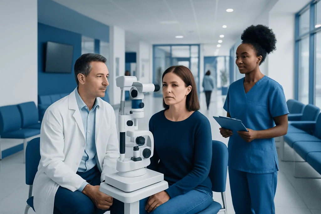
An eye biopsy is usually performed when examination and imaging cannot provide a definite diagnosis. The biopsy site may be on the eyelid, conjunctiva, eye surface, orbit or, less commonly, inside the eye.
Key Takeaways
- An eye biopsy is usually performed when examination and imaging cannot provide a definite diagnosis.
- The biopsy site may be on the eyelid, conjunctiva, eye surface, orbit or, less commonly, inside the eye.
- Local anesthesia is commonly used for surface or eyelid biopsies, while deeper procedures may require sedation or general anesthesia.
- Mild soreness, redness and swelling are common during early recovery, but severe pain or worsening vision needs urgent assessment.
- Results often take several days to two weeks, depending on the type of laboratory testing required.
An eye biopsy is a procedure in which a specialist removes a small sample of tissue from the eye, eyelid, orbit or surrounding structures for laboratory examination. It can help identify inflammation, infection, benign growths or cancer and guide the next steps in care.
Overview: What Is an Eye Biopsy?
An eye biopsy is a diagnostic procedure that removes a small amount of tissue or fluid from the eye or nearby structures. A pathologist examines the sample under a microscope and may use additional laboratory tests to identify abnormal cells, inflammation, infection or other disease processes. The procedure is not performed routinely; it is recommended when the result is likely to change diagnosis or treatment.
The term may describe biopsies from different locations, including the eyelid, conjunctiva (the clear membrane covering the white of the eye), cornea, lacrimal gland, orbit or tissues inside the eye. The precise technique, anesthesia and recovery expectations depend greatly on the location of the sample.
An eye specialist, often an ophthalmologist with expertise in ocular oncology, oculoplastic surgery, cornea disease or retina care, considers the safest method of obtaining useful tissue. In some cases, photographs, scans and close follow-up may be appropriate instead of immediate biopsy.
Why an Eye Biopsy May Be Recommended
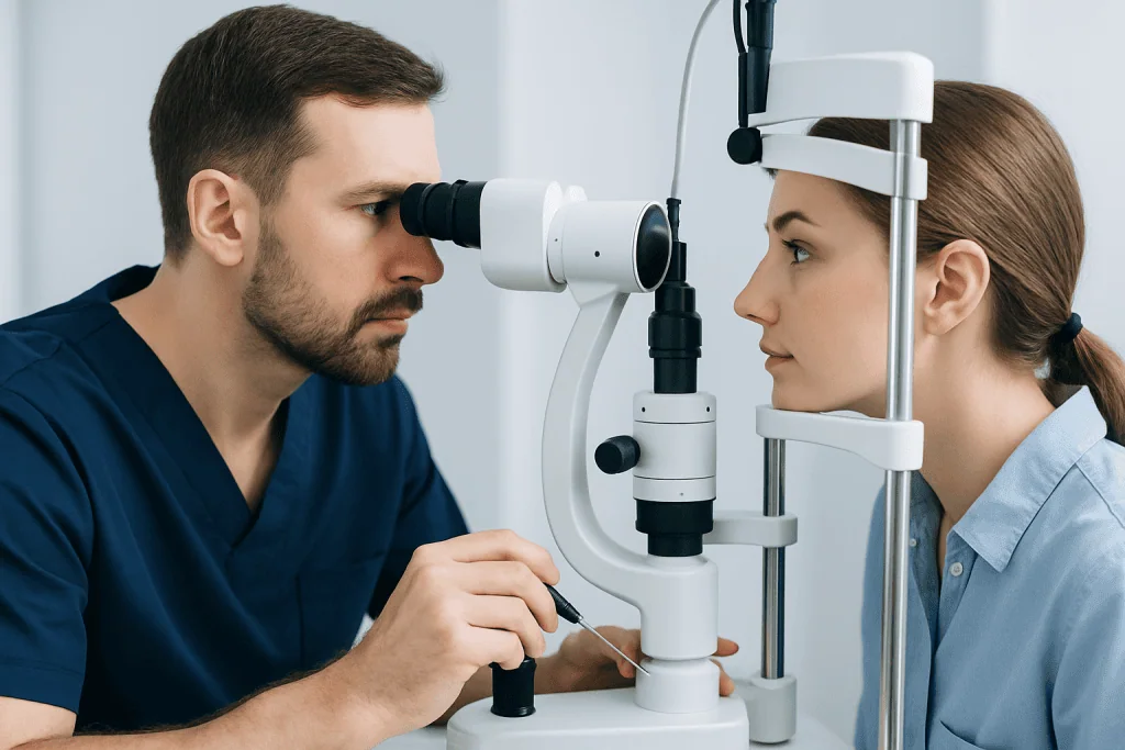
A clinician may recommend an eye biopsy for a lesion, lump, persistent irritation or unexplained change that cannot be diagnosed confidently through examination alone. It may help distinguish a harmless condition from one that needs treatment and can identify the specific type of inflammation or infection when these are suspected.
Common reasons include a persistent eyelid lesion, an unusual pigmented spot, a conjunctival growth, an orbital mass, recurrent inflammation that does not respond as expected, or concern for a tumor. Not every visible mark requires a biopsy. The decision is based on its appearance, location, growth pattern, symptoms and the person’s overall health history.
Before proceeding, the specialist may use slit-lamp examination, dilated eye examination, ultrasound, optical imaging, CT or MRI scans, and blood tests where relevant. These assessments help determine whether a biopsy is necessary and allow the team to plan the approach carefully.
- Diagnostic benefit: confirms or clarifies the cause of an abnormality.
- Treatment planning: helps select observation, medicine, surgery, radiotherapy or other care.
- Prognostic information: some tissue findings can help clinicians understand how a condition may behave.
How the Procedure Works: Preparation and Step-by-Step Care
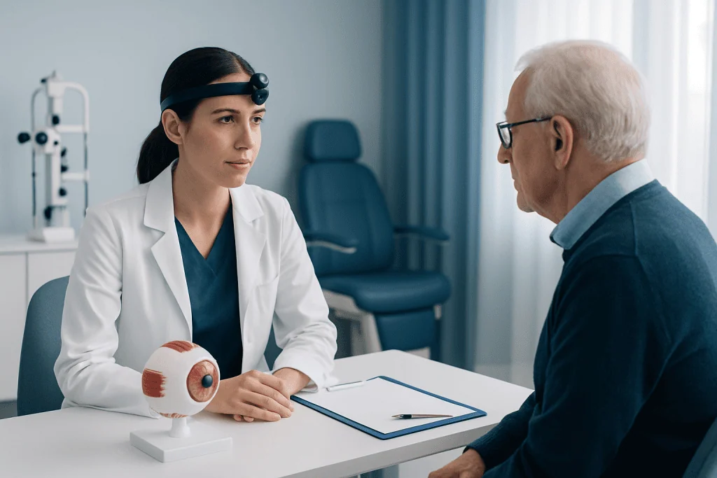
Preparation begins with a review of symptoms, medications, allergies, eye history and general medical conditions. Patients should tell the clinical team about blood-thinning medicines, diabetes medicines, previous anesthesia problems and any possibility of pregnancy. The team will provide individualized instructions on whether medicines should be continued or temporarily adjusted; patients should not stop prescribed medication without medical advice.
For an eyelid, conjunctival or surface lesion, the procedure may be performed in an outpatient clinic or day-surgery setting. Numbing drops, local anesthetic injection or both may be used. The surgeon removes a small piece of tissue, or sometimes the entire small lesion, and may close the area with fine stitches. A protective dressing or eye patch may occasionally be used.
Biopsies involving the orbit or deeper eye structures are more complex and are generally performed in an operating room. Depending on the location, sedation or general anesthesia may be needed. The surgeon may use imaging guidance or microsurgical instruments to access the area while protecting important eye structures.
After sampling, the tissue is placed in a suitable container and sent to a pathology laboratory. Special stains, microbiology testing, molecular testing or expert pathology review may be requested when needed. The clinician explains what to expect after the procedure, including eye drops, pain relief, wound care and follow-up arrangements.
Is an Eye Biopsy Painful?
An eye biopsy should not be painful during the procedure because anesthesia is used. People may feel pressure, gentle pulling or brief discomfort from the anesthetic injection, especially for eyelid or surface biopsies. If discomfort occurs during the procedure, the person should tell the surgical team promptly so that anesthesia can be adjusted.
After the anesthesia wears off, mild soreness, a gritty sensation, tearing, redness or eyelid tenderness can occur. These symptoms are usually temporary and can often be managed with the clinician’s recommended pain relief and prescribed eye medication. Avoid using over-the-counter eye drops or pain medicines unless the care team confirms they are suitable.
Significant or increasing pain is not something to ignore. Severe pain, a sudden reduction in vision, marked light sensitivity, nausea with eye pain, or increasing swelling should be reported urgently because these symptoms can indicate a complication that needs prompt assessment.
Recovery Timeline and Aftercare
Recovery varies according to the biopsy location and size, whether stitches were used, and whether a deeper surgical approach was necessary. Following a small eyelid or conjunctival biopsy, many people return home the same day and experience mild swelling or bruising for several days. A superficial eye-surface area may remain red or feel irritated while it heals.
Aftercare instructions may include using prescribed antibiotic, lubricating or anti-inflammatory drops or ointment; keeping the area clean; avoiding rubbing the eye; and attending the planned follow-up visit. Contact lenses, eye makeup, swimming and strenuous activity may need to be avoided for a period specified by the surgeon. Driving should be avoided until vision is clear and the person feels safe to do so.
For an orbital or intraocular biopsy, recovery can be longer and follow-up is particularly important. The specialist may monitor vision, eye pressure, wound healing and signs of infection. Individual recovery advice takes priority because restrictions differ between procedures.
Acibadem International’s multidisciplinary specialists and JCI-accredited hospitals support diagnosis and treatment planning for international patients who need coordinated ophthalmology and pathology care.
How Long Does It Take for an Eye Biopsy to Heal?
Small eyelid or conjunctival biopsy sites often begin to feel noticeably better within several days, while visible redness, bruising or swelling may take one to two weeks to settle. If stitches are used, they may dissolve on their own or be removed at a follow-up visit, depending on the type of suture and the biopsy location.
Healing after a deeper orbital or intraocular biopsy can take longer, sometimes several weeks, because the procedure involves tissues farther from the eye surface. The clinician will assess healing at follow-up and advise when normal activities, exercise, contact lens use and eye makeup can safely resume.
Healing does not always mean that all appearance changes resolve immediately. Mild redness or a small scar can persist for a time, particularly after eyelid surgery. Any worsening rather than gradual improvement should be discussed with the treating team.
How Many Days Should You Rest After a Biopsy?
After a simple outpatient eye or eyelid biopsy, many people need to rest for the remainder of the procedure day and may return to light daily activities within one or two days. However, the appropriate rest period depends on the biopsy site, the anesthesia used, visual symptoms and the type of work a person does.
It is often sensible to avoid heavy lifting, vigorous exercise, bending that increases pressure around the eye, swimming and activities that could expose the wound to dirt or impact until the surgeon gives clearance. People whose work requires sharp vision, driving, physical exertion or exposure to dust may need more time away from work.
Following sedation or general anesthesia, a responsible adult may need to accompany the patient home, and driving or important decision-making should be avoided for the period advised by the anesthesia team. The surgeon’s specific postoperative guidance should always be followed over general timelines.
Results, Risks and When to Seek Medical Care
Initial pathology results may be available within several days, but final results often take about one to two weeks. The timing depends on the sample, laboratory workload and whether special stains, cultures, molecular tests or an expert review are required. The specialist will explain what the result means and whether further tests, monitoring or treatment are needed.
Do biopsy results take longer if it is cancer? Not necessarily, but samples that may involve cancer can require additional testing to identify the exact cell type and relevant features. These steps help make treatment decisions more accurate, so the final report may take longer than a straightforward benign result. A delayed result does not by itself indicate cancer.
Eye biopsy complications are uncommon but can include bleeding, infection, scarring, dry eye symptoms, temporary blurred vision, eyelid shape changes or damage to nearby structures. The risk is influenced by the biopsy location and the underlying eye condition. The specialist discusses the relevant risks and benefits before obtaining consent.
When to seek medical care: Patients should contact their eye team promptly for worsening pain, increasing redness or swelling, pus-like discharge, fever, new floaters or flashes, persistent bleeding, or reduced vision. Sudden vision loss, severe eye pain or a major eye injury requires urgent medical assessment.
Frequently asked questions
What is the difference between an eye biopsy and an eyelid biopsy?
An eye biopsy is a broad term for sampling tissue from the eye or surrounding structures. An eyelid biopsy specifically removes tissue from the eyelid and is commonly used to evaluate a persistent lump, rash or suspicious skin lesion. The anesthesia, surgical technique and recovery depend on the exact site.
Will I be awake during an eye biopsy?
Many surface and eyelid biopsies are performed with local anesthesia, so the patient is awake but the area is numb. Sedation or general anesthesia may be used for children, anxious patients, or biopsies involving deeper tissues. The ophthalmologist and anesthesia team will explain the planned approach beforehand.
Can an eye biopsy affect vision?
Temporary blurred vision can occur from ointment, dilation drops, swelling or a patch after some procedures. Serious vision effects are uncommon but may be a concern with biopsies close to or inside the eye, which is why these procedures require careful planning and follow-up. New or worsening visual symptoms should be reported promptly.
Do biopsy results take longer if it's cancer?
Cancer-related samples may need extra laboratory testing to determine the precise type of cells and other features that guide treatment. This can extend the time to a final report, but a longer wait does not mean that cancer has been found. The care team can explain the expected timing for the individual sample.
How should someone prepare for an eye biopsy?
Patients should provide a complete list of medicines, supplements, allergies and medical conditions, especially medicines that affect bleeding. They should follow instructions about eating, drinking, transport and medication changes, particularly if sedation or general anesthesia is planned. It is also helpful to arrange support at home if vision may be blurred after the procedure.
What happens if the eye biopsy is inconclusive?
An inconclusive result means that the sample did not provide a definite explanation. The specialist may review imaging and clinical findings, request additional pathology testing, monitor the area, or recommend another biopsy if it is safe and necessary. Decisions are individualized to balance the value of more information with procedural risk.
References
- American Academy of Ophthalmology
- National Eye Institute
- American Cancer Society
- Royal College of Ophthalmologists
This article is for general information only and is not a substitute for professional medical advice. Please consult a qualified doctor about your individual situation.
Oncology care in Turkey — second opinion and treatment plan
JCI-accredited · board-certified surgeons · reply within 24h
Add us as a Preferred Source to see more of our trusted health content across Google Search, AI Overviews and Discover.

More from the Health Library


Related Specialists

Prof. Dr. Filiz Akyüz (m)
Gastroenterology
Dr. Burcu Yilmaz
Gynecology & Obstetrics
Asst. Prof. Dr. Hamdi Toköz
Cardiovascular Surgery
Prof. Dr. Ertap Akoğlu
Otorhinolaryngology