Glaucoma: Eye Pressure, Vision Loss, and Treatment
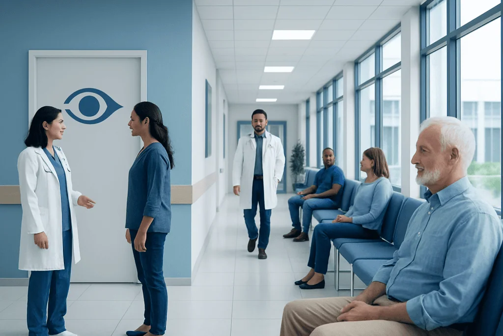
Glaucoma often develops slowly and may not cause symptoms in the early stages. High eye pressure is an important risk factor, but glaucoma can also occur with normal eye pressure.
Key Takeaways
- Glaucoma often develops slowly and may not cause symptoms in the early stages.
- High eye pressure is an important risk factor, but glaucoma can also occur with normal eye pressure.
- Regular eye examinations are the best way to detect glaucoma before noticeable vision loss occurs.
- Treatment may include eye drops, laser procedures, or surgery to lower eye pressure and protect the optic nerve.
- Vision already lost from glaucoma usually cannot be restored, but treatment can slow or prevent further damage.
- Sudden eye pain, blurred vision, halos around lights, headache, nausea, or a red eye requires urgent medical attention.
Glaucoma is a group of eye diseases that can damage the optic nerve, often related to pressure inside the eye. Early diagnosis and ongoing treatment can help protect vision and support long-term eye health.
Overview
Glaucoma is a group of eye conditions that damage the optic nerve, the structure that carries visual information from the eye to the brain. The optic nerve is essential for clear vision. When it is gradually injured, blind spots can develop, usually starting in the side vision. Without diagnosis and treatment, glaucoma may progress and affect central vision as well.
Many types of glaucoma are linked to increased pressure inside the eye, called intraocular pressure. This pressure is not the same as blood pressure. It depends on the balance between the production and drainage of a clear fluid inside the eye called aqueous humor. If this fluid does not drain properly, pressure can rise and place stress on the optic nerve.
Glaucoma is sometimes called a silent condition because the most common forms can develop over years without pain or obvious symptoms. For this reason, regular eye checks are important, especially for people with risk factors. Although vision loss from glaucoma is usually permanent, timely treatment can reduce eye pressure and help protect remaining vision.
Types of Glaucoma
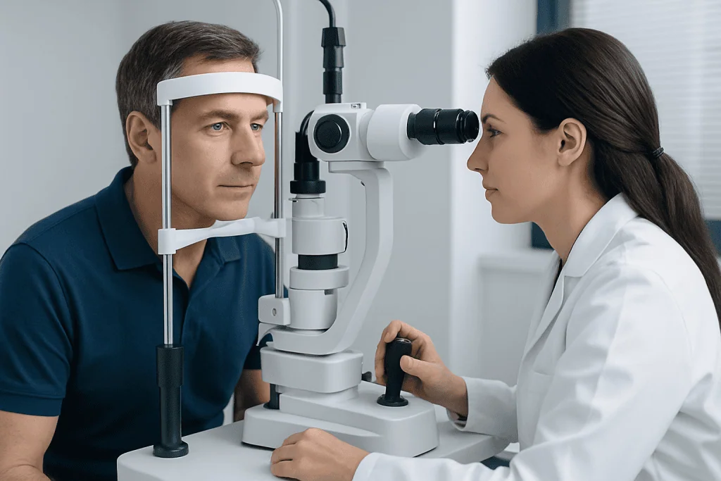
The most common type is primary open-angle glaucoma. In this form, the eye’s drainage angle remains open, but fluid does not drain efficiently enough. Pressure may rise slowly, and optic nerve damage can occur gradually. Many people feel well and see normally in daily life until the condition is more advanced, because the brain can compensate for small areas of peripheral vision loss.
Angle-closure glaucoma occurs when the drainage angle becomes blocked or very narrow. In some people, this can happen suddenly and cause a rapid rise in eye pressure. Acute angle-closure glaucoma is an emergency because it can threaten vision quickly. There is also a chronic form that develops more slowly and may be detected during a routine eye examination.
Other forms include normal-tension glaucoma, in which optic nerve damage occurs even though eye pressure is within the usual range, and secondary glaucoma, which develops due to another cause such as eye inflammation, injury, certain medications, advanced cataract, or some eye surgeries. Congenital and childhood glaucoma are less common and require specialized assessment by an ophthalmologist.
Symptoms and Warning Signs
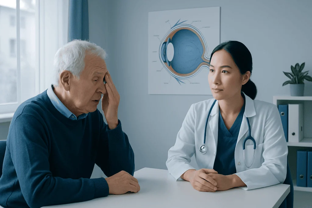
In early open-angle glaucoma, there may be no symptoms. Vision can seem normal, and there is usually no pain. As the disease progresses, a person may notice missing areas in side vision, difficulty seeing objects at the edges of the visual field, or problems adjusting in dim light. Because these changes are often subtle, they may not be recognized until significant optic nerve damage has occurred.
Acute angle-closure glaucoma is different and can produce sudden, noticeable symptoms. These may include severe eye pain, redness, blurred vision, headache, nausea, vomiting, and halos around lights. The affected eye may feel hard, and vision can decrease rapidly. These symptoms need immediate medical evaluation, preferably by an eye specialist or emergency service.
People should not wait for symptoms before having an eye examination if they are at higher risk. A comprehensive eye exam can detect raised eye pressure, optic nerve changes, and visual field loss before the person notices a problem. Early detection is especially valuable because glaucoma treatment aims to prevent future vision loss rather than reverse damage that has already occurred.
Causes and Risk Factors
Glaucoma develops when the optic nerve becomes damaged. In many cases, increased eye pressure plays a key role. The eye continually produces aqueous humor, which normally drains through a tissue network near the front of the eye. If drainage is reduced, fluid can build up and increase intraocular pressure. Over time, this pressure may contribute to optic nerve injury.
However, eye pressure is only part of the picture. Some people with high eye pressure never develop glaucoma, while others develop glaucoma even when pressure is not elevated. Blood flow to the optic nerve, individual nerve sensitivity, anatomy of the drainage angle, and genetic factors may all be involved. This is why glaucoma diagnosis requires more than a simple pressure measurement.
Risk factors include increasing age, a family history of glaucoma, high eye pressure, thin corneas, severe nearsightedness or farsightedness, previous eye injury, long-term corticosteroid use, and certain medical conditions such as diabetes, high blood pressure, or migraine. People of African, Asian, or Hispanic ancestry may have a higher risk of some forms of glaucoma. Anyone with these risk factors should ask an eye doctor how often screening is appropriate.
Diagnosis
Glaucoma is diagnosed through a comprehensive eye examination performed by an ophthalmologist or trained eye care professional. The exam usually includes measurement of intraocular pressure, inspection of the optic nerve, and evaluation of the drainage angle. Eye pressure testing is useful, but it cannot confirm or exclude glaucoma on its own.
Several tests may be used to assess the optic nerve and visual function. Visual field testing checks for missing areas in peripheral vision. Optical coherence tomography, often called OCT, creates detailed images of the optic nerve and retinal nerve fiber layer. Pachymetry measures corneal thickness, which can influence eye pressure readings. Gonioscopy helps the doctor see whether the drainage angle is open, narrow, or closed.
Because glaucoma often changes slowly, diagnosis may involve comparing results over time. Photographs of the optic nerve, repeated visual field tests, and OCT scans can help show whether the condition is stable or progressing. Regular follow-up is essential after diagnosis, because treatment plans may need adjustment if pressure targets are not met or if optic nerve changes continue.
Treatment Options
The main goal of glaucoma treatment is to lower eye pressure to a level that reduces the risk of further optic nerve damage. This target pressure is individualized. It depends on the type of glaucoma, the degree of optic nerve damage, baseline eye pressure, age, overall health, and whether the disease appears stable or progressing.
Prescription eye drops are often the first treatment for open-angle glaucoma. They work by reducing fluid production in the eye or improving fluid drainage. Different classes of medications may be used alone or together. It is important to use drops exactly as prescribed, because missed doses can allow pressure to rise. Patients should tell their doctor about side effects, allergies, breathing problems, heart conditions, pregnancy, or other medications before starting or changing eye drops.
Laser treatment may be recommended when eye drops are not enough, are difficult to use, or are not well tolerated. Selective laser trabeculoplasty can help improve drainage in open-angle glaucoma. Laser peripheral iridotomy may be used for narrow angles or angle-closure risk by creating a small opening in the iris to improve fluid movement. These procedures are usually brief, but follow-up care remains important.
Surgery may be considered for more advanced glaucoma or when pressure cannot be controlled with medication and laser treatment. Options include trabeculectomy, glaucoma drainage devices, and minimally invasive glaucoma surgery in selected patients. Each approach has benefits and risks, and the best choice depends on the eye’s anatomy, disease severity, previous treatments, and the patient’s overall situation.
Prevention, Self-care, and Living With Glaucoma
Glaucoma cannot always be prevented, but its impact can often be reduced through early detection and consistent care. People with risk factors should follow the eye examination schedule recommended by their doctor. Those already diagnosed should attend follow-up visits even when vision seems stable, because glaucoma can progress without noticeable symptoms.
Good treatment habits make a meaningful difference. Patients who use eye drops can benefit from a daily routine, reminders, and correct technique. After placing a drop, gently closing the eye and pressing near the inner corner for a short time may reduce drainage into the nose and improve comfort. If more than one drop is prescribed, the doctor or pharmacist can explain how to space them safely.
Healthy lifestyle habits support general eye and vascular health, although they do not replace glaucoma treatment. These include regular physical activity approved by a physician, not smoking, balanced nutrition, and managing conditions such as diabetes and high blood pressure. Patients should also tell all healthcare providers that they have glaucoma, especially before using steroid medications or treatments that may affect eye pressure.
Living with glaucoma may require practical adjustments. Good lighting, reducing trip hazards at home, using contrast markings on stairs, and having updated glasses can help with safety if peripheral vision is reduced. Emotional support is also important. Many people continue active, independent lives with glaucoma when it is monitored carefully and treated consistently.
When to See a Doctor
A person should schedule an eye examination if they have a family history of glaucoma, have been told they have high eye pressure, notice changes in side vision, or have risk factors such as older age, previous eye injury, or long-term steroid use. People who already have glaucoma should contact their eye doctor if vision changes, if medications cause side effects, or if they have difficulty using drops as prescribed.
Urgent medical care is needed for sudden eye pain, a red eye with blurred vision, halos around lights, severe headache, nausea, or vomiting. These symptoms may indicate acute angle-closure glaucoma or another serious eye condition. Prompt treatment can help reduce the risk of lasting vision damage.
International patients who need evaluation or ongoing care can seek support from qualified ophthalmology teams. Acibadem International’s multidisciplinary specialists and JCI-accredited hospitals diagnose and treat glaucoma and related eye conditions for international patients, with care planning based on individual medical needs. Any person concerned about glaucoma should consult a qualified eye doctor for a personalized assessment.
Frequently asked questions
Is glaucoma the same as high eye pressure?
No. High eye pressure is an important risk factor for glaucoma, but it is not the same as glaucoma. Glaucoma means there is damage to the optic nerve, and this can sometimes occur even when eye pressure is within the usual range.
Can vision loss from glaucoma be reversed?
Vision loss caused by glaucoma is usually permanent because optic nerve damage cannot typically be repaired. Treatment focuses on lowering eye pressure and slowing or preventing further damage. This is why early diagnosis and regular monitoring are so important.
How often should someone be checked for glaucoma?
The right schedule depends on age, eye pressure, family history, optic nerve appearance, and other risk factors. People at higher risk may need more frequent examinations than those at low risk. An ophthalmologist can recommend an appropriate follow-up plan after a comprehensive eye exam.
Do glaucoma eye drops need to be used forever?
Many people need long-term treatment because glaucoma is usually a chronic condition. Eye drops may be continued, changed, combined with laser treatment, or replaced by another approach depending on how the eye responds. Patients should not stop glaucoma medication without speaking to their eye doctor.
Is glaucoma surgery dangerous?
All eye procedures have potential benefits and risks, and the safest choice depends on the individual case. Glaucoma surgery is considered when the expected benefit of lowering pressure outweighs the risks of the procedure. The ophthalmologist should explain the options, recovery, follow-up needs, and possible complications before treatment.
Can lifestyle changes cure glaucoma?
Lifestyle changes cannot cure glaucoma and should not replace medical treatment. However, healthy habits such as not smoking, exercising safely, managing blood pressure and diabetes, and attending regular eye visits can support overall eye health. The most important step is to follow the treatment plan prescribed by an eye specialist.
References
- American Academy of Ophthalmology
- National Eye Institute
- World Glaucoma Association
- European Glaucoma Society
- Mayo Clinic
This article is for general information only and is not a substitute for professional medical advice. Please consult a qualified doctor about your individual situation.
Glaucoma in Turkey — costs, top hospitals & a free quote
JCI-accredited · board-certified surgeons · reply within 24h
Check your numbers in seconds
BMI, calories, due date, blood pressure and 30+ more clinical calculators — free, instant, doctor-reviewed ranges.