

Artificial Intelligence in Radiology: How It Supports Diagnosis
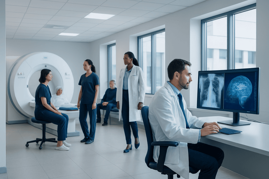
AI can help detect patterns in X-rays, CT, MRI, ultrasound and mammography images. Radiologists remain responsible for interpreting results and making clinical recommendations.
Key Takeaways
- AI can help detect patterns in X-rays, CT, MRI, ultrasound and mammography images.
- Radiologists remain responsible for interpreting results and making clinical recommendations.
- AI may support faster triage, consistency and comparison of imaging findings over time.
- Safe use depends on data quality, clinical validation, privacy protection and human oversight.
- Patients should view AI-assisted imaging as an added support layer, not an independent diagnosis.
Artificial intelligence in radiology is designed to support radiologists by analyzing medical images, highlighting findings and improving workflow efficiency. It does not replace medical expertise; rather, it acts as a decision-support tool within a supervised clinical process.
Overview: What Is Artificial Intelligence in Radiology?
Artificial intelligence in radiology refers to computer systems that are trained to recognize patterns in medical images and related clinical data. These systems may examine X-rays, computed tomography (CT), magnetic resonance imaging (MRI), ultrasound, mammography or other imaging studies. Their goal is to support radiologists by drawing attention to possible abnormalities, measuring structures, comparing images or organizing urgent cases.
Most modern radiology AI tools use machine learning, a type of artificial intelligence that learns from large collections of labeled medical images. For example, a system may be trained with many chest X-rays that have been reviewed by expert radiologists, allowing it to identify features that may suggest pneumonia, lung nodules or fluid around the lungs. In practice, the AI output is reviewed by a qualified clinician rather than accepted automatically.
For patients, the most important point is that AI is not a separate doctor and does not replace the radiologist. It is a form of decision support. The radiologist considers the image, the patient’s symptoms, medical history, previous scans and the question asked by the referring doctor before issuing a report.
How AI Supports Diagnosis in Medical Imaging
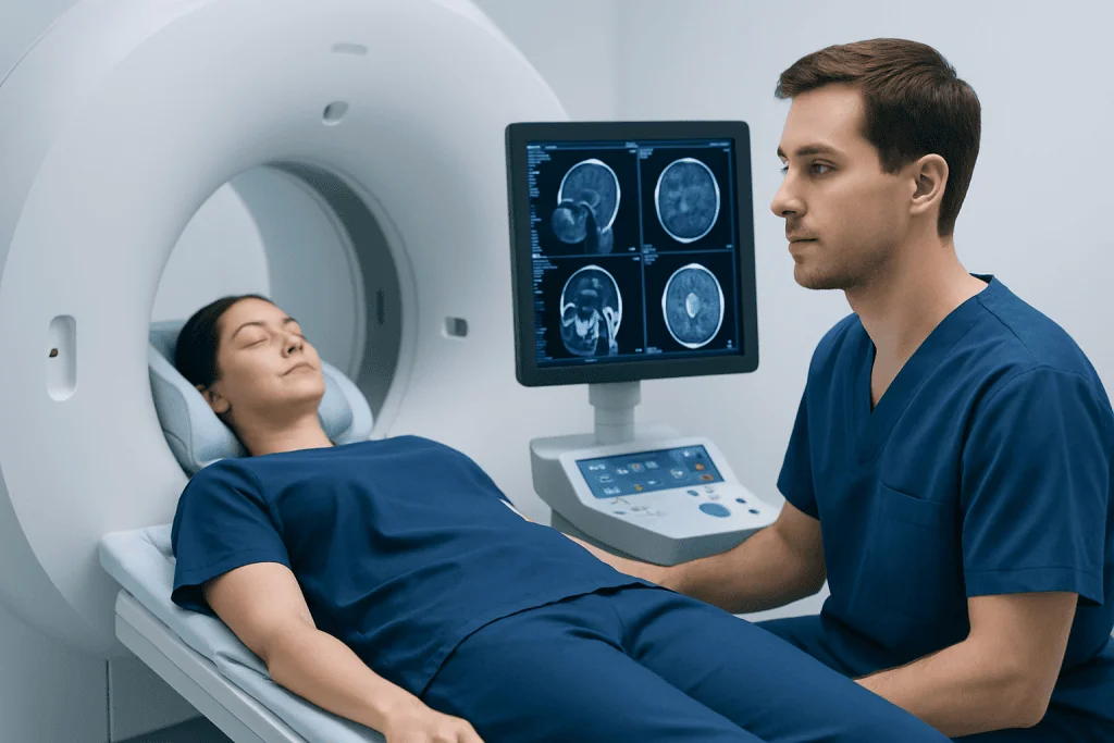
AI can assist diagnosis by helping radiologists identify findings that may be subtle, time-sensitive or easy to overlook in a busy workflow. In some settings, software can highlight suspicious areas on an image, such as a possible fracture on an X-ray, bleeding on a brain CT scan or a small lesion in a lung scan. The radiologist then evaluates whether the highlighted area is clinically meaningful.
Another important use is measurement and comparison. AI tools may help calculate the size of a tumor, estimate organ volumes, quantify calcium in the coronary arteries or compare changes between current and previous scans. This can support more consistent follow-up, especially when patients are monitored over months or years.
AI can also help prioritize imaging studies. If a scan contains features that may require urgent review, such as suspected brain hemorrhage or pulmonary embolism, the system may flag it for earlier attention. This triage function is intended to support workflow organization while keeping final interpretation under medical supervision.
Common areas where AI is being studied or used include chest imaging, breast imaging, stroke imaging, musculoskeletal radiology, cardiovascular imaging and oncology follow-up. The exact use depends on the hospital, the software, local regulations and whether the tool has been validated for that specific clinical purpose.
Common Types of AI Tools Used in Radiology
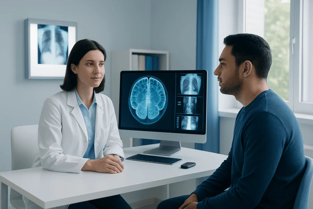
Radiology AI is not a single technology. Different tools are developed for different tasks, and each tool should be assessed for its intended use. Some systems are designed to detect a specific finding, while others support workflow, reporting or quality control.
Common categories include:
- Detection tools: highlight possible abnormalities such as nodules, fractures, bleeding, clots or suspicious breast lesions.
- Segmentation tools: outline organs, tumors or blood vessels to support measurement and treatment planning.
- Quantification tools: calculate volumes, densities, scores or other imaging markers.
- Triage tools: prioritize studies that may contain urgent findings for faster radiologist review.
- Workflow and reporting tools: help organize cases, compare prior imaging or structure reports.
Some AI tools are integrated directly into imaging equipment or radiology reporting systems, while others operate as separate software connected to hospital image archives. In all cases, safe implementation requires trained staff, clear protocols and ongoing monitoring of performance.
Benefits for Patients and Healthcare Teams
When used appropriately, AI may help radiology teams work more efficiently and consistently. Medical imaging volumes continue to grow in many healthcare systems, and radiologists often review complex studies with hundreds or thousands of images. AI can support this process by filtering, organizing and highlighting information that may deserve attention.
For patients, potential benefits may include more efficient image review, improved consistency in measurements and additional support for detecting certain findings. For example, a tool that compares a current CT scan with a previous one may help track whether a lesion has changed. A tool that measures bone alignment or organ volume may reduce variation between repeated evaluations.
For healthcare teams, AI may help standardize reporting in selected areas and support communication between radiologists, referring doctors and treatment teams. In cancer care, cardiovascular care or emergency medicine, clear and timely imaging information can help guide next steps such as additional tests, monitoring or treatment planning.
It is important to understand that benefits depend on careful use. An AI tool that performs well in one hospital, patient group or imaging protocol may not perform the same way in another. This is why radiologists and medical physicists evaluate the technology in real clinical settings before relying on it.
Safety, Accuracy and Human Oversight
AI systems can be highly useful, but they are not perfect. They may miss a finding, mark a normal structure as suspicious or perform less accurately if the image quality is poor. They may also be affected by differences in scanner type, imaging technique, patient anatomy or the population used to train the system.
Human oversight is therefore essential. Radiologists are trained to interpret the whole image in context, not just one highlighted region. They also consider clinical questions, laboratory results, previous reports and whether additional imaging is needed. AI output is one piece of information, not the final medical conclusion.
Safe use also requires attention to privacy and data protection. Medical images contain sensitive health information, so hospitals must follow legal and ethical standards for storage, transfer and analysis. AI systems should be validated, monitored and updated responsibly, with clear accountability for clinical decisions.
Patients may ask whether AI reviewed their images, but they do not usually need to make special preparations for AI-assisted radiology. The imaging examination itself is performed in the usual way. If AI is used, it works in the background as part of the radiology workflow.
What Patients Should Expect From an AI-Assisted Imaging Report
An AI-assisted imaging report is still a radiology report prepared and approved by a radiologist. The report may not always mention AI, because the technology may be used as part of internal workflow support. What matters clinically is that the final interpretation is made by a qualified medical professional.
The report usually describes the type of examination, important findings, comparison with previous studies when available and an impression or conclusion. If an abnormality is found, the radiologist may recommend follow-up imaging, further tests or referral to a specialist. These recommendations should be discussed with the referring doctor, who can connect the imaging result with symptoms and overall health.
Patients can support accurate interpretation by providing relevant information before the scan. This may include previous imaging studies, known diagnoses, surgeries, implants, allergies, pregnancy status when relevant and the reason for the examination. Prior scans are especially helpful because radiologists and AI tools may both benefit from comparison over time.
Limitations and Ethical Considerations
AI in radiology raises important ethical and clinical questions. One concern is bias: if an AI system is trained mainly on images from one population, it may not perform equally well for all age groups, ethnic backgrounds, body types or disease patterns. Responsible implementation requires testing across diverse patient groups and continuous quality review.
Another issue is transparency. Some AI systems are complex, and it may not always be clear why the software produced a particular result. Radiologists must understand the strengths and limitations of the tool and avoid overreliance. Good clinical practice requires balancing AI findings with professional judgment.
There are also practical limitations. AI cannot take a medical history, examine a patient, understand all symptoms or make treatment decisions independently. It cannot replace discussion between the patient and the healthcare team. Its role is best understood as supportive: improving the way information from images is identified, measured and communicated.
When to Speak With a Doctor or Radiology Team
Patients should speak with their referring doctor or radiology team if they have questions about an imaging result, follow-up recommendation or the use of AI in their care. It is reasonable to ask who will review the scan, when the report will be available and whether previous images should be shared for comparison.
Medical imaging should always be requested for a clinical reason. If a patient has symptoms such as persistent pain, unexplained swelling, neurological changes, breathing difficulty or a concerning new lump, a qualified doctor can decide whether imaging is appropriate and which test is most suitable. AI does not determine whether a scan is needed; that decision depends on medical evaluation.
For international patients, Acibadem International provides access to multidisciplinary specialists and JCI-accredited hospitals where radiology and related services are used to diagnose and manage many conditions. As with any medical decision, patients should seek individualized advice from a qualified healthcare professional.
Frequently asked questions
Does artificial intelligence replace the radiologist?
No. AI is used as a support tool, while the radiologist remains responsible for the final interpretation. The radiologist reviews the images in the context of the patient’s history, symptoms and previous examinations.
Can AI diagnose cancer from a scan?
AI may help identify suspicious findings or measure lesions on imaging studies, but it does not make a cancer diagnosis by itself. A diagnosis may require radiologist interpretation, clinical examination, laboratory tests, biopsy or specialist evaluation.
Will patients know if AI was used in their imaging?
In many hospitals, AI works in the background as part of the radiology workflow, and the report may not always state that it was used. Patients can ask their doctor or radiology department whether AI tools are part of the image review process.
Is AI-assisted radiology safe?
AI-assisted radiology can be safe when tools are clinically validated, properly integrated and supervised by qualified professionals. Safety depends on human oversight, data protection, quality control and using the software only for its intended purpose.
Does AI change how an MRI, CT scan or X-ray is performed?
Usually, no. The scan is performed in the standard way, and AI analysis may occur after images are acquired. Patients should follow the same preparation instructions given by the imaging center.
What should patients bring to an imaging appointment?
Patients should bring referral information, previous imaging studies or reports if available, and details about relevant medical conditions, surgeries, implants or allergies. Previous images can be especially helpful for comparison and accurate follow-up.
References
- World Health Organization
- American College of Radiology
- Radiological Society of North America
- European Society of Radiology
- U.S. Food and Drug Administration
This article is for general information only and is not a substitute for professional medical advice. Please consult a qualified doctor about your individual situation.
Explore treatments in Turkey — costs, top hospitals & a free quote
JCI-accredited · board-certified surgeons · reply within 24h
Add us as a Preferred Source to see more of our trusted health content across Google Search, AI Overviews and Discover.

More from the Health Library


Related Specialists

Dt. Gözde Anıl Gürbüz
Oral & Dental Health
Prof. Dr. Nesliar Eser Kutsal
Gastroenterology
Assoc. Prof. Dr. Ersoy Hazneci
Dermatology
Dr. Funda Aybar Susur
Gynecology & Obstetrics