Digital Pathology: How Slide Scanning Supports Faster Diagnostic Review
Digital pathology uses whole slide scanners to create detailed images of tissue, cells or biopsy samples. The diagnosis is still made by a qualified pathologist; technology supports, but does not replace, medical expertise.
Key Takeaways
- Digital pathology uses whole slide scanners to create detailed images of tissue, cells or biopsy samples.
- The diagnosis is still made by a qualified pathologist; technology supports, but does not replace, medical expertise.
- Slide scanning can reduce delays caused by transporting glass slides and can make multidisciplinary review easier.
- Quality control, image security and proper validation are essential for safe clinical use.
- Patients may benefit through more coordinated care, especially when complex cases require second opinions or tumor board discussion.
Digital pathology turns traditional glass microscope slides into high-resolution digital images that can be reviewed securely on specialized screens. This technology can support faster diagnostic review by improving access, collaboration, workflow organization and second opinions.
Overview
Digital pathology is the use of digital technology to capture, store, view and share pathology slides. In traditional pathology, a tissue sample from a biopsy or surgery is processed in a laboratory, placed on a glass slide, stained, and reviewed under a microscope by a pathologist. With digital pathology, that same glass slide can be scanned at very high resolution to create a digital image called a whole slide image.
These images allow pathologists to examine tissue on a secure computer workstation rather than only through a microscope. The pathologist can zoom in and out, move across the tissue, compare slides side by side, and in some settings use software tools that help organize the review. The medical interpretation remains the responsibility of the pathologist, who combines the slide findings with clinical information and other tests.
For patients, digital pathology is important because pathology results often guide major treatment decisions. A biopsy report may help confirm whether a growth is benign or malignant, identify the type of cancer, grade disease activity, or support decisions about surgery, medication, radiotherapy or follow-up. By making slides easier to access and share, digital pathology can support more efficient diagnostic review and better coordination among care teams.
How Slide Scanning Works
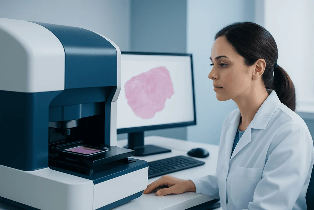
Slide scanning begins after the usual laboratory steps are completed. A sample is fixed, processed, embedded, thinly sliced, placed on a glass slide and stained so that the cells and tissue structures can be seen clearly. The slide is then loaded into a whole slide scanner, a specialized device that captures the slide in many small fields and combines them into a single detailed digital file.
The scanning process is designed to preserve the visual information that a pathologist would normally see through a microscope. Depending on the type of specimen and the clinical question, slides may be scanned at different magnifications. The digital file is then stored in a secure image management system, where authorized users can open it through approved viewing software.
A digital pathology workflow often includes several linked systems. These may include the laboratory information system, the scanner, image storage, the pathologist’s digital viewer, and the hospital’s patient record system. When these systems work together well, the pathologist can access the correct slides, patient information and prior reports more efficiently.
Quality checks are an essential part of scanning. Laboratory staff may review whether the slide is in focus, whether all relevant tissue has been captured, and whether the image is correctly matched to the patient and specimen. If a scan is incomplete or unclear, the slide can be rescanned or reviewed directly under a microscope.
How Digital Pathology Supports Faster Review
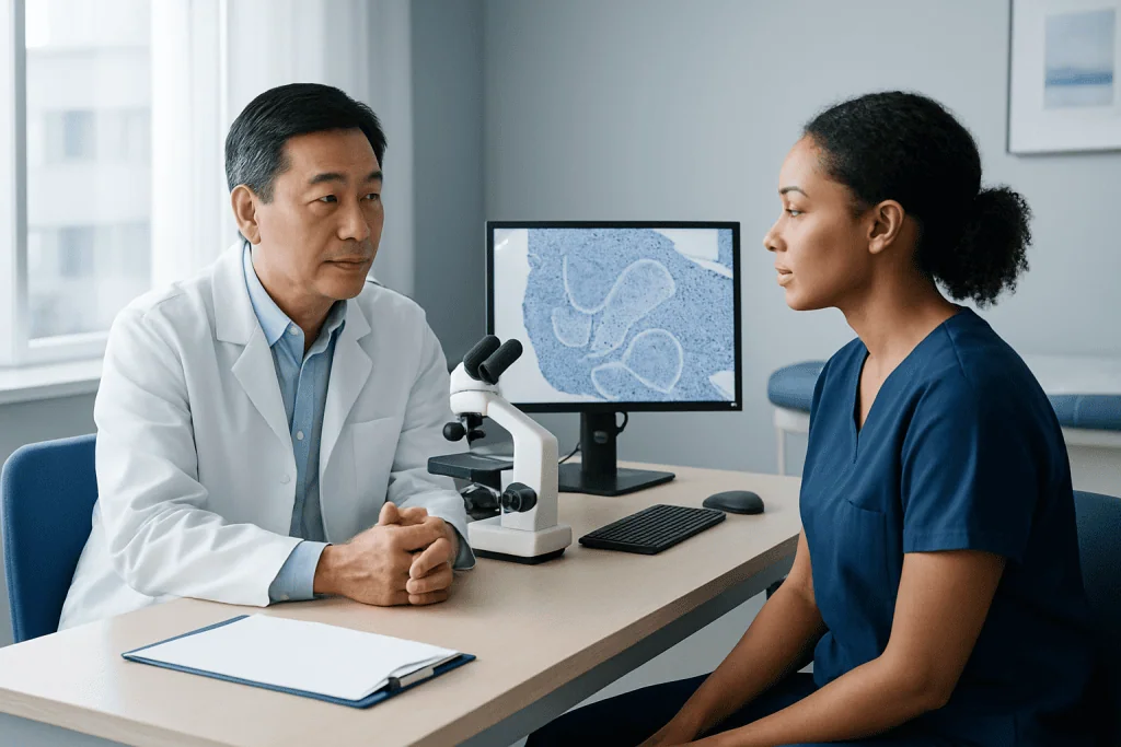
Digital pathology can support faster diagnostic review in several practical ways. One of the most important is access. A glass slide is a physical object that can be in only one place at a time, while a secure digital image can be viewed by authorized specialists in different locations. This may reduce delays when a case needs expert input, multidisciplinary discussion or review by a subspecialist pathologist.
Slide scanning can also improve workflow organization inside the pathology department. Digital systems can help prioritize urgent cases, display case lists, connect slides with clinical details, and allow pathologists to move between cases without waiting for trays of glass slides to be delivered. In large hospitals or networks, this can help distribute cases to the most appropriate available pathologist.
Common ways digital pathology may speed or streamline review include:
- Reducing the need to physically transport slides between buildings or hospitals.
- Allowing remote review by pathologists when permitted by local regulations and validated workflows.
- Supporting rapid second opinions for complex or rare cases.
- Making tumor board and multidisciplinary meeting preparation more efficient.
- Enabling easier comparison with previous biopsies or related specimens.
It is important to understand that digital pathology does not remove the time needed for safe laboratory processing. Tissue preparation, staining, special tests and careful interpretation still take time. The benefit is mainly in reducing avoidable delays in access, coordination and review after the slide is ready.
Accuracy, Safety and Quality Control
Digital pathology must meet strict standards before it is used for clinical diagnosis. Hospitals and laboratories typically validate the scanning system, digital viewer, monitors, workflows and user training before pathologists rely on digital slides for patient care. Validation helps confirm that diagnoses made using digital images are comparable to diagnoses made with traditional microscopy for the intended type of specimens.
Safety depends on more than the scanner alone. The entire process must protect the identity of the patient, ensure the right image is linked to the right case, maintain image quality, and allow pathologists to access glass slides when needed. Some cases may still require microscope review, additional levels of tissue, special stains, immunohistochemistry, molecular testing or other laboratory methods.
Pathologists are trained to recognize when a digital image is not sufficient for a final interpretation. For example, an image may be out of focus in a small area, tissue may be folded, or the stain may need to be repeated. In these situations, the laboratory can repeat the scan, prepare additional slides or use traditional microscopy. This layered approach helps keep patient safety at the center of the diagnostic process.
Digital tools may also include image analysis or artificial intelligence features in selected settings. These tools can assist with tasks such as highlighting areas of interest or supporting certain measurements, but they require careful validation and clinical oversight. They do not replace the pathologist’s judgment or the full clinical context.
Patient Benefits and Common Use Cases
Patients usually do not notice the scanning process directly, but it can influence how their care team works. When digital slides are available, specialists can more easily review the same tissue findings during treatment planning. This is especially useful in cancer care, where pathology may determine tumor type, grade, margins, biomarker testing and whether additional treatment is needed.
Digital pathology can also support second opinions. In some situations, a patient’s doctor may request review by a pathologist with expertise in a specific organ system, such as breast, prostate, lung, gastrointestinal, skin, bone marrow or brain pathology. Instead of sending the only glass slide set by courier, the laboratory may be able to share secure digital images, depending on institutional policy and local regulations.
Multidisciplinary teams may benefit as well. During tumor boards or complex case meetings, radiologists, surgeons, oncologists, pathologists and other specialists can discuss imaging, laboratory results and digital slide findings together. This shared view can make communication clearer and help ensure that treatment decisions are based on a complete understanding of the diagnosis.
Digital archives may also make future comparisons easier. If a patient has a new biopsy months or years later, the pathologist may be able to compare current digital slides with previous images. This can be helpful when doctors need to understand whether a finding is new, recurring, progressing or related to an earlier condition.
Limitations, Privacy and What Patients Should Know
Although digital pathology is a powerful technology, it has limitations. Large image files require secure storage, fast networks and reliable software. Not every laboratory has the same level of digital infrastructure, and some institutions may use digital pathology for selected case types rather than for all diagnoses. Regulations may also differ between countries and healthcare systems.
Privacy is a key concern in any digital health system. Digital pathology images are part of a patient’s medical record and must be protected with appropriate access controls, secure storage and professional confidentiality. Authorized sharing for clinical care should follow hospital policies, consent requirements where applicable, and legal data protection standards.
Patients may ask whether a digital slide is as reliable as a glass slide. In properly validated systems, digital slides can be used safely for many diagnostic purposes, but the suitability depends on the specimen type, image quality, clinical question and local approvals. If a pathologist believes that microscope review or additional testing is needed, that step remains available.
Another important point is that faster review does not always mean an immediate result. Some diagnoses require extra stains, molecular tests, correlation with imaging, or consultation between specialists. Digital pathology can help reduce logistical delays, but careful diagnosis is still a detailed medical process.
What Patients Can Expect and When to Ask Questions
From a patient’s perspective, the biopsy or surgery experience is usually the same whether the pathology department uses microscopes, digital slides or both. After the sample is collected, the laboratory prepares it and the pathologist reviews it. The final report is sent to the treating doctor, who explains the result and discusses the next steps.
Patients may wish to ask their doctor when the pathology report is expected, whether additional tests are being performed, and whether a second opinion is recommended. If treatment decisions are complex, it is reasonable to ask whether the case will be discussed in a multidisciplinary meeting. For international patients, asking how slides and reports can be shared securely between hospitals may also be helpful.
Patients should contact their healthcare team if results are delayed beyond the expected timeframe or if they do not understand the wording of a pathology report. Medical terms such as “atypia,” “dysplasia,” “grade,” “margin,” “invasive,” or “benign” can be confusing, and the treating doctor can explain what they mean in the context of the patient’s condition.
Acibadem International’s multidisciplinary specialists and JCI-accredited hospitals diagnose and treat many conditions that require pathology review for international patients. When digital pathology is part of the care pathway, it is used to support accurate communication, coordinated review and safe clinical decision-making.
Frequently asked questions
What is digital pathology?
Digital pathology is the use of technology to scan traditional glass pathology slides and create high-resolution digital images. These images can be viewed on secure computer systems by trained pathologists. The diagnosis is still made by a medical specialist, not by the scanner alone.
Does digital pathology make biopsy results faster?
It can help reduce delays related to slide transport, case distribution and specialist review. However, the sample still needs proper laboratory processing, staining and careful interpretation. Some cases also require additional tests, so timing depends on the complexity of the diagnosis.
Is a scanned slide as accurate as a microscope slide?
When a digital pathology system is properly validated and used for appropriate case types, it can support reliable diagnostic review. Quality control is essential, including checking focus, image completeness and correct case identification. If needed, the pathologist can still review the original glass slide under a microscope.
Can digital pathology be used for a second opinion?
Yes, in many settings digital slides can make second opinions easier because images can be shared securely with authorized specialists. This depends on hospital policy, local regulations and the quality of the scanned images. The final opinion should always come from a qualified pathologist.
Does artificial intelligence diagnose disease in digital pathology?
Artificial intelligence may assist with selected tasks, such as measurements or highlighting areas that need attention. It does not replace the pathologist’s medical judgment. Any AI tool used in clinical care should be validated and supervised within a safe diagnostic workflow.
Are digital pathology images private?
Digital pathology images are part of the patient’s medical information and should be protected like other health records. Hospitals use access controls, secure storage and confidentiality rules to limit who can view them. Patients can ask their care team how slides and reports are shared, especially when care involves more than one hospital.
References
- College of American Pathologists
- Royal College of Pathologists
- U.S. Food and Drug Administration
- Digital Pathology Association
- World Health Organization
This article is for general information only and is not a substitute for professional medical advice. Please consult a qualified doctor about your individual situation.
Oncology care in Turkey — second opinion and treatment plan
JCI-accredited · board-certified surgeons · reply within 24h
Add us as a Preferred Source to see more of our trusted health content across Google Search, AI Overviews and Discover.