

Evoked Potential Tests: How Nerve Pathways Are Evaluated

Evoked potential tests measure the nervous system response to controlled stimulation, such as light patterns, sounds or mild electrical pulses. The main types are visual evoked potentials, brainstem auditory evoked potentials, somatosensory evoked potentials and motor evoked potentials.
Key Takeaways
- Evoked potential tests measure the nervous system response to controlled stimulation, such as light patterns, sounds or mild electrical pulses.
- The main types are visual evoked potentials, brainstem auditory evoked potentials, somatosensory evoked potentials and motor evoked potentials.
- These tests are usually painless, do not use radiation and are often performed with small recording electrodes placed on the scalp or skin.
- Results help show whether nerve signals are delayed, reduced or interrupted, but they are usually interpreted together with symptoms, examination findings and imaging.
- Preparation is simple, but patients should tell the medical team about implanted devices, seizure history, medications, hearing or vision problems and skin sensitivity.
Evoked potential tests are noninvasive neurophysiology tests that record how quickly and effectively signals travel through specific nerve pathways. They can help doctors investigate symptoms affecting vision, hearing, sensation, movement, the spinal cord or the brain.
Overview
Evoked potential tests are diagnostic tests that evaluate how the brain, spinal cord and peripheral nerves respond to specific stimulation. The word evoked means that the response is produced by a stimulus, such as a flashing visual pattern, a clicking sound or a mild electrical pulse applied to the skin. Sensitive recording electrodes then detect the tiny electrical signals generated by the nervous system.
Doctors use these tests to assess the function of nerve pathways, especially when symptoms suggest that signals may not be traveling normally. A pathway can be affected by inflammation, compression, injury, demyelination, reduced blood flow, tumors or other neurological conditions. Evoked potential testing can sometimes detect changes that are not obvious on a routine neurological examination.
These tests are generally safe and noninvasive. Most are performed in a neurophysiology or neurology laboratory and do not require hospital admission. The information they provide is functional, meaning it shows how well a pathway is working, while tests such as MRI show detailed anatomy.
Types of Evoked Potential Tests
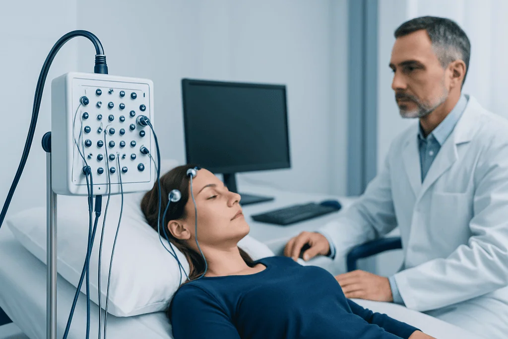
Different evoked potential tests evaluate different parts of the nervous system. The choice depends on the patient’s symptoms, medical history and the clinical question the doctor needs to answer. Sometimes more than one type is performed during the same visit to compare several pathways.
- Visual evoked potential (VEP): evaluates the pathway from the eyes through the optic nerves to the visual areas of the brain. It is commonly performed while the patient looks at a changing checkerboard pattern or flashing light.
- Brainstem auditory evoked potential (BAEP or BAER): evaluates the hearing nerve and brainstem pathways using clicking sounds delivered through earphones.
- Somatosensory evoked potential (SSEP): evaluates sensory pathways from the arms or legs through the spinal cord to the brain. A mild electrical stimulus is usually applied at the wrist, ankle or another peripheral nerve location.
- Motor evoked potential (MEP): evaluates motor pathways that carry signals from the brain to muscles. It may use magnetic or electrical stimulation and is often used in specialized settings, including some surgical monitoring.
In some cases, evoked potentials are used during surgery to monitor nerve pathway function in real time. This is called intraoperative neurophysiological monitoring. It can help surgical teams observe important pathways while operating near the brain, spinal cord or major nerves.
When Evoked Potential Tests May Be Recommended

A doctor may recommend evoked potential testing when there are symptoms that could reflect a problem along a nerve pathway. These may include blurred or reduced vision, episodes of vision loss, unexplained hearing symptoms, numbness, tingling, weakness, balance problems, walking difficulty or symptoms suggesting spinal cord involvement. The test may also be useful when a patient cannot clearly describe symptoms, such as in very young children or people with communication difficulties.
Visual evoked potentials may be used when optic nerve function needs to be assessed, including after suspected optic neuritis or in the evaluation of demyelinating diseases such as multiple sclerosis. Brainstem auditory evoked potentials can help evaluate the hearing nerve and brainstem, especially when standard hearing tests are incomplete or when a lesion along the auditory pathway is suspected. Somatosensory evoked potentials can help assess spinal cord and sensory pathway function in selected neurological and spine conditions.
Evoked potential tests do not diagnose every neurological condition by themselves. Instead, they provide one piece of evidence. Doctors interpret the findings alongside the person’s symptoms, neurological examination, blood tests, imaging studies and other tests such as electroencephalography or nerve conduction studies when appropriate.
How to Prepare for the Test
Preparation is usually simple. Patients are often advised to wash their hair before the appointment and avoid hair oils, gels or sprays because these can interfere with electrode contact. Comfortable clothing is helpful, especially if electrodes need to be placed on the arms, legs, neck or back. The medical team will provide specific instructions if the test is being performed as part of surgical monitoring or in a hospital setting.
Patients should bring glasses or contact lenses if they normally use them, because visual evoked potential testing often requires the best possible corrected vision. For auditory testing, the team should be told about hearing aids, ear infections, earwax problems or known hearing loss. These details help the examiner choose appropriate test settings and interpret the results accurately.
It is important to tell the doctor or technologist about implanted medical devices such as pacemakers, cochlear implants, deep brain stimulators or spinal cord stimulators. A history of seizures, pregnancy, metal implants, recent surgery, skin conditions or medication changes should also be discussed. These details are especially important for tests that use magnetic or electrical stimulation, such as motor evoked potentials.
What Happens During Evoked Potential Testing
During the test, the patient usually sits in a chair or lies on an examination couch. Small electrodes are placed on the scalp and sometimes on the skin over the spine, neck, arms or legs. The skin may be gently cleaned first to improve recording quality. The electrodes record electrical activity; they do not deliver strong current during most recording steps.
For a visual evoked potential test, the patient is asked to focus on a screen showing a reversing checkerboard pattern or to look at flashing lights. Each eye may be tested separately. For a brainstem auditory evoked potential test, clicking sounds are played through earphones while the responses are recorded. For a somatosensory evoked potential test, mild repetitive electrical pulses are delivered to a nerve in the wrist, ankle or another area; the sensation may feel like tapping or tingling.
The length of the appointment varies depending on how many pathways are being tested, but many studies take about 30 to 90 minutes. Patients are usually awake and can return to normal activities afterward unless they have received sedation, which is uncommon and mainly considered for some children or patients who cannot remain still. The technologist may repeat parts of the test to obtain clear, reliable recordings.
Understanding Results and Limitations
Evoked potential results are analyzed by measuring timing and signal size. Timing is often described as latency, which means how long it takes for the nervous system response to appear after the stimulus. A delayed response may suggest slowed signal conduction, which can occur when myelin, the protective coating around nerve fibers, is affected. A reduced or absent response may suggest more significant pathway dysfunction, although technical factors must also be considered.
Doctors compare the patient’s results with expected values and may also compare the right and left sides of the body. For example, a delay in one optic nerve pathway may support the presence of optic nerve involvement. In SSEP testing, abnormal responses can help localize whether a sensory pathway problem may be in the peripheral nerve, spinal cord, brainstem or brain, depending on the pattern of findings.
Like all diagnostic tests, evoked potentials have limitations. A normal result does not always rule out disease, especially if symptoms are intermittent, mild or outside the pathway being tested. An abnormal result does not automatically identify the exact cause. This is why the result should be explained by a qualified clinician who understands the patient’s full medical picture.
Safety, Aftercare and When to See a Doctor
Evoked potential tests are generally low risk. Standard VEP, BAEP and SSEP tests do not use radiation and do not involve injections. Some people may notice mild skin irritation where electrodes were placed, temporary fatigue from concentrating, or brief discomfort from the mild electrical stimulation used in SSEP testing. These effects usually pass quickly.
Patients should seek medical advice promptly if they experience new or worsening neurological symptoms such as sudden vision loss, new weakness, difficulty speaking, severe imbalance, loss of bladder or bowel control, or rapidly spreading numbness. These symptoms require clinical evaluation and should not wait for a routine test appointment. People who already have a neurologist should also ask when follow-up is needed after results are available.
At Acibadem International, multidisciplinary specialists in JCI-accredited hospitals diagnose and treat neurological and sensory pathway disorders for international patients, using evoked potential testing when it is clinically appropriate. Patients should discuss their individual results with their treating doctor, who can explain what the findings mean and whether additional tests or treatment are needed.
Frequently asked questions
Are evoked potential tests painful?
Most evoked potential tests are not painful. Visual and auditory tests involve looking at patterns or listening to clicks, while somatosensory testing uses mild electrical pulses that may feel like tapping or tingling. Patients should tell the technologist if they feel uncomfortable during the test.
What is the difference between an evoked potential test and an EEG?
An EEG records spontaneous electrical activity of the brain, often to evaluate seizures or changes in consciousness. Evoked potential tests record the nervous system response to a specific stimulus, such as light, sound or mild electrical stimulation. Both use electrodes, but they answer different clinical questions.
Can evoked potential tests diagnose multiple sclerosis?
Evoked potential tests can show slowed signal conduction that may support the evaluation of multiple sclerosis, especially when optic nerve or spinal cord pathways are involved. However, they do not diagnose multiple sclerosis on their own. Doctors usually combine symptoms, neurological examination, MRI findings and laboratory tests before making a diagnosis.
How long does it take to get results?
The recording may be completed during the appointment, but the results usually need to be reviewed and interpreted by a specialist. Timing varies by clinic and by the complexity of the test. The doctor who requested the test will typically explain the findings and next steps.
Can children have evoked potential tests?
Yes, children can have evoked potential tests when there is a medical reason to evaluate vision, hearing, sensory pathways or neurological function. The test is adapted to the child’s age and ability to cooperate. In some cases, sedation may be considered, but this is decided carefully by the medical team.
Do patients need to stop medications before the test?
Patients should not stop prescribed medications unless their doctor specifically tells them to do so. Some medications, sleepiness or medical conditions can influence certain recordings, so the team should be informed about all regular medicines and supplements. The ordering doctor will provide individualized instructions if any changes are needed.
References
- American Clinical Neurophysiology Society
- International Federation of Clinical Neurophysiology
- National Institute of Neurological Disorders and Stroke
- American Academy of Neurology
- Mayo Clinic
This article is for general information only and is not a substitute for professional medical advice. Please consult a qualified doctor about your individual situation.
Joint and spine care in Turkey — expert assessment & treatment
JCI-accredited · board-certified surgeons · reply within 24h

More from the Health Library



Related Specialists

Prof. Dr. Ercan Karaarslan
Radiology
Prof. Dr. Bora Peynircioğlu
Interventional Radiology
Prof. Dr. Koray Güven
Interventional Radiology
Assoc. Prof. Dr. Deniz Can Aliş
Radiology