

Cerebral Angiography: What to Expect During Brain Vessel Imaging

Cerebral angiography uses a thin catheter, contrast dye, and X-ray imaging to create detailed pictures of blood flow in the brain. The test may be recommended when CT, MRI, or ultrasound findings need confirmation or when treatment planning requires highly detailed vessel images.
Key Takeaways
- Cerebral angiography uses a thin catheter, contrast dye, and X-ray imaging to create detailed pictures of blood flow in the brain.
- The test may be recommended when CT, MRI, or ultrasound findings need confirmation or when treatment planning requires highly detailed vessel images.
- Most people receive local anesthesia and mild sedation, remain awake but comfortable, and go home the same day or after short observation.
- Preparation includes reviewing medicines, allergies, kidney function, pregnancy status, and instructions about eating or drinking before the procedure.
- Serious complications are uncommon, but patients should know warning signs such as worsening headache, weakness, speech changes, bleeding, or fever.
Cerebral angiography is a detailed imaging test that helps doctors see the arteries and veins of the brain. It is often used when precise information is needed to diagnose or plan treatment for conditions such as aneurysms, narrowed vessels, clots, or abnormal blood vessel connections.
Overview
Cerebral angiography, also called a brain angiogram or cerebral catheter angiography, is a medical imaging procedure that shows blood vessels in and around the brain. During the test, a doctor guides a very thin flexible tube, called a catheter, through an artery and injects a contrast dye. X-ray images are then taken as the dye moves through the brain’s arteries and veins.
This procedure is often performed with a technique called digital subtraction angiography, or DSA. DSA uses computer processing to remove background structures from the image, allowing the blood vessels to stand out clearly. Because it provides very fine detail, cerebral angiography is sometimes considered the reference test for evaluating certain brain vessel conditions.
Unlike a CT angiogram or MR angiogram, which are noninvasive scans, catheter angiography places a catheter inside a blood vessel. This makes it more invasive, but it also allows doctors to see blood flow in real time and, in selected cases, perform treatment during the same session. The decision to recommend the procedure is based on the patient’s symptoms, previous imaging, medical history, and the information needed for safe care planning.
Why Cerebral Angiography Is Performed
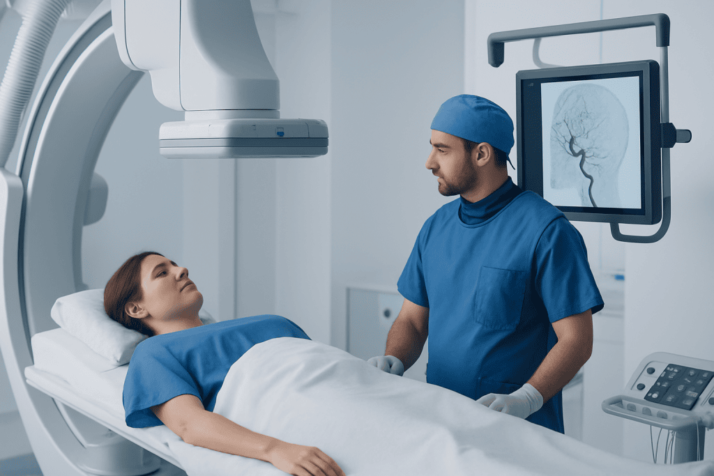
A doctor may recommend cerebral angiography when detailed information is needed about the size, shape, location, or blood flow pattern of brain blood vessels. It can help clarify findings from CT, MRI, ultrasound, or other tests. It may also be used before surgery or endovascular treatment to map vessels carefully and reduce uncertainty.
Common reasons for a brain angiogram include suspected or known aneurysm, arteriovenous malformation, arteriovenous fistula, narrowing of an artery, blockage, blood clot, inflammation of blood vessels, or unusual bleeding in or around the brain. In some patients, it is used to investigate symptoms such as certain types of stroke, transient ischemic attack, unexplained bleeding, or complex vascular headaches when other tests suggest a vessel-related cause.
Cerebral angiography can also support treatment decisions. For example, if an aneurysm is found, the images may help determine whether monitoring, surgery, or an endovascular approach is most appropriate. If a vessel is narrowed or blocked, angiography may help doctors assess the location and severity and decide whether medication, monitoring, or a procedure should be considered.
Preparation Before the Procedure

Before cerebral angiography, the care team reviews the patient’s medical history, current medicines, allergies, kidney function, and previous reactions to contrast dye. Patients should tell the team if they take blood thinners, diabetes medicines, anti-inflammatory medicines, or supplements, because some may need to be adjusted. No medication should be stopped unless the prescribing doctor or procedure team advises it.
Patients are usually asked not to eat or drink for a period of time before the test, especially if sedation will be used. Blood tests may be ordered to check kidney function and blood clotting. The team may also ask about pregnancy or breastfeeding, because imaging and contrast decisions may need special planning.
It is helpful to arrange transportation home, since sedation and post-procedure observation can make driving unsafe. Patients may be asked to remove jewelry, glasses, dentures, or metal items before entering the angiography suite. A nurse or doctor will explain the consent form, including the reason for the procedure, expected benefits, and possible risks, and patients are encouraged to ask questions before signing.
- Bring a list of medicines and allergies.
- Follow instructions about fasting and morning medicines.
- Tell the team about kidney disease, asthma, prior contrast reactions, bleeding problems, or recent illness.
- Plan for several hours at the hospital, including preparation and recovery time.
What Happens During Cerebral Angiography
Cerebral angiography is performed in a specialized imaging room by an interventional neuroradiologist, neurosurgeon, or other trained vascular specialist, supported by nurses and technologists. The patient lies on an X-ray table, and monitors are used to check heart rate, blood pressure, oxygen level, and sometimes heart rhythm. An intravenous line is placed, and mild sedation may be given to help the patient relax while remaining able to follow instructions.
The catheter is most commonly inserted through an artery in the groin or wrist. The skin is cleaned carefully, sterile drapes are placed, and local anesthetic is injected to numb the area. The patient may feel pressure at the puncture site, but sharp pain should be reported immediately. The doctor then advances the catheter through the blood vessels toward the arteries supplying the brain, using X-ray guidance.
When the catheter is in position, contrast dye is injected and rapid X-ray images are taken. Some patients feel a brief warm sensation, flushing, metallic taste, or pressure in the head or neck during the injection; these feelings usually pass quickly. The patient may be asked to stay very still or hold their breath briefly so the images are clear. Several injections may be needed from different catheter positions to view all relevant vessels.
If the angiogram is diagnostic only, the catheter is removed after imaging is complete. Pressure, a closure device, or both may be used to seal the puncture site. In selected situations, the angiography may be combined with treatment, such as placing coils, stents, or other devices, but this is discussed in advance unless urgent circumstances require immediate action.
After the Test and Recovery
After the procedure, the patient is moved to a recovery area for observation. Nurses check the puncture site, pulse, blood pressure, and neurological status, including speech, strength, and alertness. If the catheter was placed through the groin, the patient may need to lie flat for a period of time to reduce the risk of bleeding. If the wrist was used, a compression band may be applied and gradually loosened.
Most patients can eat and drink once fully awake, unless another procedure or medical condition requires different instructions. Drinking fluids may help the body clear the contrast dye, but patients with kidney or heart conditions should follow their doctor’s specific guidance. Mild soreness, bruising, or tenderness at the puncture site can occur and usually improves over the next few days.
Before discharge, the care team explains activity restrictions and warning signs. Patients are commonly advised to avoid heavy lifting, strenuous exercise, or soaking the puncture site for a short time. They should keep the site clean and dry as directed. A responsible adult should accompany the patient home if sedation was used, and important decisions, alcohol, and driving are usually avoided for the rest of the day.
Benefits, Risks, and Safety Considerations
The main benefit of cerebral angiography is the high level of detail it provides. It can show small aneurysms, vessel narrowing, abnormal connections, and blood flow patterns that may not be fully visible on other imaging tests. This information can be especially valuable when doctors are deciding whether treatment is needed and which approach may be safest.
As with any invasive procedure, cerebral angiography has possible risks. These include bleeding or bruising at the catheter site, infection, injury to a blood vessel, allergic reaction to contrast dye, kidney strain from contrast, and, rarely, stroke or seizure. The overall risk depends on factors such as age, vascular condition, kidney function, medications, and the complexity of the procedure.
Safety steps are taken before, during, and after the procedure to reduce these risks. The team screens for contrast allergies and kidney problems, uses sterile technique, monitors the patient closely, and applies careful pressure or closure at the access site. Radiation exposure is kept as low as reasonably possible while still obtaining the images needed for diagnosis and treatment planning.
Patients can support safety by sharing complete medical information and reporting symptoms promptly during or after the test. Sudden severe headache, new weakness or numbness, trouble speaking, vision changes, chest pain, shortness of breath, increasing swelling at the puncture site, ongoing bleeding, fever, or worsening pain should be treated as urgent medical concerns.
Understanding Results and Next Steps
In many cases, the doctor performing the angiogram can describe the main findings shortly after the procedure. A formal report is then prepared and shared with the referring doctor. The report may describe whether blood vessels are normal or whether there is an aneurysm, narrowing, blockage, malformation, fistula, dissection, vasculitis pattern, or other abnormality.
Next steps depend on the results and the patient’s overall health. Some findings may only need observation and follow-up imaging. Others may require medication, lifestyle risk reduction, surgery, endovascular treatment, or urgent care if the condition is actively causing symptoms. The care team will explain the options, expected benefits, possible risks, and alternatives in a way that supports shared decision-making.
Patients should ask for clear instructions about when they will receive final results, which doctor will review them, and whether follow-up appointments or additional imaging are needed. International patients may benefit from having imaging files and reports translated or shared digitally with their medical team. Acibadem International’s multidisciplinary specialists and JCI-accredited hospitals diagnose and treat brain vascular conditions for international patients, including when advanced imaging and coordinated follow-up are needed.
When to See a Doctor
A doctor should be consulted if a patient has been told that brain vessel imaging is needed, or if previous CT or MRI results mention an aneurysm, vascular malformation, vessel narrowing, or unexplained bleeding. Cerebral angiography is not performed for routine screening in most people; it is chosen when the expected diagnostic value is important for care decisions.
Urgent medical attention is needed for symptoms that may suggest stroke or bleeding in the brain, such as sudden weakness or numbness on one side of the body, difficulty speaking, sudden vision loss, confusion, loss of balance, a seizure, or a sudden severe headache unlike previous headaches. These symptoms do not always mean that angiography will be required, but they do require immediate medical evaluation.
After cerebral angiography, patients should contact their care team or seek urgent care if they develop new neurological symptoms, persistent vomiting, fever, increasing redness or drainage at the puncture site, a growing lump, uncontrolled bleeding, coldness or color change in the affected limb, or pain that is worsening instead of improving. Prompt assessment helps the team manage complications early if they occur.
Frequently asked questions
Is cerebral angiography painful?
Most patients do not describe the procedure as painful. Local anesthetic is used to numb the catheter insertion area, and mild sedation may be given for comfort. Patients may feel pressure at the access site or brief warmth when contrast dye is injected.
How long does a brain angiogram take?
The imaging portion often takes less than one to two hours, but the total hospital visit is longer because of preparation and recovery. Patients should expect to spend several hours in the medical facility. More complex cases or procedures combined with treatment may take longer.
What is the difference between CT angiography and cerebral catheter angiography?
CT angiography is a noninvasive scan performed through an intravenous contrast injection, while cerebral catheter angiography uses a catheter placed inside an artery. Catheter angiography provides very detailed, real-time images of blood flow and may allow treatment during the same session. The best test depends on the clinical question and the patient’s condition.
Can a patient go home the same day after cerebral angiography?
Many patients go home the same day after a diagnostic cerebral angiogram, once they are stable and the puncture site is safe. Some patients may need longer observation, especially if treatment was performed, if symptoms are present, or if there are medical risk factors. The care team gives individualized discharge instructions.
Is contrast dye safe for everyone?
Contrast dye is safely used in many patients, but it requires extra caution in people with kidney disease, prior contrast reactions, certain allergies, or specific medical conditions. Before the procedure, the team reviews risk factors and may order blood tests or take preventive steps. Patients should report any previous reaction to contrast dye.
What should patients avoid after cerebral angiography?
Patients are usually advised to avoid heavy lifting, strenuous activity, and soaking the puncture site for a short period. If sedation was used, driving, alcohol, and important decisions are typically avoided for the rest of the day. The exact restrictions depend on the access site and the doctor’s instructions.
When are cerebral angiography results available?
Preliminary findings may be discussed soon after the procedure, especially if they affect immediate care. A formal report is usually prepared after the images are reviewed in detail. Patients should ask who will explain the final results and when follow-up is needed.
References
- American College of Radiology
- Radiological Society of North America
- Society of NeuroInterventional Surgery
- American Heart Association
- National Institute of Neurological Disorders and Stroke
This article is for general information only and is not a substitute for professional medical advice. Please consult a qualified doctor about your individual situation.
Explore treatments in Turkey — costs, top hospitals & a free quote
JCI-accredited · board-certified surgeons · reply within 24h
Check your numbers in seconds
BMI, calories, due date, blood pressure and 30+ more clinical calculators — free, instant, doctor-reviewed ranges.

More from the Health Library



Related Specialists

Prof. Dr. Ercan Karaarslan
Radiology
Prof. Dr. Bora Peynircioğlu
Interventional Radiology
Prof. Dr. Koray Güven
Interventional Radiology
Assoc. Prof. Dr. Deniz Can Aliş
Radiology