Cerebral Angiography for Diagnosis: Step by Step

Cerebral angiography uses contrast dye and X-ray imaging to show brain blood vessels clearly. It is often used when doctors need more detailed information than CT angiography or MR angiography can provide.
Key Takeaways
- Cerebral angiography uses contrast dye and X-ray imaging to show brain blood vessels clearly.
- It is often used when doctors need more detailed information than CT angiography or MR angiography can provide.
- The test is usually done through a thin catheter inserted into an artery, commonly in the wrist or groin.
- Most people go home the same day, but follow-up instructions and short-term activity limits are important.
- Although generally safe, cerebral angiography carries small risks such as bleeding, allergy to contrast, stroke, or kidney strain.
Cerebral angiography is a specialized imaging procedure used to examine the blood vessels of the brain in great detail. It can help doctors diagnose aneurysms, vessel narrowing, arteriovenous malformations, and other conditions that may not be fully visible on standard scans.
Overview: What Cerebral Angiography Is
Cerebral angiography, sometimes called a cerebral angiogram or brain angiography, is a diagnostic procedure that creates detailed images of the blood vessels in the brain and neck. It is considered one of the most precise ways to evaluate how blood flows through arteries and veins that supply the brain.
During the procedure, a doctor guides a thin tube called a catheter into a blood vessel and injects contrast dye. X-ray images are then taken in rapid sequence, allowing specialists to see the shape of the vessels, areas of narrowing or blockage, abnormal connections, or balloon-like weak spots called aneurysms.
This test may be recommended after symptoms such as sudden severe headache, stroke-like symptoms, unexplained bleeding in the brain, or when another scan suggests a blood vessel problem. It is also commonly used to plan treatment for conditions such as brain aneurysm or arteriovenous malformation.
Why Doctors Recommend It
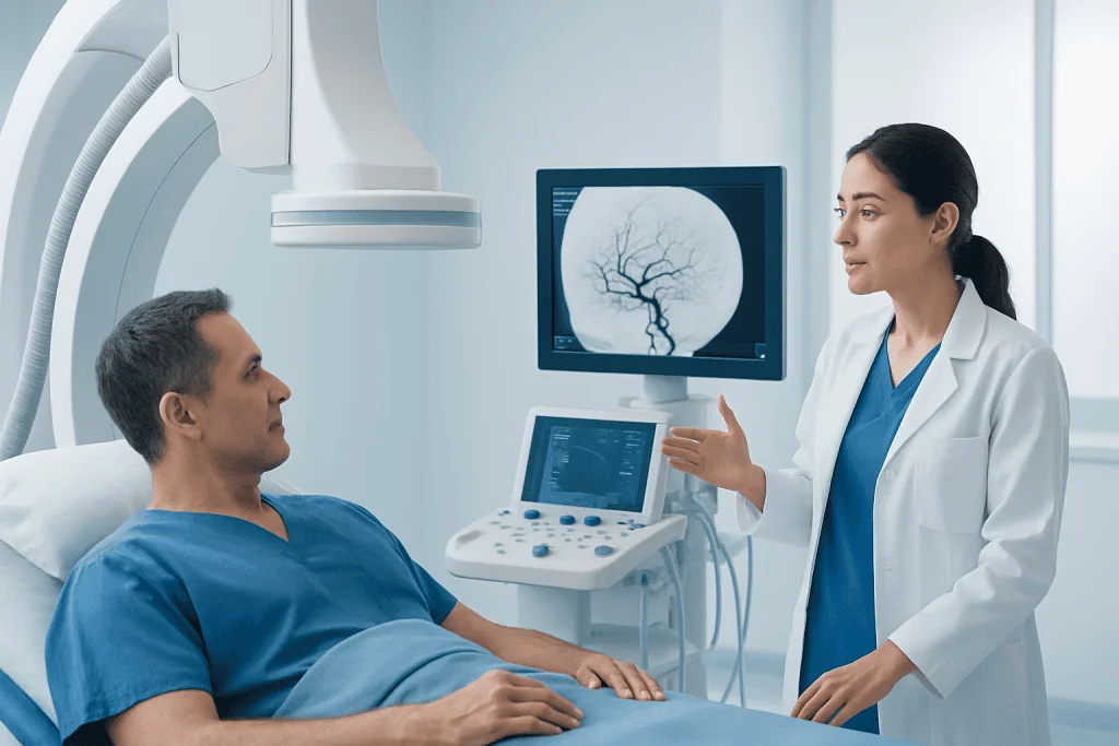
Doctors usually order cerebral angiography when they need more exact information than noninvasive imaging can provide. CT angiography and MR angiography are very useful, but catheter-based angiography can show smaller vessels and more subtle abnormalities with greater detail.
The test may help diagnose several conditions affecting the brain’s circulation. These include aneurysms, arteriovenous malformations, vessel narrowing from atherosclerosis, vasculitis, dissections, and some causes of stroke or transient ischemic attack.
In some situations, cerebral angiography is not only diagnostic but also the first step toward treatment. If an abnormality is found, the same session may sometimes lead into an interventional procedure such as aneurysm treatment or embolization when this has been planned in advance and is clinically appropriate.
- To investigate suspected aneurysm or vascular malformation
- To identify the source of brain bleeding
- To evaluate vessel narrowing or blockage
- To clarify the cause of certain strokes
- To help plan surgery or endovascular treatment
How to Prepare Before the Procedure

Preparation begins with a review of medical history, current symptoms, allergies, and medicines. Patients should tell their care team if they have had a reaction to contrast dye before, if they have kidney disease, diabetes, a bleeding disorder, or if they may be pregnant.
Blood tests may be ordered to check kidney function and clotting. The medical team may advise stopping or adjusting certain medicines, especially blood thinners, before the procedure. It is important not to stop prescription medication without clear instructions from the treating doctor.
Most patients are asked not to eat or drink for several hours beforehand. On the day of the test, they typically change into a hospital gown, have an intravenous line placed, and discuss the procedure again with the doctor, including its benefits, possible risks, and what recovery will involve.
Cerebral Angiography Step by Step
The procedure is usually performed in a specialized angiography suite by an interventional neuroradiologist, interventional neurologist, or another trained physician. The patient lies on an X-ray table while the team monitors heart rate, blood pressure, and oxygen levels. Sedation may be given to help with comfort, though many patients remain awake and able to follow instructions.
After cleaning the skin, the doctor numbs the access site, most often at the wrist or groin. A small sheath is placed into the artery, and a catheter is carefully guided through the blood vessels toward the arteries that supply the brain. This part is usually felt as pressure rather than pain.
Contrast dye is then injected through the catheter while a series of X-ray images is taken. Some people notice a brief warm sensation, a metallic taste, or a flushing feeling when the dye is injected. The doctor may ask the patient to stay very still or hold their breath for a moment during image capture.
When all necessary images are obtained, the catheter is removed and pressure or a closure device is used to reduce bleeding at the insertion site. In selected cases, if a treatable vascular problem is confirmed, the team may proceed with a planned endovascular procedure such as stroke treatment or another catheter-based therapy.
What the Results Can Show
Cerebral angiography provides a detailed map of the brain’s circulation. It can show whether blood vessels are narrowed, blocked, enlarged, abnormally connected, or leaking. This level of detail helps doctors decide whether monitoring, medication, surgery, or endovascular therapy is the best next step.
Findings may include aneurysms, vascular malformations, vasospasm after bleeding, carotid or intracranial stenosis, vessel dissection, or unusual tumor blood supply. In some patients, the test helps explain symptoms even when earlier imaging has been inconclusive.
The results are interpreted together with the patient’s history, neurological examination, and other imaging studies. A normal angiogram can also be helpful because it may rule out serious vascular conditions and guide the care team toward other possible causes of symptoms.
Risks, Safety, and Recovery
Cerebral angiography is widely used and is generally safe when performed by experienced teams. Still, like any invasive test, it has possible risks. These include bruising or bleeding at the catheter site, allergic reaction to contrast dye, temporary kidney strain, blood vessel injury, infection, and, rarely, stroke.
After the procedure, patients are monitored for several hours. The team checks the access site, blood pressure, and neurological status. Drinking fluids may be encouraged if appropriate, as this can help the body clear the contrast dye. Some people go home the same day, while others stay longer depending on their overall condition and the reason for the test.
Recovery instructions often include avoiding heavy lifting, strenuous exercise, and soaking the access site for a short period. Mild soreness or bruising can happen, but worsening swelling, persistent bleeding, severe headache, weakness, numbness, chest pain, or trouble speaking should be reported urgently.
When to See a Doctor and Questions to Ask
A doctor may recommend cerebral angiography when symptoms or earlier scans suggest a vascular problem in the brain that needs closer evaluation. Patients should seek medical attention promptly for sudden severe headache, facial drooping, weakness on one side, trouble speaking, vision changes, seizures, or unexplained loss of consciousness, as these may point to a neurological emergency.
Before the procedure, it is reasonable to ask why this test is needed, whether CT or MRI-based angiography is enough, what risks apply in the individual case, and whether treatment might be performed in the same session. Patients may also want to ask how to manage regular medicines and how long recovery is likely to take.
For people seeking coordinated care, Acibadem International’s multidisciplinary specialists in JCI-accredited hospitals diagnose and treat brain vascular conditions for international patients. A clear discussion with a qualified specialist can help patients understand whether cerebral angiography is the best next step and what the findings may mean for treatment planning.
Frequently asked questions
Is cerebral angiography painful?
Most people do not describe cerebral angiography as painful, although there can be brief discomfort. The skin is numbed before the catheter is inserted, and patients may feel pressure at the access site or a warm sensation when contrast dye is injected.
How long does a cerebral angiogram take?
The procedure itself often takes about one to a few hours, depending on how complex the study is. Extra time is also needed before and after the test for preparation, monitoring, and recovery.
What is the difference between cerebral angiography and CT or MR angiography?
CT and MR angiography are noninvasive imaging tests that can provide valuable information about brain blood vessels. Cerebral angiography is more invasive, but it often gives sharper, more detailed images and may allow treatment to begin during the same session in selected cases.
Can a person go home the same day?
Many patients go home the same day after a period of observation, especially if the procedure was done only for diagnosis. Some people may need a longer stay if they have other medical issues, had treatment performed, or need closer neurological monitoring.
Are there risks from the contrast dye?
Contrast dye is usually well tolerated, but some people can have an allergic reaction or temporary kidney strain. This is why the care team asks about allergies, kidney disease, and previous reactions before the procedure.
Why might a doctor choose cerebral angiography instead of another scan?
A doctor may choose cerebral angiography when the blood vessels need to be seen in greater detail or when earlier imaging does not fully explain symptoms. It is also useful when doctors are planning endovascular treatment and need very precise information about the vessel anatomy.
References
- American Heart Association
- National Institute of Neurological Disorders and Stroke
- Radiological Society of North America
- Society of Interventional Radiology
- National Health Service
This article is for general information only and is not a substitute for professional medical advice. Please consult a qualified doctor about your individual situation.
Explore treatments in Turkey — costs, top hospitals & a free quote
JCI-accredited · board-certified surgeons · reply within 24h
Add us as a Preferred Source to see more of our trusted health content across Google Search, AI Overviews and Discover.
Check your numbers in seconds
BMI, calories, due date, blood pressure and 30+ more clinical calculators — free, instant, doctor-reviewed ranges.