

Ahead of Eye Angiography: Allergies, Pregnancy, Kidney History and Bringing a Driver

Key Takeaways
- Fluorescein is injected into an arm vein and reaches the eye within roughly 10–15 seconds, so the crucial photographs are taken in the first minute after injection.
- Dilating drops take 15–30 minutes to work and leave pupils wide for about 4–6 hours, which is why a driver is required rather than suggested.
- Fluorescein contains no iodine and is not known to injure kidneys; kidney disease mainly prolongs the orange urine and skin tint beyond the usual 24–48 hours.
- Shellfish allergy does not predict a dye reaction, but any previous reaction to fluorescein, indocyanine green or iodine should be disclosed because ICG contains iodide.
- Fluorescein crosses the placenta and enters breast milk, so pregnant or breastfeeding people should raise this before the drops go in so alternatives or timing can be discussed.
- Eating a light meal beforehand is usually encouraged, because arriving hungry increases the chance of the brief nausea and fainting that can follow the injection.
Eye angiography preparation is mostly about information, not fasting. Before the test, tell your eye team about any allergy or past dye reaction, whether you are pregnant or breastfeeding, any kidney or liver condition, and every medicine you take. Because your pupils will be dilated for several hours and the dye briefly tints skin and urine, arrange a driver and a light meal in advance.
The appointment letter arrives with a single line that stops most people: “Please bring someone to drive you home.” Then a form asks about allergies, pregnancy, kidneys and a list of medicines. Suddenly a routine-sounding retina check feels like something else, and the questions pile up on the kitchen table. Do I skip breakfast? Is the dye like the one my brother had for his heart scan? Why would anyone need to know about my kidneys to photograph my eye?
Good eye angiography preparation is far less dramatic than the form suggests. The test involves a small injection of a colored dye and a series of quick photographs of the back of the eye, and the questions exist to make a short, well-understood procedure as safe and comfortable as possible.
What follows is the honest version: what actually happens in the room, why each pre-test question matters, and which worries are real and which are borrowed from other kinds of scans.
Why eye angiography preparation starts with a conversation, not a fast
Eye angiography, also called retinal or fluorescein angiography, is a photographic test of the blood vessels at the back of the eye. A dye is injected into a vein in the arm, and as it travels through the circulation a special camera captures how it flows through the retina, the light-sensing layer lining the inside of the eye, and the macula, the small central area responsible for fine detail (MedlinePlus).
Two dyes are in common use. Fluorescein is a yellow-orange dye that glows under blue light and shows the retinal vessels. Indocyanine green, often shortened to ICG, is a darker dye imaged with infrared light that shows the deeper choroidal vessels beneath the retina. Which one you receive, or whether both are used, depends on the question your ophthalmologist is trying to answer.
That is why the preparation is built around a conversation. The dye is not the kind of contrast used for CT scans or heart catheterization, so the classic instructions about fasting and kidney blood tests mostly do not apply. What does matter is a clear, complete picture of your history: allergies, past reactions to any dye, pregnancy or breastfeeding, kidney or liver disease, and your current medicines.
Most people find the practical side of eye angiography preparation surprisingly light. You can usually eat normally, take your usual medicines unless told otherwise, and expect to be at the clinic for well under a couple of hours. The parts that genuinely need planning are the dilated pupils that make driving unsafe afterward, and the honest disclosure that lets the team decide whether the test is right for you today.
How eye angiography actually works, step by step
The visit begins with dilating drops. These relax the muscle that narrows the pupil, widening it so the camera can see the whole retina. Dilation typically takes 15–30 minutes to develop (MedlinePlus), and while you wait a technician may take baseline photographs of both eyes without any dye.

You sit at a camera that looks much like the one used for a standard retinal photograph, chin on a rest, forehead against a bar. A small cannula, a thin plastic tube, is placed in a vein in your arm or hand. The dye is injected quickly, over a few seconds, and the photography starts immediately.
Here is the part most people do not expect: the interesting information is in the timing. Fluorescein reaches the eye within roughly 10–15 seconds of injection. The camera fires in rapid bursts during the first minute as the dye fills the arteries, then the capillaries, then the veins. Further images follow at intervals over the next several minutes, sometimes up to 10 minutes or more, to catch dye that leaks from damaged vessels or pools under the retina (Cleveland Clinic).
Leakage is the clue clinicians are looking for. Healthy retinal vessels keep fluorescein inside them. Vessels damaged by diabetes, blockage or abnormal new growth let it seep out, and areas with poor blood flow show up dark because no dye arrives. The photographs turn an invisible plumbing problem into a map.
Once the late images are done, the cannula comes out, a small dressing goes on, and you move to a waiting area while your eyes stay dilated. The ophthalmologist usually reviews the images the same day or at a follow-up visit, and any decision about treatment rests entirely with that clinician and you.
How long does an eye angiogram take?
The injection-to-final-image portion of a fluorescein angiogram usually lasts 10–20 minutes. The whole visit runs longer, because dilation needs time to work and because staff monitor you briefly afterward. Most clinics quote an appointment of around 30–60 minutes from arrival to leaving the imaging room (Cleveland Clinic; MedlinePlus).
Three things stretch the visit. If both fluorescein and indocyanine green are planned, the second dye adds its own imaging sequence, and ICG images are often taken later than fluorescein ones because the deeper vessels fill and clear more slowly. If your pupils dilate slowly, which is more common with darker irises and in older adults, the wait before photography grows. And if you feel briefly nauseated after the injection, the technician will pause until you are settled.
The dilation itself lasts far longer than the test. Pupils typically stay wide for 4–6 hours, and in some people longer (MedlinePlus). Reading, screens and bright sunlight are uncomfortable during that window, which is why the driver question in the appointment letter is not optional.
A realistic timeline for a typical morning appointment looks like this: arrive and check in, drops placed within 10 minutes, dilation over the next half hour, baseline photographs, cannula and dye, 10–20 minutes of imaging, a short observation period, then home with someone else at the wheel. Plan for the rest of the morning to be low-demand rather than trying to slot the test between meetings.
None of these numbers are promises. Clinic workflow, the type of camera, and your own response to the drops all vary. If the timing matters for a caregiver’s schedule, ask the booking team how long their patients usually stay.
Who eye angiography is usually for, and who is usually asked to wait
Eye angiography is a diagnostic test, not a treatment. It is ordered when a clinician needs to see blood flow rather than just structure. The most common reasons include diabetic retinopathy, where high blood sugar damages retinal vessels and can cause leakage or abnormal new vessels (Mayo Clinic); neovascular or “wet” age-related macular degeneration, where fragile new vessels grow under the macula (NIH National Eye Institute); retinal vein or artery occlusions; unexplained swelling of the macula; and certain inflammatory or inherited retinal conditions.

In practice, many of these conditions can now be assessed with optical coherence tomography, a dye-free scan that produces cross-sectional images of the retina, and with OCT angiography, which maps blood flow without any injection. Dye angiography is still valuable when leakage, timing of filling, or the deeper choroidal circulation needs to be seen directly. Your ophthalmologist will weigh which test answers the specific question.
Who is usually asked to wait, or offered an alternative, depends on the disclosures in that pre-test conversation:
- People with a previous serious reaction to fluorescein or indocyanine green.
- People with an iodine allergy, when ICG is planned, because ICG contains iodide (MedlinePlus).
- People who are pregnant, unless the clinical need is pressing, since the dye crosses the placenta.
- People who are acutely unwell, dehydrated or unable to sit at the camera for the required minutes.
Kidney disease on its own is not a reason to withhold fluorescein, and neither is age. Children can have the test when the indication is clear. Being asked to delay is not a judgment about your health; it is the team choosing the safest route to the same answer, and that decision sits with them together with you.
Allergies: what to tell the team before the dye goes in
The allergy question on the form is the single most important line of eye angiography preparation. Fluorescein is well tolerated by the vast majority of people, but it can provoke allergic reactions ranging from itching and hives to, rarely, anaphylaxis, a severe whole-body reaction that affects breathing and blood pressure (MedlinePlus). The clinic wants to know your history so it can prepare, adjust, or choose a different test.
Be specific. “I once got a rash from a dye” is useful; “I had fluorescein for my eyes twenty years ago and was fine” is also useful, because a previous uneventful angiogram is reassuring. Mention reactions to iodinated CT contrast, MRI contrast, or medicines, and describe what happened rather than just naming the trigger. Hives, swelling of the lips or throat, wheezing, and fainting mean different things to the team.
Two myths need clearing away. First, a shellfish allergy does not predict a reaction to eye dyes. The old belief linking shellfish to iodine contrast has not held up; shellfish allergy is a response to muscle proteins, not iodine. Second, an allergy to iodinated CT contrast does not mean you will react to fluorescein, which contains no iodine. It is, however, relevant if ICG is planned, since ICG contains sodium iodide.
Tell the team about asthma, a history of anaphylaxis from any cause, and any medicine you take for allergies. Do not stop a prescribed antihistamine or asthma medicine on your own before the test. If the clinician wants a medicine held or added beforehand, they will say so.
Every angiography suite is expected to have emergency equipment and staff trained to respond. That is standard practice, not a sign that trouble is expected. It is the reason a full allergy history matters more than any fasting rule.
Pregnancy and breastfeeding: why the team pauses to think
Fluorescein crosses the placenta. That single fact explains why the pre-test form asks whether you are or might be pregnant. Reported experience with fluorescein angiography in pregnancy has not shown a pattern of harm to the fetus, but the number of well-documented cases is small, and no one can promise the absence of risk on that basis. Most eye clinics therefore defer non-urgent angiography during pregnancy and reach for dye-free imaging, such as optical coherence tomography, instead.
There are situations where the test still goes ahead. A pregnant person with a sight-threatening problem that cannot be assessed any other way may reasonably choose, with their ophthalmologist and obstetric team, to proceed. The point of disclosing pregnancy is not to be refused; it is to make sure that trade-off is discussed openly rather than discovered after the fact.
If there is any chance you could be pregnant, say so before the drops go in. The dilating drops themselves are generally considered acceptable, but they belong in the same conversation.
Breastfeeding raises a related question. Fluorescein is excreted into breast milk for a period after the injection. Practices vary: some teams suggest expressing and discarding milk for a set interval, others consider the amount reaching an infant negligible. Because the advice differs and the evidence is limited, ask your own team what they recommend, and if you plan to express and store milk beforehand, build that into your eye angiography preparation.
Indocyanine green is generally considered to pass into milk in very small amounts, but again, the data are thin. Neutral honesty is the right tone here: the risk appears low, the evidence is limited, and the decision belongs to you and your clinicians (MedlinePlus).
Kidney history: fluorescein is not the contrast you may be thinking of
Many people arrive worried about their kidneys because a relative needed blood tests and fluids before a CT or a heart angiogram. That concern is about iodinated contrast, which in higher volumes can strain kidney function. Fluorescein is a different substance, given in a small volume, and it is not known to cause kidney injury. People with chronic kidney disease, including those on dialysis, can have fluorescein angiography.
So why does the form ask? Two practical reasons. Fluorescein is cleared from the body largely by the kidneys. When kidney function is reduced, the dye lingers, which means the yellowish skin tint and bright orange urine last longer than the usual 24–48 hours (MedlinePlus). Knowing your kidney history lets the team warn you accurately instead of leaving you alarmed on day three.
Second, fluorescein can interfere with certain laboratory tests, including some urine and blood measurements, for a day or two afterward. If you have kidney monitoring scheduled, or blood work planned for another condition, tell both teams so results are not misread.
Indocyanine green behaves differently. It is removed by the liver and excreted into bile, not through the kidneys. Liver disease therefore matters more for ICG than kidney disease does, and it is another reason the form asks about both organs.
People with diabetes often have both eye and kidney disease, which is why this question comes up so frequently. The reassurance is straightforward: your kidney condition is not a reason to avoid the retinal test that may protect your sight. Bring your most recent kidney results if you have them, mention dialysis timing if relevant, and let the team fold that information into the plan (Mayo Clinic; NIH National Eye Institute).
What should you not do before an angiogram of the eye?
The honest answer is: very little, and less than most people assume. Eye angiography is not surgery, involves no sedation, and does not require an empty stomach. In fact, arriving hungry can make the brief nausea that some people feel after the injection worse, and it raises the chance of a vasovagal episode, the light-headed, sweaty faint that can follow any needle. A light meal and normal fluids beforehand are usually encouraged unless your clinic instructs otherwise.
What to avoid or arrange:
- Do not plan to drive yourself home. Dilated pupils blur near vision and flood the eyes with glare for several hours (MedlinePlus).
- Do not stop or skip prescribed medicines on your own, including blood thinners, blood pressure tablets or diabetes medicines. If a change is needed, the ordering clinician will tell you.
- Do not wear contact lenses to the appointment; bring glasses instead, since drops and lenses do not mix well.
- Do not withhold information because it seems unrelated. Asthma, a past faint with blood draws, or a recent reaction to any medicine all belong on the form.
Skin makeup around the eyes and heavy mascara can interfere with photography and with the camera’s contact with your face, so arrive with a clean face. Bring sunglasses for the journey home; the world is uncomfortably bright through a wide pupil.
People with diabetes should eat and take their usual medicines as normal, then let the team know if a long wait is affecting how they feel. Because the timing of dilation is variable, carrying a small snack is sensible.
None of this replaces the instructions your own clinic sends. If their sheet differs from what you read here, their sheet wins; they know their equipment, their dye choice, and your chart.
Bringing a driver: dilated pupils and the road home
The request for a driver is not a formality. Dilating drops paralyze the muscles that focus the lens and narrow the pupil. The result is blurred near vision, difficulty reading a dashboard or a phone, and marked light sensitivity. Those effects typically last 4–6 hours and can persist longer, particularly in people with lighter irises or after stronger drops (MedlinePlus).
Driving in that state is unsafe and, in many places, may not meet legal standards for fitness to drive. Public transport is workable for some people, but bright platforms and small printed timetables are hard to manage. A friend, family member or booked taxi service is the practical answer, and the appointment letter asks so that you can arrange it in advance rather than scramble in the car park.
Plan the companion’s role. They should expect to wait 45–90 minutes in total, help you navigate bright outdoor light, and be available for the rest of the day if you feel washed out. The dye does not impair judgment or coordination, so you will not need physical support unless you felt faint during the injection.
A few things make the journey easier:
- Wraparound sunglasses, even on a cloudy day.
- A hat with a brim for the walk to the car.
- Pre-set directions or a driver who already knows the way, so no one is reading a screen.
Once home, rest your eyes, keep lighting soft, and accept that reading and detailed work will be frustrating until the drops wear off. There is no need to lie down or avoid activity; simply avoid tasks that depend on sharp near vision.
If you live alone and cannot find a companion, tell the clinic when booking. Many will help you think through options rather than leave you to cancel a test that matters for your sight.
Is eye angiography painful?
For most people the answer is a brief sting from the cannula and little else. The photography is painless, though the flashes are bright and, with a dilated pupil, dazzling. Many describe a few minutes of dark or purple afterimages once the burst sequence ends, which fade as the retina recovers from the light.
The injection produces two sensations worth knowing about in advance so they do not frighten you. A wave of warmth or flushing can pass through the body in the first seconds as the dye circulates. A short spell of nausea follows in a minority of people, usually within 30–60 seconds of the injection and typically settling within a minute or two (Cleveland Clinic; MedlinePlus). Technicians see it often and will pause, hand you a bowl, and continue when you are ready. Actual vomiting is uncommon and generally brief.
Some people feel light-headed, especially if anxious about needles or if they arrived without eating. This is the same vasovagal response that can accompany a blood draw. Sitting quietly, breathing slowly and not skipping breakfast reduce the chance.
The one genuinely painful complication is extravasation, where dye leaks from the vein into the surrounding tissue at the injection site. It causes a stinging, burning ache and local swelling. It is uncommon, and staff manage it with a cold compress and by stopping the injection. Tell the technician immediately if the arm hurts as the dye goes in; do not wait politely for the pictures to finish.
A dull headache in the hours afterward is reported by some people and is thought to relate to the drops, the bright flashes and dehydration rather than the dye. Fluids and rest usually help. Persistent or severe pain is not expected and should be reported.
Fluorescein angiography side effects, graded by how common they are
Neutral, graded language matters here. Most side effects are cosmetic and self-limiting; serious ones are rare but real, which is exactly why the allergy history and emergency preparedness exist. The table summarizes what mainstream references describe (MedlinePlus; Cleveland Clinic).
| Effect | How often | What it looks like | Typical course |
|---|---|---|---|
| Yellow or tan skin tint | Expected in nearly everyone | Skin looks faintly jaundiced under daylight | Fades over 6–12 hours, up to a day or two |
| Bright orange urine | Expected | Startlingly vivid color | Clears within roughly 24–36 hours, longer with reduced kidney function |
| Warmth or flushing | Common | Passing wave in first seconds | Seconds |
| Nausea | A minority | Brief queasiness after injection | Usually under 1–2 minutes |
| Vomiting | Uncommon | Single episode | Brief |
| Hives, itching | Uncommon | Raised itchy welts | Treated in clinic if needed |
| Extravasation | Uncommon | Pain and swelling at injection site | Days; cold compress |
| Fainting (vasovagal) | Uncommon | Light-headed, pale, sweaty | Minutes with rest |
| Anaphylaxis | Rare | Breathing difficulty, throat swelling, collapse | Emergency treatment in clinic |
Exact percentages vary between studies and dye preparations, and the sources above deliberately describe frequencies in words rather than promising a number. What the evidence consistently shows is a large gap between the nuisance effects, which are nearly universal, and the dangerous ones, which are rare and are the reason trained staff and resuscitation equipment are present.
Indocyanine green has a similar profile, with a somewhat lower reported rate of nausea and no skin or urine discoloration, but it carries the additional consideration of iodide content for people with iodine allergy.
What the following days usually look like
The hours after the test are defined by the drops, not the dye. Expect blur, glare and difficulty with close work for the rest of the day. By evening most people can read again, and near vision is normally back to baseline by the next morning (MedlinePlus). Screens are fine to use; they are simply frustrating until focus returns.
The yellowish cast to your skin is usually gone by the following day. Urine stays orange for roughly a day, occasionally a little longer, and this is worth warning household members about so no one panics at the bathroom. Drinking normally helps flush the dye; there is no need to force fluids. If you have reduced kidney function, allow an extra day or two for the color to clear.
The injection site may be mildly tender or show a small bruise. A firm, painful, swollen area or spreading redness is not typical and should be checked.
Results follow their own timeline. Some ophthalmologists review images immediately and discuss findings the same day; others schedule a separate consultation so the images can be studied alongside OCT scans and your history. Ask before you leave which to expect. If a treatment is being considered, such as injections into the eye for macular swelling or laser for diabetic retinopathy, that discussion belongs to the consultation, and no article can tell you which option fits your eye (Mayo Clinic; NIH National Eye Institute).
Angiography is sometimes repeated over months or years to monitor a condition, particularly diabetic retinopathy or vein occlusions. Each repeat involves the same preparation, the same driver, and the same disclosure of anything that has changed, including new allergies, a new pregnancy, or a change in kidney function.
Within a couple of days the test is, for most people, a memory and a striking set of photographs in the chart.
What people often get wrong about eye angiography preparation
“I have to fast.” No. There is no sedation and no operation. A light meal reduces nausea and fainting. Follow your clinic’s specific instructions, but do not assume the rules of a surgical admission apply.
“The dye goes into my eye.” It goes into a vein in your arm and travels to the eye with your blood. Nothing is injected into the eyeball for this test.
“My shellfish allergy means I cannot have it.” Shellfish allergy is a reaction to fish proteins and has no established link to fluorescein or to iodine-containing dyes. Tell the team anyway; just do not cancel on that basis.
“My kidneys will be damaged like they can be with CT contrast.” Fluorescein is not known to injure kidneys. Reduced kidney function mainly means the skin and urine color lasts longer.
“I can drive if I feel fine.” Feeling fine is not the issue; dilated pupils blur near vision and cause glare for hours, regardless of how alert you are (MedlinePlus).
“Orange urine means something went wrong.” It means the dye is leaving your body exactly as expected.
“It is radiation.” Eye angiography uses ordinary light, including blue and infrared, and a camera. There are no X-rays involved.
“It is the same as an OCT scan.” OCT and OCT angiography are dye-free and image structure and flow without an injection. Dye angiography shows leakage and timing that those scans cannot. They answer different questions and are often used together.
“If I had it once without trouble, I never need to mention allergies again.” A previous uneventful test is reassuring, but reactions can occur on later exposures. The history is asked every time for a reason.
Correcting these ideas is the quiet heart of good eye angiography preparation: fewer needless cancellations, fewer people arriving hungry and frightened, and better disclosures where they actually matter.
Questions to ask your care team
A short list, brought to the booking call or the pre-test conversation, turns a form-filling exercise into a real exchange. Choose the ones that fit your situation.
- Which dye will be used, fluorescein, indocyanine green or both, and why that choice for my eye?
- Is there a dye-free scan that could answer the same question in my case, and what would be gained or lost by using it instead?
- Given my allergy history, what precautions will you take, and what should I watch for afterward?
- I am pregnant, may be pregnant, or am breastfeeding: how urgent is this test, and what would you advise about timing or expressing milk?
- I have kidney disease or am on dialysis: does that change anything about the test or how long the dye will take to clear?
- Should I take all my usual medicines the morning of the test, including blood thinners and diabetes medicines?
- Should I eat beforehand?
- How long will I be at the clinic, and how long should my driver expect to wait?
- Do I have other blood or urine tests scheduled in the next two days that the dye could affect?
- When and how will I hear the results, and will treatment options be discussed at the same visit?
- Who do I contact, and at what number, if I develop a rash, arm pain or other symptoms once I am home?
Write the answers down or ask your companion to. Dilated eyes make it hard to read notes later, and details about dye clearance or follow-up timing are easy to lose in the glare of the walk to the car.
The answers you receive should be specific to you. Where this article gives typical ranges, your team will give you their own expectations based on their equipment and your chart, and theirs take precedence.
When to call your doctor
Most people go home with nothing more than blurred vision and orange urine. A small number develop problems that need prompt attention. Knowing the difference is the final piece of eye angiography preparation.
Call emergency services immediately if, during or after the test, you or your companion notice difficulty breathing, wheezing, swelling of the lips, tongue or throat, a widespread rash with faintness, chest tightness, or collapse. These are signs of anaphylaxis, a severe allergic reaction, and they are treated as an emergency wherever they occur (MedlinePlus). Reactions most often begin within minutes of the injection, which is why you are observed in the clinic, but delayed reactions over the following hours are possible.
Contact the clinic or your doctor the same day for:
- Hives or itching that appears or spreads after you get home.
- Increasing pain, firm swelling, blistering or spreading redness at the injection site, which may indicate extravasation or a developing skin problem.
- Nausea or vomiting that does not settle within a few hours.
- Severe or persistent headache.
- Fever or feeling generally unwell in the day or two afterward.
Contact your eye team promptly if your vision has not returned toward normal by the following morning, if you develop new eye pain, new flashes, a shower of floaters, or a curtain across part of your vision. These are not expected effects of the dye or the drops, and they need to be assessed regardless of the recent test.
Skin tint and orange urine that fade over one to two days, a few hours of blur and glare, a small bruise on the arm and a brief queasy spell at the time of injection are all within the expected range and do not need a call.
If in doubt, ring. Clinics would rather hear about a symptom that turns out to be nothing than miss one that is not.
Frequently asked questions
How long does an eye angiogram take from start to finish?
The dye imaging itself usually takes 10–20 minutes, but the full visit is typically 30–60 minutes and can stretch toward 90 when both fluorescein and indocyanine green are used. Dilating drops need 15–30 minutes to take effect first, and a short observation period follows the injection. Pupils then stay dilated for around 4–6 hours, so the practical time commitment is most of a morning or afternoon.
What should you not do before an angiogram of the eye?
Do not plan to drive yourself home, do not stop prescribed medicines without being told to, do not wear contact lenses, and do not skip meals unless your clinic specifically asks you to. Eye angiography needs no fasting or sedation. Arrive with a clean face free of eye makeup, bring sunglasses and glasses, and disclose allergies, pregnancy, kidney or liver disease and every medicine you take.
Is eye angiography painful?
For most people it is not painful beyond the brief sting of the cannula in the arm. The camera flashes are bright and leave short-lived afterimages. Some people feel a wave of warmth or a minute of nausea right after the injection. The one uncomfortable complication is dye leaking into tissue at the injection site, which stings and swells; tell the technician immediately if your arm hurts as the dye goes in.
Who should not have fluorescein angiography?
People with a previous serious reaction to fluorescein are usually offered a dye-free alternative, and those with iodine allergy should not receive indocyanine green, which contains iodide. Pregnancy is a reason to defer non-urgent testing because the dye crosses the placenta. Kidney disease, age and diabetes are not reasons to avoid the test. The final decision rests with the ophthalmologist after reviewing your history.
What are the most common fluorescein angiography side effects?
Nearly everyone notices a yellowish skin tint for several hours and bright orange urine for about a day. A minority feel brief nausea within a minute of the injection. Less common effects include hives, fainting related to the needle, and pain from dye leaking at the injection site. Severe allergic reactions are rare, which is why staff monitor you briefly and keep emergency equipment on hand.
Can I have eye angiography if I have kidney disease or am on dialysis?
Yes, in general. Fluorescein is not known to damage kidneys and is given in a small volume, unlike the iodinated contrast used for CT scans. Reduced kidney function does slow how quickly the dye leaves the body, so skin and urine discoloration may last a day or two longer. Tell the team about your kidney history and any dialysis schedule so they can advise you accurately.
Is retinal angiography safe in pregnancy?
Fluorescein crosses the placenta, and although reported experience has not shown a clear pattern of harm, the evidence is limited. Most clinics defer non-urgent angiography during pregnancy and use dye-free imaging such as optical coherence tomography instead. If a sight-threatening problem cannot be assessed any other way, the decision to proceed is made jointly with your ophthalmologist and obstetric team.
Can I breastfeed after fluorescein angiography?
Fluorescein is excreted into breast milk for a period after the injection. Advice varies: some teams suggest expressing and discarding milk for a set interval, others consider the amount reaching an infant negligible. Because the evidence is limited, ask your own team what they recommend and, if you prefer to pause, express and store milk before the appointment as part of your preparation.
Does a shellfish or iodine allergy mean I cannot have the dye?
A shellfish allergy is a reaction to fish proteins and has no established link to eye dyes, so it is not a reason to cancel. An iodine allergy matters only if indocyanine green is planned, because that dye contains iodide; fluorescein contains no iodine. Report both anyway. The team will decide which dye, if any, is appropriate based on your full history.
Why do I need someone to drive me home after retinal angiography?
Because the dilating drops, not the dye, blur near vision and cause intense light sensitivity for around 4–6 hours, sometimes longer. Reading a dashboard, judging distances in glare and reacting to bright light are all impaired, which makes driving unsafe and may fall below legal standards for fitness to drive. Arrange a companion or booked transport, bring sunglasses, and plan a low-demand rest of the day.
References
- MedlinePlus: Fluorescein angiography
- NIH National Eye Institute: Diabetic Retinopathy
- NIH National Eye Institute: Age-Related Macular Degeneration
This article is for general information only and is not a substitute for professional medical advice. Please consult a qualified doctor about your individual situation.

More from the Blog

Getting Ready for Cataract Treatment: Medication Review, Drops and Arranging a Companion
Preparing for cataract treatment usually means attending a pre-assessment visit, giving the care team a complete list of your medicines (including blood thinners, prostate…
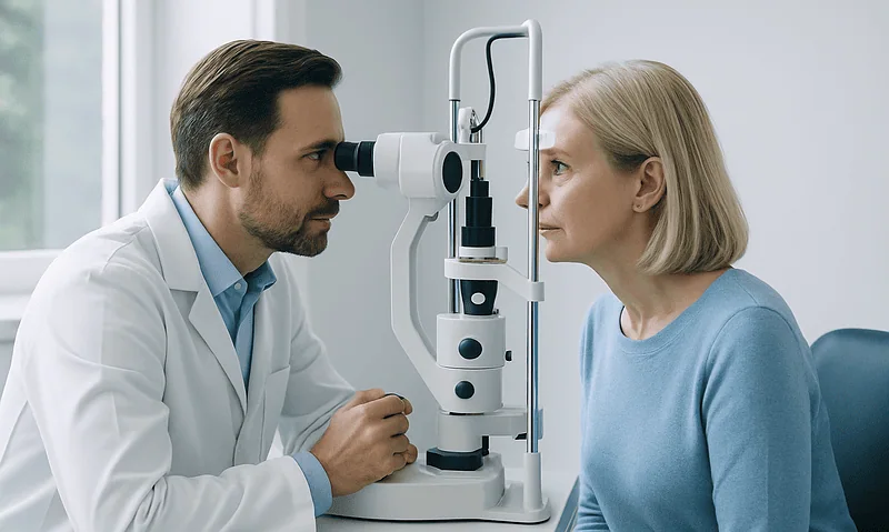
Which Tests Diagnose Ocular Surface Disease? Tear Film Checks, Staining and Eyelid Exams
Ocular surface disease, including dry eye, is diagnosed with a combination of symptom questionnaires and slit-lamp tests rather than a single test. Clinicians typically…

The Week Before LASIK: Contact Lens Breaks, Makeup, Perfume and the Ride Home
Before LASIK, most surgical teams ask you to switch from contact lenses to glasses for a period of days to weeks so the cornea…

Who Is a Candidate for Presbyopia Surgery? Lens Clarity, Eye Health and Daily Habits
A good presbyopia surgery candidate is usually an adult whose glasses prescription has been stable, whose natural lens is still clear or is already…

Glare, Dryness and Ghosting After Astigmatism Correction: Normal Healing or a Call to Make?
Glare, halos, dry eyes and faint ghost images are among the most common astigmatism surgery side effects, and in most people they fade as…

How Patching and Glasses Retrain a Lazy Eye: What Amblyopia Treatment Actually Does
Patching treats a lazy eye by covering the stronger eye for a set number of hours each day, which forces the brain to use…