

Which Tests Diagnose Ocular Surface Disease? Tear Film Checks, Staining and Eyelid Exams
Key Takeaways
- No single test diagnoses dry eye; guidelines pair a symptom questionnaire with tear film measurements and staining because symptoms and signs often disagree.
- A tear break-up time under 10 seconds is generally considered abnormal and points toward evaporative dry eye, the most common subtype.
- The Schirmer test measures tear volume over five minutes, with more than 10 millimeters of wetting considered normal, and it cannot detect poor tear quality.
- Tear osmolarity of 308 mOsm/L or higher, or a difference above 8 mOsm/L between eyes, is a recognized marker of an unstable tear film.
- Lissamine green stains the conjunctiva and the eyelid's inner wiping edge, revealing damage that fluorescein on the cornea can miss.
- Meibography can show permanent loss of eyelid oil glands, which explains why lubricating drops alone sometimes feel inadequate.
Ocular surface disease, including dry eye, is diagnosed with a combination of symptom questionnaires and slit-lamp tests rather than a single test. Clinicians typically measure how quickly tears break up, how much tear fluid the eye makes (Schirmer test), tear concentration (osmolarity), surface damage with fluorescein or lissamine green dye, and eyelid oil gland function. Results are interpreted together by the examining eye care professional.
By mid-afternoon the numbers on her spreadsheet had begun to swim. She blinked hard, reached for the lubricating drops in her desk drawer for the fourth time that day, and wondered whether it was simply screen fatigue or something with a name. When she finally booked an eye appointment, her real question was not whether her eyes felt dry. It was whether anyone could measure it.
That is a fair question, because dry eye tests are unlike most laboratory work. There is no single blood value, no scan that lights up. Instead the clinician assembles evidence from several small measurements: a stopwatch, a strip of paper, a drop of dye, a careful look at the eyelids under magnification.
Each test answers a different question about a tear film thinner than a sheet of tissue paper. Put together, they turn a vague complaint into a specific diagnosis, and that specificity is what steers everything that follows.
What actually happens during dry eye tests?
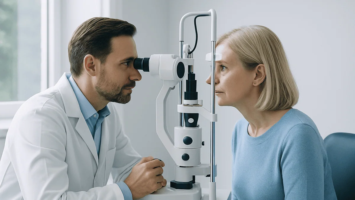
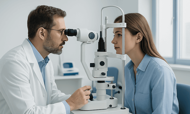
Most dry eye tests fit inside an ordinary eye appointment, and none of them should hurt. You sit at a slit lamp, the upright microscope with a chin rest that every eye clinic owns, while the clinician passes a narrow beam of light across the front of your eye. That beam is the workhorse of the visit. Under magnification it reveals the tear layer, the eyelid margins, the tiny oil gland openings along the lash line and the clear front window of the eye, the cornea.
Before any instrument touches you, expect questions. Symptom questionnaires come first because dry eye is defined partly by how it feels, not only by what a device reads. Then come the measurements, usually in a deliberate order: the gentlest and least disruptive first, the ones that involve touching the eye or adding dye last. The sequence matters. Pressing a paper strip against the lower lid before timing how quickly tears evaporate would make the tears themselves unreliable, a point stressed in the international TFOS DEWS II diagnostic methodology report.
A few tests need a drop of orange or green dye. The dye clings to cells that are damaged or unprotected and glows under blue light, so the clinician can see exactly where the surface has been exposed. Others need only a camera or a handheld device that samples a bead of tears far smaller than a raindrop. The eyelids get their own examination: the clinician may press gently on the lid to see what emerges from the oil glands, or photograph the glands through the lid with infrared light.
You leave with a picture rather than a single score: how much tear fluid you make, how long it stays in place, whether the oily layer is doing its job, and whether the surface has begun to pay a price.
How is dry eye diagnosed? Why symptoms come first
Dry eye has an awkward habit: symptoms and test results often disagree. Some people report burning, grittiness or fluctuating vision with tear measurements that look nearly normal, while others show visible surface damage and barely notice it. The TFOS DEWS II report describes this mismatch as one reason no single test can diagnose the condition on its own.

That is why a modern workup begins with a validated questionnaire. Tools such as the Ocular Surface Disease Index or the five-item Dry Eye Questionnaire ask how often your eyes feel dry, how light sensitivity affects you, and whether reading, driving at night or using a screen has become harder. The answers are turned into a score that can be tracked over time. The DEWS II diagnostic algorithm treats a positive symptom score as the entry point; only then do objective tests confirm the diagnosis and sort it into a subtype.
The history matters as much as the score. Clinicians will ask about contact lens wear, previous eye surgery, autoimmune conditions such as Sjögren’s disease or rheumatoid arthritis, and medicines that can reduce tear production. Antihistamines, certain antidepressants, some blood pressure drugs and hormone therapies appear on the Mayo Clinic and National Eye Institute lists. Nobody is asked to stop anything on the spot; the information simply explains what the tests may find.
Environment counts too. Air conditioning, long flights, wind and prolonged screen use all increase evaporation, and Mayo Clinic notes that people blink less when concentrating on a screen. Ten minutes of conversation before the first drop of dye is not padding. In dry eye, it is half the diagnosis.
Tear break-up time test: a stopwatch for your tear film
Tear break-up time, usually shortened to TBUT, measures how long the tear film stays smooth and unbroken between blinks. Think of a freshly washed windshield in light rain: for a few seconds the water forms an even sheet, then dry patches appear. Your tear film does the same thing, and the speed at which those dry spots form tells the clinician whether the film is stable enough to protect the eye.
In the classic version, a small amount of fluorescein dye is placed on the eye. You blink a few times, then hold your eyes open while the clinician watches through the slit lamp under blue light and counts the seconds until the first dark spot appears in the glowing yellow film. According to the DEWS II report, a break-up time under 10 seconds is generally considered abnormal, and Cleveland Clinic uses the same threshold in its patient guidance.
Newer instruments measure non-invasive break-up time, or NIBUT, without dye. A ring of light is projected onto the cornea and a camera detects the moment the reflected pattern distorts. Because no drop is added, the reading reflects your natural tears more faithfully, which is why DEWS II lists NIBUT as a preferred marker of tear film stability where the equipment is available.
Short break-up times point toward evaporative dry eye, the pattern in which tears are produced in adequate volume but disappear too fast, usually because the oily outer layer is deficient. That distinction is useful: it shifts attention from the tear glands to the eyelids, where the oil is made.
One caveat worth knowing. The test is sensitive to room humidity, how long you held your eyes open beforehand, and even how much dye was used. Clinicians usually take more than one reading for that reason.
Schirmer test for dry eye: what the paper strips measure
The Schirmer test is the oldest dry eye test still in routine use, and it answers a blunt question: how much tear fluid does the eye produce over five minutes? A thin strip of sterile filter paper, folded at one end, is hooked over the edge of the lower eyelid toward the outer corner. You close your eyes, or sometimes look slightly upward, and wait. The paper wicks up tears and the clinician measures how far the wet edge has travelled.
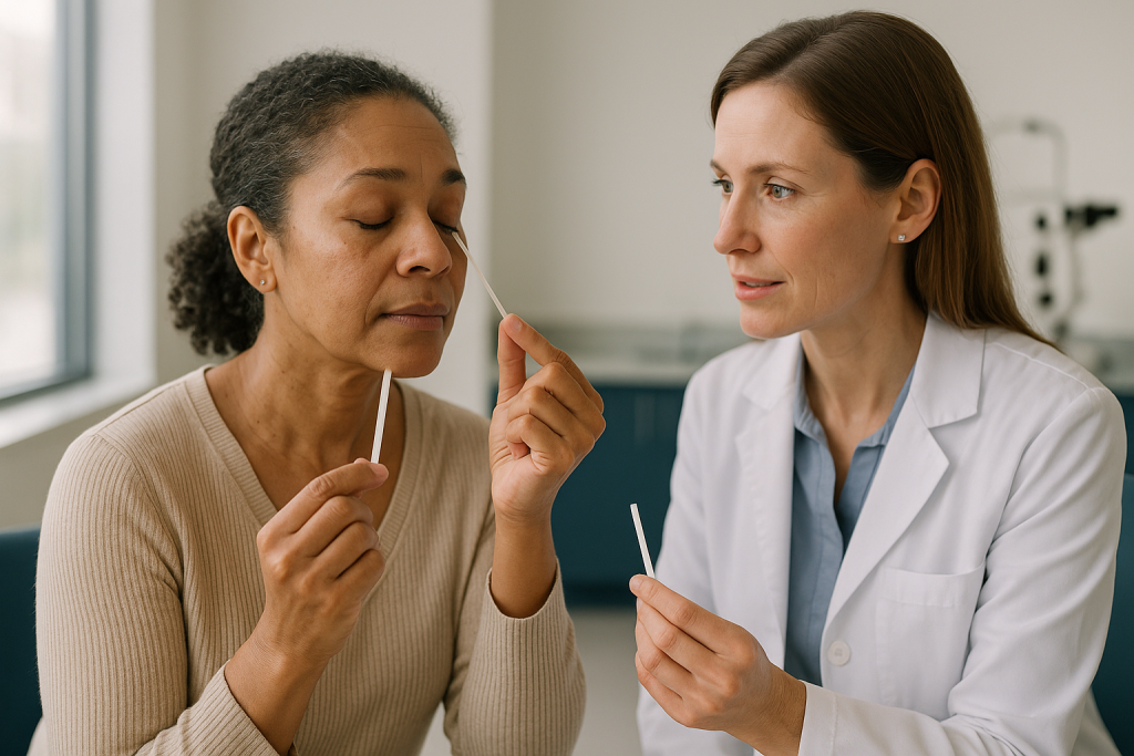
MedlinePlus describes the reading in plain terms: more than 10 millimeters of wetting in five minutes is considered normal, while lower values suggest the eye may not be producing enough aqueous tears. The test can be done with or without a numbing drop. Without anesthetic it measures total secretion, including the reflex tearing triggered by the paper itself. With anesthetic it comes closer to measuring basal, resting tear production, though results are more variable than the simple description implies.
Low Schirmer readings point toward aqueous-deficient dry eye, the pattern in which the lacrimal glands under the outer upper lid do not make enough watery tears. That subtype is strongly associated with Sjögren’s disease and other autoimmune conditions, which is why a markedly low result may prompt questions about dry mouth, joint pain or fatigue and sometimes a referral for blood tests. The National Eye Institute lists autoimmune disease among the recognized causes.
A related test, phenol red thread, uses a fine cotton thread impregnated with pH-sensitive dye and takes only about 15 seconds, per the DEWS II report. It is less irritating but less widely used.
The Schirmer strip feels odd, like an eyelash you cannot blink away. Most people describe it as strange rather than painful. Because the paper stimulates the eye, this test usually comes last.
Tear osmolarity, tear meniscus and other tear film checks
Tears are salty, and when there is too little water in the film the salt concentrates. Tear osmolarity measures exactly that. A handheld instrument touches the edge of the lower lid, draws up a tear sample smaller than a grain of sand, and returns a reading within seconds. The DEWS II report proposes a cut-off of 308 milliosmoles per liter or higher, or a difference of more than 8 milliosmoles per liter between the two eyes, as suggestive of dry eye. The between-eye difference is telling because healthy tear films are remarkably consistent, whereas an unstable film swings from one blink to the next.
Osmolarity does not say whether the problem is low production or fast evaporation; both raise the salt concentration. Its value lies in confirming that the tear film is genuinely disturbed, particularly when symptoms and other tests disagree.
The tear meniscus is the thin crescent of fluid that pools along the lower eyelid margin. Under the slit lamp, or with a scanning imaging device, the clinician estimates its height. A shallow meniscus suggests a low tear reservoir. It is a quick, non-invasive indicator that supports, rather than replaces, the Schirmer reading.
Some clinics also test tear film markers of inflammation. A test for elevated matrix metalloproteinase-9, an enzyme released when the ocular surface is inflamed, uses a small collection strip and gives a positive or negative result. The DEWS II report lists inflammatory markers as supportive rather than diagnostic, since inflammation accompanies several surface conditions.
Interferometry, which photographs the rainbow-colored oily layer on top of the tears, estimates the thickness of the lipid layer. Thin oil means faster evaporation. None of these tests stands alone, and not every clinic owns every device. A good examiner uses what is available and interprets the pattern, not a single number.
Fluorescein and lissamine green staining: mapping the damage
Staining tests show consequences. Where the tear film has failed to protect the surface, cells become damaged, dead or exposed, and certain dyes stick to them. Under the slit lamp the pattern of dots and patches becomes a map of where and how badly the eye has suffered.
Fluorescein, the orange dye also used for break-up time, highlights the cornea. Under blue light, damaged corneal cells glow as fine green pinpoints. A handful of scattered dots after a long day at a screen is common; a dense band across the lower cornea, where the eye is most exposed between blinks, signals a more significant problem. Clinicians grade the pattern using standardized scales so that the same eye can be compared at a later visit.
Lissamine green is a blue-green dye that behaves differently. It preferentially stains the conjunctiva, the thin membrane covering the white of the eye and the inside of the lids, along with any cells that have lost their protective mucus coating. Staining in a wedge shape on the nasal and temporal conjunctiva is characteristic of dry eye, and the DEWS II report treats both corneal and conjunctival staining as key markers of ocular surface damage.
Lissamine green also reveals a finding many people have never heard of: lid wiper epitheliopathy. The lid wiper is the strip of tissue on the inner edge of the eyelid that sweeps across the eye with each blink. When the surface lacks lubrication, that strip stains, the way a squeegee dragged across a dry window wears at its edge.
Staining is painless and the color washes out with your own tears shortly afterward, though fluorescein can briefly tint the skin at the corner of the eye. Contact lenses are usually kept out until the dye has fully cleared.
Meibomian gland dysfunction test: why the eyelid exam matters most
If one part of the examination deserves the most attention, it is the eyelids. Along the margin of each lid, just behind the lashes, sit roughly 25 to 40 meibomian glands in the upper lid and fewer in the lower, according to the DEWS II report. They produce the oil that spreads over the tears with each blink and slows evaporation. When these glands become blocked, thickened or shrunken, the condition is called meibomian gland dysfunction, and the same report identifies evaporative dry eye driven by this problem as the most common form of the disease. Asked what the biggest cause of dry eyes is, an evidence-based answer usually begins here.
The eyelid exam has several parts. First the clinician inspects the lid margin under magnification for redness, notched edges, capped or plugged gland openings, and the frothy tear debris that often accompanies gland dysfunction. Then comes gentle expression: pressing the lid with a fingertip or a small paddle to see what the glands release. Healthy oil flows clear and thin, like olive oil. Dysfunctional glands yield cloudy, toothpaste-thick material, or nothing at all.
Meibography goes further. An infrared camera photographs the glands through the everted or transilluminated lid, showing them as pale vertical stripes. Shortened, twisted or missing stripes mean glands have dropped out, and once lost they do not regrow. Seeing the images can be sobering, but it also explains why lubricating drops alone sometimes feel insufficient: the drops add water, and the problem is oil.
The lid exam also picks up related conditions. Blepharitis, inflammation of the lid margin often linked to bacterial overgrowth or tiny Demodex mites, shows up as crusting and cylindrical collars around the lashes. Incomplete lid closure during sleep, checked by asking you to close your eyes gently, leaves a tell-tale band of staining across the lower cornea.
Which dry eye tests matter most? A side-by-side comparison
Every test in the workup answers a slightly different question, and understanding which is which makes the results far easier to follow. The table below summarizes the core tests, what each measures, how it feels and where its limits lie. Thresholds are drawn from the DEWS II report and MedlinePlus; individual clinics may use slightly different cut-offs.
| Test | What it measures | How it feels | Commonly cited threshold | Main limitation |
|---|---|---|---|---|
| Symptom questionnaire | Frequency and impact of symptoms | Paper or tablet form | Validated score cut-offs vary by tool | Symptoms often do not match signs |
| Tear break-up time | Tear film stability | Dye drop, hold eyes open briefly | Under 10 seconds abnormal | Affected by humidity and dye volume |
| Tear osmolarity | Salt concentration of tears | Brief touch at lid edge | 308 mOsm/L or higher, or more than 8 mOsm/L difference between eyes | Does not identify the subtype |
| Schirmer test | Tear production volume | Paper strip in lower lid for 5 minutes | 10 mm or less in 5 minutes suggests deficiency | Variable; irritating; done last |
| Fluorescein staining | Corneal surface damage | Dye drop, painless | Graded on standardized scales | Shows damage, not cause |
| Lissamine green staining | Conjunctival and lid wiper damage | Dye drop, painless | Graded on standardized scales | Less widely stocked |
| Meibomian gland expression and meibography | Oil quality and gland structure | Gentle lid pressure; infrared photo | Descriptive grading | Imaging not available everywhere |
Read across the rows and a logic emerges. Questionnaires and osmolarity confirm that dry eye is present. Break-up time, the Schirmer test and the eyelid exam sort it into evaporative, aqueous-deficient or mixed. Staining shows how much harm has already occurred. Most people have a mixed picture, which is why the treating clinician weighs the whole table rather than any single cell.
Who is usually offered dry eye testing, and who is asked to wait
Formal testing is usually offered to anyone whose symptoms persist despite simple measures, whose vision fluctuates through the day, who wears contact lenses uncomfortably, or who has a condition that raises the risk of severe disease. The National Eye Institute estimates that nearly 16 million people in the United States have dry eye, and the numbers rise with age, after menopause, and in people with diabetes, thyroid disease or autoimmune conditions. Anyone in those groups who reports dryness is a reasonable candidate for a full workup.
People preparing for eye surgery are another common group. Cataract and laser vision correction both depend on a stable tear film for accurate measurements and a smooth recovery, so surgeons frequently screen for ocular surface disease beforehand and may treat it first. The exam is the same; the reason is different.
Who is asked to wait? Timing rather than eligibility is the usual reason. Someone with an acutely red, painful eye needs an urgent examination for infection or injury, not a dry eye battery. A person who used lubricating drops minutes before arriving may be asked to return another day, because the drops temporarily alter break-up time, osmolarity and staining. Contact lens wearers are often asked to leave lenses out for a period before testing, since lenses disturb the tear film and can themselves cause staining. Recent eye infection or allergy flares can also cloud the results.
Children are tested less often, partly because dry eye is less common in childhood and partly because sitting still at a slit lamp for a timed test is a lot to ask. When testing is needed, clinicians favor the non-invasive options, questionnaires adapted for age, and a thorough look at the lids.
Nobody is excluded permanently. The aim is simply to test the tear film in its natural state.
What is stage 3 dry eye? Severity grading explained
People searching for stage 3 dry eye are usually looking at a grading scale that appears on many clinic handouts. It descends from an earlier international consensus report, which sorted dry eye into four severity levels by combining symptoms, staining, break-up time, Schirmer results and eyelid findings. Level 1 described occasional, mild symptoms with little or no staining. Level 2 added frequent symptoms and mild corneal staining. Level 3 described severe or constant symptoms, marked staining, break-up time of only a few seconds and possible fine strands of mucus on the cornea. Level 4 described disabling, constant symptoms with severe surface damage.
Two things are worth understanding about that scale. First, it was designed as a framework for clinicians, not a diagnosis to be given to patients as a label. Second, the more recent DEWS II report moved away from rigid staging toward describing subtype and severity separately, because a person can have a badly damaged surface with modest symptoms, or the reverse. A single stage number cannot capture that.
In practice, being told your findings sit in a moderate-to-severe range means the examiner has seen more than occasional dryness: measurable surface damage, a very short break-up time, and possibly reduced tear production. It usually prompts a more thorough search for the cause, closer follow-up, and consideration of treatments beyond over-the-counter lubricants. It does not mean vision loss is imminent. Dry eye at any stage is, for most people, a chronic condition to be managed rather than a countdown.
If a clinician uses a stage or grade, it is reasonable to ask which scale it comes from and which specific findings placed you there. Those findings, not the number, are what will be tracked at the next visit.
What the weeks after diagnosis usually look like
Dry eye rarely responds overnight, and the tests explain why. If break-up time was short and the meibomian glands were blocked, the early plan often centers on the eyelids: warm compresses to soften stagnant oil, lid hygiene to reduce crusting, and lubricating drops for comfort. NHS guidance suggests seeking review if self-care and pharmacy products have not helped after a few weeks, a useful marker of when to check back rather than a promise of improvement by then.
If the Schirmer result was low, the early weeks may include a search for underlying causes, sometimes blood tests for autoimmune disease, and discussion of ways to keep the tears you make on the eye longer. That can include tiny plugs placed in the tear drainage ducts, a brief in-office procedure described by both Mayo Clinic and the National Eye Institute. Where staining showed significant inflammation, the clinician may discuss prescription anti-inflammatory drops. These work by calming the immune response on the ocular surface rather than adding moisture, and they are typically judged over weeks to months rather than days. Whether any of these options is appropriate, and in what order, is a decision for the treating clinician.
Follow-up usually repeats a subset of the same tests. That is the quiet advantage of a thorough baseline: break-up time, staining grade and questionnaire score can be compared directly. Symptoms often lag behind measurable change, so a surface that looks better under the slit lamp may not yet feel better.
Expect some adjustment. Preservative-free drops may replace preserved ones if staining persists, since preservatives can irritate a fragile surface. Environmental changes, humidifiers, screen breaks and conscious blinking are often revisited. Progress tends to be measured in fewer bad days rather than a single good one.
What people often get wrong about dry eye tests
A normal Schirmer test rules out dry eye. It does not. The Schirmer strip measures tear volume, and the most common form of dry eye is evaporative: normal volume, poor quality. Someone with blocked oil glands can wet the strip perfectly well and still have a tear film that collapses in four seconds.
Watery eyes mean the opposite of dry eye. Reflex tearing is one of the most frequent complaints in dry eye. When the surface becomes irritated, the lacrimal gland floods the eye with watery tears that lack the oil and mucus needed to stay put, so they spill over. Mayo Clinic lists watery eyes among the symptoms for exactly this reason.
Dry eye is a screen problem that only affects office workers. Screens reduce blink rate and worsen evaporation, but the biggest risk factors identified by the National Eye Institute are age, hormonal change, autoimmune disease, medications and eyelid gland dysfunction. Retired people and people who never use a computer are diagnosed every day.
The tests are uncomfortable or risky. The dyes are painless and clear quickly. The Schirmer strip feels odd for five minutes. Nothing in a standard dry eye workup involves needles, pressure on the eye or dilating drops, though a broader eye exam on the same day might.
If the machine says I am normal, my symptoms are imaginary. The DEWS II report explicitly acknowledges that symptoms and signs correlate poorly. Persistent discomfort with clean test results is a recognized presentation, and it sometimes points to nerve sensitivity of the ocular surface rather than a tear problem. It deserves a considered explanation, not dismissal.
One good test is enough. No single measurement diagnoses ocular surface disease. Anyone offering a diagnosis from a single reading, or from no examination at all, is skipping the part that determines what treatment makes sense.
Questions to ask your care team
A dry eye examination generates a lot of numbers, and it is easy to leave without knowing what they meant. Writing down a few questions beforehand keeps the conversation focused on what will change your day-to-day comfort.
- Which type of dry eye do my results suggest: too little tear production, too much evaporation, or a mix of both?
- What did the eyelid exam show? Are my oil glands blocked, and did imaging show any gland loss?
- How much surface staining did you see, and where? Does the pattern suggest incomplete eyelid closure or another specific cause?
- Do any of my current medicines or health conditions plausibly contribute, and is it worth discussing that with the prescriber?
- Should I be checked for an autoimmune cause such as Sjögren’s disease, and what would that involve?
- Which of today’s tests will you repeat at follow-up, and roughly when should that visit happen?
- Are there habits, such as contact lens wear or drop use before appointments, that could make future readings unreliable?
- If my symptoms are worse than my test results suggest, what other explanations are you considering?
Bring a list of every eye drop you use, including over-the-counter products, and note how many times a day you reach for them. Frequency of use is a practical severity marker that clinicians find helpful. If you use screens for long stretches, mention when in the day symptoms peak; morning burning and evening blur can point in different directions.
The best answers will be specific to your findings. If a response sounds like a generic pamphlet, it is reasonable to ask which of your own results it is based on. The purpose of testing, after all, is to make the plan personal.
When to call your doctor
Dry eye is uncomfortable but rarely dangerous, and most of its symptoms wax and wane without alarm. Some signs, however, do not belong to ordinary dry eye and need prompt assessment, ideally the same day. Both the NHS and Mayo Clinic list warning features that should not be waited out.
- Sudden or significant loss of vision, or a new blurred patch that does not clear with blinking.
- Severe eye pain, especially with nausea, a cloudy cornea or a fixed, dilated pupil.
- A red, painful eye in a contact lens wearer, which can indicate infection of the cornea.
- Yellow or green discharge, marked swelling of the eyelid, or fever alongside eye symptoms.
- A recent injury, chemical splash or foreign body sensation that persists after rinsing.
- Extreme sensitivity to light that has appeared suddenly.
- A white or gray spot visible on the cornea.
Less urgent but still worth a scheduled call: symptoms that keep worsening despite the plan you agreed, a new medicine that seems to have made dryness sharply worse, or dry mouth, joint pain and fatigue appearing alongside dry eyes, since that combination can signal an autoimmune cause. If drops seem to sting more over time, or if a dye test at a previous visit showed significant staining and you have not been re-examined, mention it.
None of the tests described in this article are designed to detect infection, glaucoma or retinal problems, so a normal dry eye workup does not exclude other causes of a painful or red eye. When in doubt, an urgent examination is the safer path, and the clinician who examined you can decide whether the symptom fits the dry eye picture or needs a different kind of test.
Frequently asked questions
How is dry eye diagnosed if there is no single test?
Dry eye is diagnosed by combining a validated symptom questionnaire with at least one abnormal objective finding, such as a short tear break-up time, raised tear osmolarity or surface staining. The international DEWS II report sets out this approach because symptoms and measurements frequently disagree. Once the diagnosis is confirmed, further tests including the Schirmer test and an eyelid gland exam sort it into aqueous-deficient, evaporative or mixed subtypes to guide management.
Is it better to see an optometrist or ophthalmologist for dry eye?
Both can diagnose and manage dry eye, and the National Eye Institute simply advises seeing an eye care professional. Optometrists routinely perform the full range of dry eye tests and manage most cases. Ophthalmologists, who are medical doctors, are typically involved when surgery is being considered, when an autoimmune or corneal disease is suspected, or when standard measures have not helped. Many people move between the two as their needs change.
What is stage 3 dry eye?
Stage 3 usually refers to the third of four severity levels in an older consensus grading scale, describing severe or constant symptoms, marked surface staining, very short tear break-up time and possibly mucus strands on the cornea. Newer guidance describes subtype and severity separately rather than using a single stage. A moderate-to-severe finding prompts closer follow-up and a wider search for causes, but dry eye at this level is still a manageable chronic condition.
What is the number one treatment for dry eyes?
There is no single best treatment, because the right approach depends on what the tests show. Lubricating eye drops are the usual first step for comfort, while eyelid warming and hygiene address blocked oil glands, and prescription anti-inflammatory drops target surface inflammation. NHS and Mayo Clinic guidance describe these as a stepped approach matched to severity and subtype. The choice and sequence belong to the examining clinician.
What is the biggest cause of dry eyes?
Meibomian gland dysfunction, in which the eyelid oil glands become blocked or shrink, is identified by the DEWS II report as the leading driver of dry eye because it causes tears to evaporate too quickly. Other frequent contributors include aging, hormonal change, autoimmune disease, certain medicines, contact lens wear and reduced blinking during screen use. Most people have more than one factor, which is why the eyelid exam is central to testing.
Does the Schirmer test for dry eye hurt?
The Schirmer test is generally described as strange rather than painful. A thin paper strip rests against the lower eyelid for five minutes, which feels like a stray eyelash you cannot blink away. Some clinicians use a numbing drop first, which reduces the sensation and measures resting tear production. Because the strip stimulates tearing and can disturb the surface, it is usually performed last in the sequence of tests.
What does the tear break-up time test show?
The tear break-up time test measures how many seconds the tear film stays intact after a blink before dry spots appear. A result under 10 seconds is generally considered abnormal and suggests the tears are evaporating too quickly, often because the oily surface layer is deficient. Non-invasive versions use a projected light pattern instead of dye. Clinicians usually take more than one reading because humidity and technique influence the result.
Should I stop using eye drops or contact lenses before dry eye tests?
Ask the clinic in advance, because lubricating drops used shortly before the appointment can temporarily change break-up time, osmolarity and staining, and contact lenses disturb the tear film and can cause their own staining. Many clinics ask that lenses be left out for a period and that drops be held on the morning of testing. Never stop a prescribed medicated drop without checking with the prescriber first.
What is a meibomian gland dysfunction test?
It is an eyelid examination that assesses the oil glands along the lid margin. The clinician inspects the gland openings under magnification, presses gently on the lid to judge whether the oil flows clear or emerges thick and cloudy, and may photograph the glands with an infrared camera, a technique called meibography. Shortened or missing glands on the image indicate gland loss, which points toward evaporative dry eye.
Why do I have dry eye symptoms if my tests came back normal?
Symptoms and test results often do not match, a mismatch the DEWS II report explicitly recognizes. Tear film measurements vary with the day, humidity and recent drop use, so a single normal set does not exclude dry eye. Persistent discomfort with clean findings can also reflect heightened nerve sensitivity of the ocular surface. Repeat testing, a review of medicines and environment, and an honest discussion with the examining clinician are reasonable next steps.
References
- National Eye Institute (NIH): Dry Eye
- MedlinePlus Medical Encyclopedia: Schirmer test
- NHS: Dry eyes
- Cleveland Clinic: Dry Eye
This article is for general information only and is not a substitute for professional medical advice. Please consult a qualified doctor about your individual situation.

More from the Blog

Getting Ready for Cataract Treatment: Medication Review, Drops and Arranging a Companion
Preparing for cataract treatment usually means attending a pre-assessment visit, giving the care team a complete list of your medicines (including blood thinners, prostate…
The Week Before LASIK: Contact Lens Breaks, Makeup, Perfume and the Ride Home
Before LASIK, most surgical teams ask you to switch from contact lenses to glasses for a period of days to weeks so the cornea…
Who Is a Candidate for Presbyopia Surgery? Lens Clarity, Eye Health and Daily Habits
A good presbyopia surgery candidate is usually an adult whose glasses prescription has been stable, whose natural lens is still clear or is already…

Glare, Dryness and Ghosting After Astigmatism Correction: Normal Healing or a Call to Make?
Glare, halos, dry eyes and faint ghost images are among the most common astigmatism surgery side effects, and in most people they fade as…
How Patching and Glasses Retrain a Lazy Eye: What Amblyopia Treatment Actually Does
Patching treats a lazy eye by covering the stronger eye for a set number of hours each day, which forces the brain to use…
Choosing an Intraocular Lens: Questions to Bring to Your Ophthalmologist
Choosing an intraocular lens means weighing how you use your eyes each day against the trade-offs of each lens design. Monofocal lenses give one…