

Can Fibroids Be Cancerous, Burst, or Be Dangerous?
Key Takeaways
- Fewer than 1 in 1,000 fibroids prove to be cancerous on examination, and having fibroids does not raise the risk of uterine cancer.
- Uterine leiomyosarcoma is thought to arise on its own from muscle cells, not by transformation of a long-standing benign fibroid.
- Ultrasound and MRI can raise or lower suspicion but cannot prove a uterine mass is benign; only tissue examination after removal can.
- A uterine mass that grows or bleeds after menopause is the single most important warning sign, because ordinary fibroids shrink once hormones fall.
- Fibroids are solid muscle and cannot burst, but they can degenerate or twist on a stalk, causing sudden severe pain that usually settles within one to two weeks.
- The most common real harm from fibroids is iron-deficiency anemia from heavy periods, which a simple blood count can detect.
Fibroids are almost always benign and do not turn into cancer; a cancerous growth in the uterine wall (leiomyosarcoma) is found in fewer than 1 in 1,000 people who have fibroid surgery. Fibroids are solid muscle and do not burst, though they can degenerate or twist and cause sudden pain. Their most common real danger is heavy bleeding that leads to iron-deficiency anemia.
The sentence usually arrives at the end of a routine scan, half over the shoulder: you have a couple of fibroids, nothing to worry about. Then the sonographer leaves, the paper gown crinkles, and the questions start. Fibroids of what? Growing where? And how does anyone know a lump is nothing to worry about without taking it out and looking?
Those are fair questions, and the internet answers them badly. Type the word into a search bar and you get one page insisting fibroids are harmless and the next hinting they hide cancer, followed by a headline about a warned-against surgical technique. No wonder people leave the clinic reassured and go to bed frightened.
The honest picture sits between those extremes, and it is more interesting than either. Fibroids are extraordinarily common, overwhelmingly benign, and occasionally genuinely troublesome for reasons that have little to do with cancer. Knowing which worries deserve your attention, and which can be set down, is the point of what follows.
How common are uterine fibroids, and who gets them?
Fibroids are so common that having one is closer to a normal finding than an abnormal one. According to the Cleveland Clinic, they affect up to 80 percent of women by age 50, though many never know because the growths cause no symptoms. The NHS puts the figure for fibroids that are noticed or diagnosed at roughly one in three women, most often between the ages of 30 and 50.
The medical name is leiomyoma, which simply means a benign tumor of smooth muscle. Each fibroid is a firm, rubbery knot of muscle and connective tissue that grows within the wall of the uterus, on its outer surface, or into the cavity. Sizes range from a lentil to a melon, and a single uterus can carry one fibroid or dozens.
Several patterns are well documented. Fibroids depend on the hormones estrogen and progesterone, so they tend to appear during the reproductive years and shrink after menopause. They run in families. They are diagnosed more often, at younger ages, and in larger sizes in Black women, a disparity the Cleveland Clinic and Mayo Clinic both note and researchers have not fully explained. Earlier first periods and higher body weight are also linked to greater risk, while pregnancy appears to lower it.
What causes a single muscle cell to start multiplying into a fibroid is still not fully understood. Genetic changes within the fibroid cells, hormone sensitivity, and growth factors that regulate tissue repair all appear to play a part. None of those mechanisms has anything to do with a fibroid being, or becoming, cancer.
Can fibroids be cancerous? What the numbers actually say
Start with the figure that matters most. The Cleveland Clinic states that fewer than 1 in 1,000 fibroids turn out to be cancerous. The Mayo Clinic puts it in plain language: fibroids are not associated with an increased risk of uterine cancer and almost never develop into cancer. Those two statements come from separate institutions reviewing the same body of evidence, and they agree.
Hold that number against the prevalence figure from the previous section. If up to 80 percent of women develop fibroids and fewer than one in a thousand of those growths is malignant, the overwhelming majority of people reading this with a diagnosed fibroid are carrying a benign lump of muscle and nothing more. That is not a soothing phrase; it is arithmetic.
Why does the question keep resurfacing, then? Partly because the word tumor unsettles people, even when preceded by benign. Partly because a rare cancer called leiomyosarcoma can arise in the same muscle layer and, on a scan, can look like a fibroid. When a sarcoma is discovered during surgery planned for fibroids, the story travels fast, and the exception starts to sound like the rule.
It helps to be precise about what the statistics do and do not say. They do not say cancer of the uterine wall never happens. They say that when a fibroid is examined under a microscope after removal, it is benign in the vast majority of cases, and that having fibroids does not raise your odds of uterine cancer above someone who has none. That distinction is the whole reassurance, and it deserves to be stated clearly rather than hedged.
Do fibroids turn into cancer, or is the cancer something different?
The prevailing view among specialists, reflected on the Cleveland Clinic page on leiomyosarcoma, is that these cancers do not usually grow out of an existing benign fibroid. They are thought to arise on their own from smooth muscle cells, carrying their own set of genetic changes from the start. A fibroid that has sat unchanged for a decade is not slowly ripening into something sinister.
This matters for how you think about risk. If fibroids were a stage on the way to cancer, the sensible advice would be to remove them all, and no reputable guideline says that. Watchful waiting for symptom-free fibroids is standard precisely because the growth itself is not a threat.
Leiomyosarcoma belongs to a small group of uterine sarcomas, cancers of the muscle and supporting tissue rather than the lining. They are rare, they behave differently from the far more common endometrial cancers, and they are typically found in women after menopause. Symptoms overlap with fibroids, which is the crux of the diagnostic problem: abnormal bleeding, pelvic pressure, and a mass that can be felt.
One more piece of context. Some observational studies have explored links between fibroids and other cancers, including breast and endometrial cancer, with mixed and modest findings. None of this research shows that a fibroid causes cancer elsewhere. Shared risk factors, such as hormone exposure over a lifetime, are a more likely explanation for any statistical association, and the major patient-information sources do not list fibroids as a cancer risk factor.
Can an ultrasound tell the difference between fibroids and cancer?
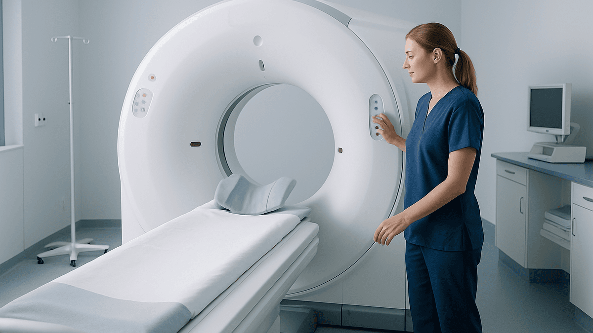
Not with certainty, and any clinician who is honest with you will say so. Ultrasound is superb at answering the first questions: is there a mass, where is it, how big, and how many? It shows the classic features of a fibroid, a well-defined round growth with a whorled texture and a clear border. It cannot see individual cells, and cells are where the difference between benign and malignant lives.
The Mayo Clinic describes ultrasound as the routine first test for fibroids, with MRI used when more detail is needed about size, position, or the character of the tissue. MRI can raise or lower suspicion. Features such as irregular borders, areas of dead tissue, unusual signal patterns, or rapid growth between scans may prompt a specialist to look harder. None of these features is proof, and benign fibroids that are degenerating can mimic several of them.
Blood tests do not settle the question either. There is no reliable screening blood marker for uterine sarcoma. A biopsy of the uterine lining can detect endometrial cancer but frequently misses a sarcoma buried in the muscle wall, because the sample does not reach it.
The only definitive answer comes from a pathologist examining the tissue after a fibroid is removed. That is why the diagnosis of leiomyosarcoma is so often made after surgery rather than before it, and why the language on your scan report will say consistent with fibroid rather than proven fibroid. The phrasing is not evasion. It is an accurate description of what imaging can and cannot do.
Which warning signs make a doctor think beyond a fibroid?
Because scans cannot rule out a sarcoma, clinicians rely on context. A handful of features shift a growth from routine to worth a closer look, and it is worth knowing what they are so you can raise them yourself.
The first is timing. Fibroids are fueled by reproductive hormones, so they typically stop growing and often shrink after menopause, as the NHS and Mayo Clinic both describe. A uterine mass that appears, enlarges, or begins to bleed after periods have stopped does not fit the usual fibroid pattern and should be evaluated promptly. This is the single most important red flag, and it applies regardless of how many fibroids someone had earlier in life.
The second is pace. Fibroids grow slowly for the most part, and growth spurts do happen during the reproductive years. Very rapid enlargement, especially alongside new pain or bleeding, is a reason for repeat imaging and specialist review rather than reassurance alone.
The third is imaging character, covered in the previous section: an MRI that shows irregular edges, internal areas of dead tissue, or an appearance that does not match a typical fibroid.
Age adds weight to the others. Uterine sarcomas are more common in women over 50, while fibroids peak in the 30s and 40s. A new mass in a 62-year-old is interpreted differently from an identical-looking one in a 34-year-old.
What does not belong on this list is the mere presence of fibroids, their number, or a size that sounds alarming. Large fibroids are usually just large fibroids. It is the combination of features, above all growth after menopause, that changes the conversation.
Can fibroids burst?
The short answer is no, not in the way the word suggests. A fibroid is a dense ball of muscle fibers, closer in texture to a rubber ball than a water balloon. It contains no fluid-filled cavity that can rupture. The image of a fibroid popping like a cyst is borrowed from ovarian cysts, which are a different structure entirely and do sometimes rupture.
That said, fibroids can produce sudden, severe events that people understandably describe as something bursting. The Mayo Clinic notes that a fibroid can outgrow its blood supply and begin to die from the center, a process called degeneration, which can cause sharp, acute pain. A fibroid attached to the uterus by a narrow stalk can twist on that stalk, cutting off its own circulation, an event known as torsion. Both feel dramatic. Neither involves anything rupturing.
Very rarely, a blood vessel running across the surface of a fibroid can tear and bleed into the abdomen. This has been described in isolated case reports over many decades and is considered a medical emergency when it happens, but it is so uncommon that it does not feature in mainstream patient guidance as a risk to plan around.
Why does the myth persist? Partly because fibroids that degenerate can release inflammatory chemicals that cause fever and a tender abdomen, mimicking an acute surgical problem. Partly because the sudden onset of pain, after months of nothing, feels like a rupture. If you experience abrupt severe pelvic pain, the right response is the same whatever the cause: seek urgent assessment. The reassurance is that the fibroid itself has not exploded, and once the acute episode settles, degenerated fibroids frequently shrink.
What is fibroid degeneration, and why does it hurt so much?
Every living tissue needs blood, and a fibroid is no exception. As it grows, it can reach a size where the vessels feeding it can no longer keep pace with demand. The center, farthest from the supply, starves first. Cells die, the tissue softens or turns to a scar-like or chalky consistency, and the body mounts an inflammatory response to clear the debris. That inflammation is the source of the pain.
Doctors describe several patterns. Hyaline degeneration is the most common and often causes no symptoms at all; the fibroid simply becomes more fibrous over time. Cystic degeneration leaves fluid-filled pockets within the muscle. Calcification, in which the dead tissue hardens with calcium, is typical of long-standing fibroids in older women and can be seen on plain X-rays. Red degeneration, so called because the fibroid becomes engorged with blood, is the variety most associated with severe pain and is seen especially in pregnancy, when rapid uterine growth stretches and disrupts the fibroid’s vessels.
The pain of acute degeneration is usually localized to the fibroid, constant rather than cramping, and may come with a low-grade fever, nausea, and tenderness when the abdomen is pressed. It typically peaks over a day or two and then eases across one to two weeks as the inflammation subsides. Imaging during an episode can look worrying, because the internal changes resemble features that also appear in sarcomas, which is one reason follow-up scans after recovery are sometimes recommended.
Degeneration is uncomfortable, occasionally frightening, and fundamentally benign. In many cases the fibroid ends up smaller than it started, because the tissue that died does not grow back.
What are the worst symptoms of fibroids?
Ask people who live with fibroids what they fear and many will say cancer. Ask what actually disrupts their lives and the answer is bleeding. This is the gap worth closing. The realistic danger of fibroids is not malignancy but the slow drain of heavy periods, and it deserves more attention than it usually gets.
The Mayo Clinic lists heavy menstrual bleeding as the leading symptom, along with periods lasting more than a week, pelvic pressure, frequent urination, difficulty emptying the bladder, constipation, and back or leg pain. The location of the fibroid predicts the symptom: growths bulging into the uterine cavity disrupt the lining and cause bleeding, while those on the outer wall press on neighboring organs.
| Symptom | Why it happens | Why it matters |
|---|---|---|
| Heavy or prolonged periods | Fibroids enlarge the lining and interfere with how the uterus clamps down on bleeding | Ongoing blood loss can outstrip iron intake and lead to anemia |
| Pelvic pressure or fullness | Bulk of the enlarged uterus | Often mistaken for weight gain or bloating; drives many consultations |
| Frequent urination | A fibroid on the front wall compresses the bladder | Disrupts sleep and daily routine |
| Constipation or backache | A fibroid on the back wall presses on the bowel or nerves | Frequently attributed to other causes for years |
| Sudden sharp pain | Degeneration or torsion | Alarming but benign; needs assessment to exclude other emergencies |
Iron-deficiency anemia, which the Mayo Clinic identifies as a direct consequence of fibroid-related bleeding, creeps up quietly: fatigue, breathlessness on stairs, a racing heart, brittle nails, a craving for ice. People adapt to it, and adaptation is the problem. A simple blood count can reveal what months of pushing through have hidden.
Can fibroids be dangerous in pregnancy?
Most pregnancies with fibroids proceed without incident, and that is the headline worth keeping. Many women learn they have fibroids only because a pregnancy scan happens to show them. Still, fibroids can complicate a pregnancy in specific ways, and the Mayo Clinic lists the main ones: placental abruption, in which the placenta separates early; fetal growth restriction; and preterm birth.
Size and location drive the risk. A large fibroid near the placenta, or one that distorts the cavity, is more likely to cause trouble than a small one on the outer wall. Fibroids low in the uterus can sit in the path of the baby, raising the odds of a breech position or a cesarean delivery. Those that project into the cavity may make it harder for an embryo to implant, which is why submucosal fibroids are the type most often discussed in fertility clinics.
Pregnancy hormones can prompt fibroids to grow, most noticeably in the first trimester, and the rapid stretching of the uterus makes red degeneration more common. The result is the sudden localized pain described earlier, sometimes with fever, usually settling within a week or two. It is distressing and generally harmless to the baby, but it should always be assessed, because the same symptoms can signal problems unrelated to fibroids.
After birth, fibroids can interfere with the uterus contracting firmly, which is one reason obstetric teams watch for heavier bleeding in the hours after delivery. Fibroids also tend to shrink back in the months after pregnancy.
If you are planning a pregnancy and know you have fibroids, the useful conversation is about where they are and how large, not whether they are dangerous in the abstract. That detail determines whether any action is worth considering beforehand, and it is a decision for you and your care team together.
Can fibroids press on other organs or cause lasting harm?
The uterus does not have much room to expand. Behind it lies the rectum, in front of it the bladder, and either side run the ureters, the tubes carrying urine from the kidneys. A fibroid the size of a grapefruit displaces whatever is next to it, and the symptoms follow the geography.
Bladder compression is the classic complaint: needing to urinate every hour, waking several times a night, or feeling unable to empty fully. Pressure on the rectum produces constipation and a sensation of incomplete emptying. Large fibroids can push up against the diaphragm, contributing to a feeling of fullness after small meals. Back and leg pain arise when a growth presses on pelvic nerves.
The one pressure effect that can cause lasting harm is ureteric compression. If a very large fibroid squeezes a ureter over a long period, urine backs up toward the kidney, a condition called hydronephrosis. Left unrecognized, this can impair kidney function on that side. The Cleveland Clinic and Mayo Clinic mention urinary obstruction as a reason a clinician might recommend treatment even in the absence of bleeding. It is uncommon and usually seen only with fibroids that have grown very large, but it is the clearest example of a fibroid being dangerous by bulk alone.
Chronic pelvic pain and pain during intercourse are also reported, though they are less specific to fibroids and often have other contributors.
None of these effects is subtle once you know to look, yet many people normalize them for years, assuming a swollen belly or a weak bladder is simply age. If the mechanics described here sound familiar, a scan can settle whether a fibroid is the explanation.
What does a fibroid workup involve, and what will the scan report say?
Most fibroids are found during a routine pelvic examination, when the uterus feels enlarged or irregular, or on an ultrasound ordered for another reason. From there the process is methodical rather than dramatic.
The first step is a history focused on bleeding, pain, pressure symptoms, and any change since menopause. A pelvic ultrasound follows, often performed both across the abdomen and through the vagina, because the internal approach gives sharper detail. The report will typically describe each fibroid by size in centimeters, by position (submucosal into the cavity, intramural within the wall, subserosal on the outside, or pedunculated on a stalk), and by appearance. The phrase consistent with leiomyoma is the standard way of saying the features fit a benign fibroid.
Blood tests commonly include a full blood count to check for anemia and, where relevant, iron studies and thyroid function to exclude other causes of heavy bleeding. If the picture is unclear, or if surgery is being planned, MRI maps the fibroids precisely and helps distinguish them from other conditions such as adenomyosis, in which lining tissue grows into the uterine muscle.
Two further tests are sometimes added. Saline infusion ultrasound, in which a little fluid is introduced into the cavity, outlines fibroids that bulge into it. Hysteroscopy passes a thin camera through the cervix to view the cavity directly and can allow removal of small submucosal fibroids at the same time.
Nothing in this workup can prove a fibroid is benign, as the section on imaging explained. What it does is build a picture, and a picture that fits the fibroid pattern across history, examination, and imaging is the basis on which clinicians offer reassurance and, for many people, the option of simply keeping an eye on things.
Is it possible to shrink fibroids naturally?
Fibroids do shrink naturally, but the natural event that shrinks them most reliably is menopause. Once estrogen and progesterone fall, most fibroids stop growing and gradually reduce in size, as both the NHS and Mayo Clinic describe. For a woman in her late 40s with tolerable symptoms, time itself is a legitimate strategy, and clinicians often frame it exactly that way.
Beyond that, the evidence for shrinking fibroids through diet, supplements, or herbal remedies is thin. Observational studies have linked higher intake of fruit and vegetables and adequate vitamin D levels with a lower likelihood of developing fibroids, and lower vitamin D has been associated with larger fibroid volume in some research. Association is not shrinkage. Small trials of green tea extract and of vitamin D supplementation have reported modest reductions in fibroid size, but these studies are few, short, and involve small numbers of participants. The honest summary is that the findings are interesting, not proven, and no major guideline recommends any supplement as a fibroid treatment.
Claims that particular herbs, castor oil packs, or elimination diets dissolve fibroids are not supported by evidence. Some products marketed for this purpose carry their own risks, particularly if they interfere with iron absorption in someone already anemic.
What lifestyle can realistically do is support the person rather than shrink the lump. Eating iron-rich foods and treating confirmed anemia as your clinician advises addresses the most common harm. Regular physical activity and maintaining a weight that feels healthy for you are linked to lower fibroid risk across populations, though nobody should read that as blame. If you want to try dietary changes, do so alongside monitoring, not instead of it, and tell your care team what you are taking.
How do medicines and procedures address fibroids?
Treatment is never mandatory for fibroids that cause no symptoms, and the decision about any intervention rests with you and your treating team. What follows describes the main approaches in general terms, so the conversation is easier to follow.
Medicines work in two broad ways. Some target the bleeding rather than the fibroid, reducing blood loss during periods and helping the body rebuild iron stores. Others act on hormones: by lowering estrogen and progesterone signaling, they can shrink fibroids over a period of months. Shrinkage from hormonal therapy is typically temporary, with regrowth once the medicine stops, so these treatments are often used to ease symptoms or reduce fibroid size before a procedure rather than as a permanent solution. Side effects mirror the hormone changes involved, and the prescribing clinician weighs those against the benefit.
Procedures aim to remove, destroy, or starve fibroids. Myomectomy removes the fibroids and preserves the uterus, an option often chosen by those who hope to become pregnant. Hysterectomy removes the uterus and is the only approach that guarantees fibroids will not return. Uterine artery embolization blocks the vessels feeding fibroids so they shrink, and focused ultrasound or radiofrequency techniques destroy fibroid tissue with heat. Each carries its own recovery time and risks, which your team will explain in relation to your circumstances.
One safety concern is worth naming plainly. Some minimally invasive operations use a device to cut a fibroid into small pieces so it can be removed through tiny incisions, a technique called morcellation. The Mayo Clinic notes that if an unsuspected cancer is present, this fragmentation could spread it within the abdomen. Because sarcomas cannot be excluded beforehand, surgical teams now discuss this risk in advance, use containment methods, or choose other approaches, particularly for women after menopause or with any features that raise suspicion. It is a reason for a careful preoperative conversation, not a reason to avoid treatment.
When should you see a doctor about fibroids?
Fibroids rarely demand urgency, which makes it easy to postpone the appointment indefinitely. A few situations should override that instinct.
Seek care promptly, the same day or at an urgent care service, if you have severe pelvic pain that comes on suddenly, especially with fever or vomiting; bleeding so heavy that you are soaking through a pad or tampon every hour for several hours; dizziness, fainting, or a racing heart alongside heavy bleeding; or an inability to pass urine. These are the red flags for acute degeneration, torsion, dangerous blood loss, and urinary obstruction, and they need assessment even though the underlying cause is usually benign.
Book a routine appointment if periods are lasting longer than a week or have become noticeably heavier; if you feel constant pelvic pressure, need to urinate far more often than before, or struggle with constipation you cannot explain; if you feel persistently tired or breathless, which may point to anemia; or if you are planning a pregnancy and know you have fibroids.
Any bleeding after menopause, or a pelvic mass that appears or grows after your periods have stopped, warrants evaluation without delay. This is the pattern that does not fit ordinary fibroids and the one clinicians take most seriously.
Bring the details with you. How many days your period lasts, how often you change protection, whether you pass clots, and how the symptoms affect work, sleep, and exercise are the facts that turn a vague worry into a plan. The overwhelming likelihood is that you will leave with confirmation of a benign condition and a clear set of options, including the option of doing nothing yet. What you should not leave with is the same unanswered question you came in carrying.
Frequently asked questions
What percentage of fibroids turn cancerous?
Fewer than 1 in 1,000 fibroids, or under 0.1 percent, turn out to be cancerous according to the Cleveland Clinic, and the Mayo Clinic states fibroids almost never develop into cancer. The rare cancer in question, leiomyosarcoma, is thought to arise independently rather than from an existing fibroid. Having fibroids does not increase your overall risk of uterine cancer compared with someone who has none.
Can an ultrasound tell the difference between fibroids and cancer?
No, not definitively. Ultrasound shows the size, number, and position of uterine growths and can identify the typical features of a benign fibroid, but it cannot see cells. MRI adds detail and can raise or lower suspicion based on borders, internal texture, and growth over time. A definite diagnosis requires a pathologist to examine the tissue after removal, which is why cancers are sometimes found only after surgery.
What are the worst symptoms of fibroids?
Heavy, prolonged menstrual bleeding is the most damaging symptom because it can cause iron-deficiency anemia, with fatigue, breathlessness, and a racing heart. Other troubling symptoms include constant pelvic pressure, frequent urination, constipation, back or leg pain, and sudden severe pain when a fibroid degenerates or twists. Rarely, a very large fibroid can compress a ureter and affect kidney drainage.
Can fibroids burst?
Fibroids do not burst. They are dense balls of muscle with no fluid cavity to rupture, unlike ovarian cysts. They can, however, degenerate when they outgrow their blood supply, or twist on a stalk and lose circulation, both of which cause sudden severe pain that people sometimes describe as something bursting. Bleeding from a vessel on a fibroid’s surface has been reported but is extraordinarily rare.
Is it possible to shrink fibroids naturally?
Fibroids usually shrink naturally after menopause as estrogen and progesterone decline. Beyond that, evidence for diet or supplements is limited. Small studies of vitamin D and green tea extract have suggested modest reductions in size, but the trials are few and short, and no major guideline recommends any supplement as a treatment. Eating iron-rich foods and treating confirmed anemia addresses the most common harm.
What are the signs a fibroid might actually be cancer?
The most important warning sign is a uterine mass that appears, grows, or bleeds after menopause, when ordinary fibroids should be shrinking. Very rapid enlargement, new bleeding or pain, and MRI features such as irregular borders or areas of dead tissue also prompt closer evaluation. Age over 50 adds weight to these signs. Size alone or the number of fibroids is not a warning sign.
Are fibroids dangerous during pregnancy?
Most pregnancies with fibroids proceed normally. Large fibroids or those near the placenta can raise the risk of placental abruption, fetal growth restriction, and preterm birth, according to the Mayo Clinic. Fibroids low in the uterus may lead to a breech position or cesarean delivery. Pregnancy hormones can prompt painful degeneration, which is distressing but usually settles within one to two weeks without harming the baby.
Do fibroids need to be removed if they are not causing symptoms?
Usually not. Because fibroids are benign and do not turn into cancer, guidelines support monitoring symptom-free fibroids rather than removing them. Treatment is considered when bleeding, pressure, pain, fertility concerns, or urinary obstruction affect health or quality of life, or when features raise suspicion of something other than a fibroid. The decision always sits with you and your treating team.
Why do fibroids hurt suddenly?
Sudden fibroid pain is most often degeneration, in which a fibroid outgrows its blood supply and its center begins to die, triggering inflammation, tenderness, and sometimes a low fever. A fibroid on a stalk can also twist and cut off its circulation. Both events peak over a day or two and ease across one to two weeks. Any abrupt severe pelvic pain should be assessed to exclude other emergencies.
Do fibroids go away after menopause?
Fibroids typically stop growing and gradually shrink after menopause because they depend on estrogen and progesterone, as the NHS and Mayo Clinic describe. They rarely disappear entirely, and older fibroids often calcify into harmless hardened knots. A fibroid that grows or causes bleeding after periods have stopped is unusual and should be evaluated promptly, since it does not fit the expected pattern.
References
This article is for general information only and is not a substitute for professional medical advice. Please consult a qualified doctor about your individual situation.

More from the Blog

Life After Robotic Hysterectomy: Scars, Bleeding That Is Expected and Returning to Work
Life after robotic hysterectomy usually involves a hospital stay of a night or less, a few small abdominal scars that fade over months, and…

Prolapse Surgery Recovery: Lifting, Constipation Prevention and When Intercourse Resumes
Prolapse surgery recovery usually unfolds over about six weeks, though heavy lifting is often restricted longer. Most surgeons advise avoiding lifting anything heavier than…

After Hysteroscopy: Heavy Bleeding, Fever or Severe Pain and When to Call Your Gynecologist
After a hysteroscopy, light spotting and period-like cramping for a few days are expected. Warning signs that need a same-day call to your gynecologist…
Why Do Pelvic Floor Assessment and Urodynamic Testing Come Before Prolapse Surgery?
Pelvic floor assessment and urodynamic testing come before prolapse surgery because a dropped bladder, uterus or vaginal wall can disguise how the bladder actually…

Is PCOS a Chronic Illness? Pain, Periods and Weight Explained
Yes. PCOS is widely classified as a chronic, lifelong hormonal and metabolic condition rather than a passing illness. It cannot be cured, but its…

Hysteroscopy Recovery: Cramping, Watery Discharge and When Normal Activity Resumes
Hysteroscopy recovery time is usually short. Most people go home the same day, and many feel able to return to ordinary activities within one…