

Cardiovascular Toxicology: How Heart Effects of Cancer Therapies and Chemicals Are Managed

Key Takeaways
- Early heart injury from cancer drugs is usually silent, which is why guideline monitoring is scheduled by risk rather than triggered by symptoms.
- The ESC cardio-oncology guideline defines significant treatment-related dysfunction as an ejection fraction drop of at least ten points to below fifty percent, measured against a baseline scan.
- HER2-targeted therapy effects often recover after a pause, while anthracycline damage is cumulative and can surface years later; the distinction shapes how each is managed.
- Immune checkpoint inhibitor myocarditis affects roughly one percent of treated patients but is treated as an emergency because it can be severe.
- Chest radiation can produce coronary, valve and pericardial disease ten to thirty years later, so survivors should keep their treatment history in every future medical record.
- Controlling blood pressure, cholesterol, smoking and alcohol multiplies protection because conventional risk factors compound the harm from cardiotoxic exposures.
Cardiovascular toxicology is the study of how medicines, cancer treatments and environmental chemicals can harm the heart and blood vessels, and how that harm is prevented, detected and managed. In practice it means risk assessment before treatment, monitoring with heart imaging, ECGs and blood tests during it, and early action by the treating team when function changes, while cancer care continues wherever possible.
The oncology nurse had almost finished explaining the infusion schedule when the patient asked the question she had been holding since the waiting room: “Someone told me this drug can weaken your heart. Is that true?” The nurse did not brush it aside. She said yes, for some medicines it can, which is exactly why an echocardiogram was already booked for the following week.
That small exchange sits at the center of a field most people have never heard named. Cardiovascular toxicology explained simply is the science of what medicines, cancer therapies and everyday chemicals do to the heart and blood vessels, and what clinicians do about it. It has grown quickly because more people are living for years after cancer, and the treatments that made that possible sometimes leave a mark on the circulation.
What follows is the honest version: which exposures genuinely matter, how the damage happens, how teams watch for it, and where the evidence is still thin.
What is cardiovascular toxicology explained in plain language?
Toxicology is the study of how substances cause harm to living tissue. Cardiovascular toxicology narrows that lens to the heart muscle, its electrical system, the valves, the coronary arteries and the wider network of blood vessels. The substances in question range from prescribed medicines to industrial solvents to the carbon monoxide leaking from a faulty boiler.
Two things make the field unusual. First, the heart has almost no capacity to grow new muscle cells, so an injury that a liver would repair in weeks can become permanent in the heart. Second, many of the most important exposures are deliberate. A person receives an anthracycline, a class of chemotherapy drug that works by damaging the DNA of dividing cells, because it treats their cancer well. The heart effect is an accepted trade, not an accident.
That is why the clinical arm of this science, often called cardio-oncology, is built around balance rather than avoidance. The European Society of Cardiology’s cardio-oncology guideline frames the goal as delivering the most effective cancer treatment with the least possible cardiovascular harm, and it describes how to stratify risk before treatment, monitor during it and follow people afterward.
The environmental arm asks a parallel question with a different cast of characters: how much fine particulate pollution, lead, arsenic, alcohol or stimulant exposure shifts a population’s risk of heart attack, arrhythmia or heart failure, and which mechanisms explain it. Both arms share the same tools, echocardiography, electrocardiograms and blood markers of heart strain, and the same limitation. Cause and effect in a single patient is rarely certain. Most conclusions come from patterns across thousands of people, which is worth remembering whenever a headline announces that one drug or one chemical “causes” heart disease.
How do cancer therapies and chemicals actually reach and injure the heart?
The heart is a busy organ. Each of its muscle cells is packed with mitochondria, the structures that turn oxygen and fuel into energy, and it never rests. That metabolic intensity is its vulnerability.
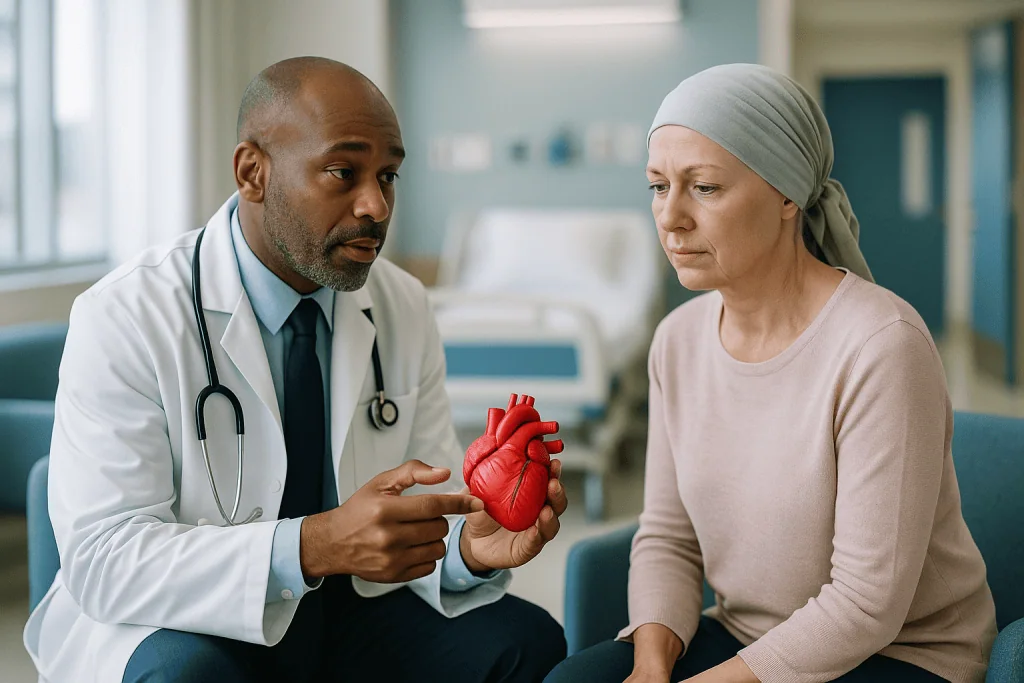
Anthracyclines illustrate the first pathway. Once inside heart cells they generate reactive oxygen species, unstable molecules that damage membranes and mitochondria, and they interfere with an enzyme called topoisomerase II beta that heart cells need to keep their DNA in order. Cells die, scar tissue forms, and the pumping chamber may gradually enlarge and weaken. Because the injury accumulates with each exposure, risk tracks the total lifetime amount received, a point the ESC guideline uses to define who needs closer surveillance.
A second pathway is interference with signaling rather than direct destruction. HER2-targeted antibodies, used in some breast and stomach cancers, block a receptor that heart cells also rely on for repair under stress. Function can fall while the drug is on board and often recovers when it is paused, which is why this type of dysfunction is described as largely reversible in guideline literature.
A third pathway runs through the blood vessels. Fluoropyrimidines can trigger coronary artery spasm, a sudden narrowing that produces chest pain even in healthy arteries. Drugs that block new blood vessel growth, the VEGF inhibitors, commonly raise blood pressure because the lining of vessels loses a key relaxing signal.
A fourth is inflammation. Immune checkpoint inhibitors release the brakes on the immune system so it attacks tumors, and in a small minority of people that unleashed immunity also attacks heart muscle. The ESC guideline reports myocarditis in roughly one percent of treated patients, uncommon but serious.
Environmental chemicals use similar routes: carbon monoxide starves the heart of oxygen, fine particles inhaled into the lungs trigger body-wide inflammation and clotting tendency, and heavy alcohol intake poisons muscle cells directly over years.
Cardiac toxic drugs examples: which treatments are most often involved?
People ask for a list, and it is fair to give one, with a caveat. Naming a class here is not a warning against it. Every drug below remains a standard treatment because its benefits are well established, and most people who receive these medicines never develop a heart problem from them.
| Drug class or exposure | Main heart effect seen | Usual pattern |
|---|---|---|
| Anthracyclines | Weakened pumping (cardiomyopathy) | Cumulative; may appear months to years later |
| HER2-targeted antibodies | Reduced ejection fraction | During treatment; frequently recovers on pausing |
| Fluoropyrimidines | Coronary spasm, chest pain | Early, often within the first cycles |
| VEGF pathway inhibitors | High blood pressure, clots | Within weeks of starting |
| Immune checkpoint inhibitors | Myocarditis (heart inflammation) | Uncommon; mostly in the first few months |
| Some tyrosine kinase inhibitors | Rhythm changes, QT prolongation | Variable; ECG-monitored |
| Chest radiation | Coronary, valve and pericardial disease | Years to decades afterward |
The timing column matters as much as the effect column. The ESC guideline groups cancer therapy-related cardiac dysfunction into forms that show up during treatment and forms that surface long after it ends, and monitoring schedules are shaped by that distinction. A person who finished anthracycline treatment years ago is not out of scope simply because the infusions are a memory.
Outside oncology, the same guideline literature and general cardiology sources flag a handful of everyday exposures: cocaine and amphetamine-type stimulants (acute vessel spasm and arrhythmia), long-term heavy alcohol (a dilated cardiomyopathy), certain antipsychotics and antibiotics (QT prolongation, a delay in the heart’s electrical reset), and some older diabetes and arthritis medicines (fluid retention or vascular events). Which of these applies to any one person is a conversation for the prescriber, not a list to self-audit.
What are the common symptoms of cardiovascular toxicity that clinicians watch for?
Here is the uncomfortable truth that shapes the whole field: the earliest heart injury from a toxic exposure usually has no symptoms at all. The pumping chamber can lose a measurable fraction of its strength while the person feels exactly as tired as they expected to feel during chemotherapy. Surveillance exists precisely because waiting for symptoms means waiting too long.

When symptoms do arrive, they tend to be the ordinary language of a struggling heart, which is why they are so easily blamed on the cancer or its treatment. Cleveland Clinic and MedlinePlus describe the pattern of heart failure as breathlessness that worsens with exertion or when lying flat, swelling of the ankles and legs, a persistent dry cough, and fatigue that feels disproportionate. A new awareness of the heartbeat, whether racing, fluttering or skipping, can point to a rhythm disturbance. Chest tightness or pressure during or shortly after an infusion is the signature of coronary spasm and is treated as urgent.
Myocarditis from immune therapy adds its own texture. It may begin with vague chest discomfort, breathlessness or light-headedness, but it can also travel with muscle weakness or inflammation elsewhere, because the same immune process is at work in several tissues. That combination is one reason teams ask about symptoms beyond the heart.
Environmental poisoning looks different again. Carbon monoxide exposure produces headache, dizziness, nausea and confusion before anything obviously cardiac, according to the CDC, and the heart strain is often discovered only on an ECG or blood test in the emergency department.
None of this is a checklist to diagnose yourself with. Breathlessness during cancer treatment has a dozen possible causes, most of them not the heart. Its purpose is to explain why your team asks the questions it asks at every visit, and why answering them precisely, including how many stairs before you stop, is genuinely useful clinical data.
What are the four main types of cardiomyopathies, and where does toxic damage fit?
Cardiomyopathy simply means disease of the heart muscle itself, as opposed to problems of the arteries, valves or electrical wiring. The NHS and Mayo Clinic describe four principal types, and knowing them makes the toxicology conversation far easier to follow.
Dilated cardiomyopathy is the one that matters most here. The main pumping chamber stretches and thins, like a balloon that has been inflated too many times, and each beat pushes out a smaller share of the blood inside. Anthracycline injury, long-term heavy alcohol use and some viral infections all produce this pattern. Ejection fraction, the percentage of blood the chamber ejects with each beat, is the number teams track.
Hypertrophic cardiomyopathy is the opposite geometry: the muscle wall thickens, usually because of an inherited gene change, and the chamber becomes stiff and sometimes obstructed. Toxic exposures are not a typical cause, though a person born with this condition may tolerate certain drugs less well.
Restrictive cardiomyopathy leaves the walls a normal thickness but rigid, so the chamber cannot relax and fill properly. Radiation fibrosis, the scarring that can follow chest radiotherapy, is one of the recognized routes to this pattern, alongside infiltrative diseases such as amyloidosis.
Arrhythmogenic cardiomyopathy, historically called arrhythmogenic right ventricular cardiomyopathy, involves muscle being replaced by fat and scar, mainly on the right side, producing dangerous rhythms. It is primarily genetic; intense endurance exercise can unmask it, but medicines rarely cause it.
Why does the classification matter to a patient? Because it shapes expectations. A dilated pattern caught early, with the exposure paused and supportive treatment started, has a meaningful chance of improvement in function, according to the ESC guideline’s summary of the evidence. A restrictive pattern from old radiation is more about managing fluid and symptoms than reversing structure. The label tells you what kind of conversation you are in.
Beyond the clinic: carbon monoxide, air pollution, metals, alcohol and stimulants
Most of the search traffic behind cardiovascular toxicology comes from cancer patients, but the largest number of affected hearts worldwide belong to people who were never prescribed anything. The World Health Organization attributes a substantial share of deaths from ischemic heart disease and stroke to ambient air pollution, and the American Heart Association has for years listed fine particulate matter as a recognized contributor to cardiovascular events.
The mechanism is not mysterious. Particles small enough to reach the deepest air sacs provoke inflammation that spills into the bloodstream, nudging arteries toward plaque rupture and blood toward clotting. Population studies see the effect as a rise in heart attacks and admissions on high-pollution days, a signal strong enough that WHO air-quality guidance is built around it. What no study can do is tell an individual whether last winter’s smog contributed to their particular event.
Carbon monoxide is the acute cousin. The gas binds to hemoglobin far more tightly than oxygen does, so the heart is asked to work hard while being starved of fuel. The CDC notes that people with existing heart disease are especially vulnerable, and that survivors of significant poisoning can carry heart injury forward. A working detector is the single most cost-free piece of cardiovascular toxicology advice anyone can give.
Heavy metals sit in the slow lane. Lead and arsenic exposure, mostly through contaminated water or occupational contact, are associated in NIH-summarized research with higher blood pressure and, over decades, more coronary disease. The associations are consistent, but the evidence remains observational, which is the honest grade.
Alcohol and stimulants round out the list. Sustained heavy drinking is a well-documented cause of dilated cardiomyopathy, and cocaine or amphetamines can trigger coronary spasm and life-threatening rhythms even in young people with clean arteries. These are not moral judgments; they are dose-and-time relationships a cardiologist will want to know about, without shame attached.
Who is usually offered cardio-oncology monitoring, and who is usually asked to wait?
Not everyone starting cancer treatment needs a cardiologist, and pretending otherwise would swamp clinics and frighten people for no gain. The ESC guideline approaches this with a baseline risk assessment before the first dose, sorting people into low, moderate, high and very high risk based on the planned treatment and the person’s own heart history.
Closer monitoring is usually offered when several of the following stack up: a treatment class with well-known cardiac effects, especially anthracyclines, HER2-targeted therapy or chest radiation; existing heart failure, cardiomyopathy or a prior heart attack; poorly controlled high blood pressure or diabetes; older age; previous exposure to the same drug class; or an abnormal baseline echocardiogram or blood marker. People in the higher tiers may be seen by cardiology before treatment starts so that blood pressure, cholesterol and any early dysfunction can be addressed while the oncology plan is being finalized.
People at low risk are usually asked to wait, in the sense that they proceed with standard oncology care and a baseline test, without a dedicated cardiology pathway unless something changes. That is not neglect. The guideline’s own evidence review shows that intensive surveillance in low-risk groups finds few problems and generates anxiety and unnecessary procedures.
A middle group exists too. Someone with, say, well-controlled hypertension starting a moderate-risk regimen might receive scheduled echocardiograms during treatment but no separate clinic. The exact cadence is a decision the oncology and cardiology teams make together, informed by the guideline and by how the person actually responds.
Two practical points. First, the assessment is only as good as the history you give, so an old diagnosis of a “heart murmur” or a relative with cardiomyopathy is worth mentioning. Second, risk tiers are not fixed. A person who begins at low risk and develops high blood pressure on treatment moves up, and monitoring moves with them.
How is cardiovascular toxicity detected? Echo, strain, biomarkers and ECG
Detection rests on four tools, each catching something the others miss.
The echocardiogram is the workhorse. An ultrasound probe on the chest produces moving images of the heart, from which the ejection fraction is calculated. The ESC guideline defines cancer therapy-related cardiac dysfunction largely in these terms: a fall in ejection fraction of ten percentage points or more to a value below fifty percent marks the moderate-to-severe range, while smaller drops, or changes in a more sensitive measure, define the mild form. Having a baseline scan before treatment is what makes any later comparison meaningful.
Global longitudinal strain is that more sensitive measure. Software tracks how much the muscle fibers shorten during each beat, and a relative decline of more than fifteen percent from baseline, per the ESC criteria, can flag injury before the ejection fraction has moved at all. Think of it as noticing a runner’s stride shortening before their lap time slows.
Blood tests add a chemical view. Troponin is a protein released when heart muscle cells are damaged; natriuretic peptides rise when the chambers are stretched under pressure. A rising troponin during anthracycline or checkpoint inhibitor treatment prompts closer imaging, and in suspected myocarditis it is one of the first tests ordered.
The electrocardiogram covers the electrical side: the QT interval for drugs that prolong it, new conduction blocks in myocarditis, and rhythm disturbances that imaging cannot see. Cardiac MRI is reserved for unclear cases, particularly suspected inflammation, where it can show swelling and scar in the muscle itself.
None of these is perfect. Ejection fraction varies a few points between scans done on different days by different operators, which is why teams look for consistent trends rather than reacting to a single reading. If your number dipped slightly and your team wants a repeat rather than a change of plan, that is measured practice, not indecision.
How are heart effects of cancer therapies managed once they are found?
Management is where the trade-off becomes explicit, and it is always a two-team decision. The oncologist knows how much a pause or switch would cost the cancer plan; the cardiologist knows how much function has been lost and how likely it is to return. Neither decides alone, and the patient’s own priorities sit at the table.
The first lever is the exposure itself. For a reversible pattern such as HER2-related dysfunction, a temporary hold while the heart is supported, followed by rechallenge once function recovers, is a recognized strategy in the ESC guideline. For anthracyclines, options include capping further exposure, switching to a different formulation or agent, or continuing with heightened surveillance when the cancer benefit clearly outweighs the risk. Coronary spasm from fluoropyrimidines may lead to stopping that drug and considering an alternative.
The second lever is standard heart failure therapy, applied earlier than it would be in the general population. Medicine classes that lower the workload on the heart and interrupt the hormonal signals that drive remodeling, such as angiotensin-converting enzyme inhibitors, angiotensin receptor blockers and beta-blockers, are the backbone. Their job is to give injured muscle the best conditions to recover and to prevent the chamber from stretching further. Blood pressure control matters just as much for VEGF inhibitor hypertension.
Myocarditis from checkpoint inhibitors is handled differently and urgently: the immune drug is stopped and high-intensity anti-inflammatory treatment is started in hospital, with monitoring for dangerous rhythms.
Radiation-related coronary or valve disease decades later is managed like any other coronary or valve disease, with the added consideration that scarred tissue can complicate procedures.
What patients most want to know, whether their own heart will recover, has an honest answer: often partly, sometimes fully, sometimes not, and earlier detection shifts the odds in the right direction. Which medicines, at what intensity and for how long, is a decision for the prescribing team and depends on the individual picture.
What do the following weeks and months usually look like?
Timelines vary enormously, so what follows is a typical shape drawn from guideline surveillance schedules, not a promise about any one person.
Before treatment, expect a baseline: a clinic history, blood pressure, an ECG, an echocardiogram and, for higher-risk regimens, troponin and natriuretic peptide levels. This usually happens in the one to three weeks between diagnosis and first infusion.
During treatment, the rhythm depends on the drug. HER2-targeted therapy typically comes with an echocardiogram roughly every three months while the drug continues, according to ESC guidance. Anthracycline regimens in moderate-risk patients often carry a repeat scan after the cumulative exposure crosses a threshold and again at the end of treatment; high-risk patients may be scanned more often with blood markers before selected cycles. Checkpoint inhibitor patients commonly have troponin and ECG checks concentrated in the first weeks, when myocarditis risk is highest.
If a change is picked up, the next few weeks usually involve a repeat scan to confirm, a cardiology review, and the start of protective heart medicines. Reassessment follows within weeks to a few months to see whether function is stabilizing or recovering. A paused cancer drug may be restarted once the picture is reassuring; that decision is made jointly and is never automatic.
After treatment ends, the ESC guideline recommends a reassessment within the first year for people who received cardiotoxic therapy, with longer-term follow-up scaled to risk. Survivors of high-dose anthracycline or chest radiation are advised to stay in periodic surveillance for years, because late effects can surface a decade or more later.
Day to day, most people notice very little of this beyond a few extra appointments. The scans are painless. The blood tests are the same draws you are already having. What changes is that someone is deliberately looking, which is the whole point.
Radiation and the long tail: why the heart is followed for decades
Radiotherapy earns its own section because its heart effects arrive on a different clock. Where a drug’s damage tends to declare itself within months or a few years, radiation to the chest sows changes that may not become clinically visible for ten, twenty or even thirty years, a range the ESC guideline draws from long-term survivor cohorts.
The mechanism is slow fibrosis. Radiation injures the lining of small blood vessels and stimulates scar formation in whatever lies in the beam: the coronary arteries, which develop premature plaque; the valves, which thicken and stiffen; the pericardium, the sac around the heart, which can become constricting; and the muscle itself, which may take on the restrictive pattern described earlier. The electrical system is not spared, and conduction blocks can appear late.
Modern techniques have changed the picture substantially. Breath-hold methods that move the heart away from the beam, more precise planning and lower total doses mean that a person treated for left-sided breast cancer today receives far less cardiac exposure than one treated a generation ago. That is a genuine advance, but the survivors of older treatment eras are still with us and still need follow-up.
Who should be on that follow-up list? The ESC guideline highlights people who received significant heart exposure from mediastinal or left breast radiation, particularly if they were young at the time, if they also received anthracyclines, or if they carry other risk factors such as smoking, diabetes or high cholesterol. Those risk factors compound the radiation effect rather than simply adding to it, which makes their control the most powerful lever a survivor has.
The practical translation is simple. If you had chest radiation in the past, make sure whoever manages your general health knows it, because a symptom that would be dismissed in most forty-year-olds deserves an earlier look in you.
What people often get wrong about cardiotoxicity of chemotherapy
Myths in this area tend to run in two opposite directions, both harmful.
“If chemotherapy can damage the heart, I should refuse it.” The trade-off is real, but for most regimens the cancer risk being treated is far greater than the cardiac risk being taken, and modern surveillance exists precisely to catch problems early. Declining effective treatment out of heart fear is a decision that should only ever follow a detailed conversation with both teams, never a search result.
“My heart was fine on the last scan, so it is fine for good.” Some effects are cumulative and some are delayed. A normal echocardiogram halfway through anthracycline treatment is reassuring about that moment, not a permanent clearance, and radiation effects can surface decades later.
“Heart damage always causes symptoms.” Early dysfunction is usually silent. That is why the monitoring is scheduled rather than symptom-triggered.
“Once the ejection fraction falls, it never recovers.” It can, particularly when the exposure is paused and protective treatment starts early, and especially for the reversible patterns seen with HER2-targeted therapy. Recovery is not guaranteed, but permanence is not either.
“Supplements can protect my heart during chemo.” There is no high-quality evidence that any over-the-counter antioxidant or herbal product prevents cardiotoxicity, and some interact with cancer drugs. The NIH Office of Dietary Supplements is candid that antioxidant supplement trials have generally failed to show benefit for heart disease. Tell your team about anything you take.
“Air pollution and chemicals only matter for lungs.” The cardiovascular burden of fine particulate pollution exceeds the respiratory burden in WHO and AHA estimates.
“Exercise is dangerous for a treated heart.” Guided, moderate activity is encouraged in guideline literature during and after cancer treatment for its cardiovascular and functional benefits; the caution is about unsupervised extremes, not movement itself.
Living well during and after treatment: what the evidence actually supports
The things a person can do themselves are unglamorous and effective, and they are the same things that protect every heart.
Blood pressure, cholesterol and blood sugar control head the list, because the ESC guideline is explicit that conventional risk factors multiply the harm from cardiotoxic treatment. Someone with well-managed hypertension entering anthracycline therapy carries a different risk from someone whose readings have drifted unchecked for years. This is the moment to take those numbers seriously, and your primary care clinician or cardiologist will guide any medicine decisions.
Physical activity is the second lever. Cardio-oncology rehabilitation programs, modeled on cardiac rehabilitation after a heart attack, have been described in AHA scientific statements as a reasonable approach for survivors at elevated cardiovascular risk. The rationale is that regular aerobic exercise improves how efficiently the heart and muscles use oxygen, blunts treatment-related fatigue and helps blood pressure. Intensity should be set with the team, particularly during active treatment, but sitting still is not the safe option it feels like.
Smoking cessation is non-negotiable in evidence terms; smoking compounds both radiation-related and chemotherapy-related vascular injury. Alcohol deserves a frank conversation, since heavy intake adds a second cardiomyopathy pathway on top of the first.
Diet evidence is broader and less specific. A pattern rich in vegetables, legumes, whole grains, fish and unsaturated fats, as summarized by the American Heart Association, is associated with lower cardiovascular risk across populations. No single food protects against cardiotoxicity, and supplements are covered above.
Finally, information continuity. Survivors change cities, doctors and health systems. A one-page summary of what you received, including drug classes, radiation fields and any heart findings, is the most valuable document you can carry, because the clinician you meet in fifteen years cannot monitor a risk they do not know exists.
Questions to ask your care team about cardiovascular toxicology explained for your case
Good questions turn a monitoring plan from something done to you into something you understand. These are the ones cardio-oncology clinicians say they most wish patients asked.
- Which of my planned treatments have known heart effects, and what kind: pumping, rhythm, blood pressure or arteries?
- How have you rated my baseline heart risk, and what pushed me into that category?
- What heart tests will I have before, during and after treatment, and roughly how often?
- Who reviews those results, and how will I hear about a change?
- What change on a scan or blood test would prompt you to alter the plan, and what would the options be at that point?
- If a heart problem appears, would it usually be expected to improve, and what determines that?
- Are there heart risk factors I should work on now, before treatment starts, and who helps me with them?
- Is exercise encouraged during my treatment, and is there a rehabilitation program available?
- Do any of my existing medicines or supplements interact with the planned cancer drugs in a way that affects the heart?
- After treatment ends, how long should heart follow-up continue, and who owns it if I move or change doctors?
- Did or will my radiation field include the heart, and should that change how future symptoms are handled?
- What symptoms should make me call the same day, and which number do I use out of hours?
Bring someone to write down the answers if you can. Oncology consultations are dense, and the cardiac details are easy to lose among everything else. Asking for a written copy of your surveillance schedule is entirely reasonable, and most teams will provide it.
One question worth avoiding is “What are my chances?” phrased as a single percentage. The honest answer is a range that depends on factors nobody can fully know in advance, and a good clinician will explain that range rather than pick a number to satisfy the question.
When to call your doctor: red-flag signs during and after cancer treatment
Most days during treatment will be unremarkable from a heart standpoint. A small number of signs, though, should never be sat on until the next scheduled appointment.
Call emergency services immediately for chest pain or pressure, especially during or within a day of an infusion; sudden severe breathlessness; fainting or near-fainting; a heartbeat that is very fast, chaotic or accompanied by dizziness; or any combination of headache, confusion and nausea in a household where a fuel-burning appliance is running, which can signal carbon monoxide poisoning.
Contact your oncology or cardiology team the same day for new breathlessness on exertion that was not there last week, needing to sleep propped up on extra pillows, new swelling of the ankles or abdomen, a weight gain of several pounds over a few days that suggests fluid, a persistent new cough, palpitations that come and go, or muscle weakness or double vision during immune checkpoint therapy, since these can accompany myocarditis.
Raise at your next visit, but do not ignore, blood pressure readings that are consistently higher than your usual, unusual fatigue out of proportion to the treatment phase, or any symptom that simply worries you. Teams would always rather hear about something that turns out to be nothing.
For survivors years out from treatment, the same list applies with one addition: mention your treatment history whenever you present with chest or breathing symptoms, so the clinician in front of you knows to consider the heart earlier than they otherwise might.
Every decision about pausing, continuing or changing treatment, and about any heart medicine, belongs to the treating team who know your full picture. The purpose of recognizing these signs is to get you in front of them quickly, not to interpret them yourself.
Frequently asked questions
What is cardiovascular toxicology?
Cardiovascular toxicology is the branch of toxicology that studies how medicines, cancer treatments and environmental chemicals damage the heart and blood vessels, and how that damage is prevented, detected and managed. It covers heart muscle weakening, rhythm disturbances, blood pressure effects, coronary artery injury and valve disease. Its clinical arm, cardio-oncology, applies these principles to people receiving cancer therapy through risk assessment and scheduled monitoring.
What are the common symptoms of cardiovascular toxicity?
The most common symptoms are breathlessness on exertion or lying flat, swelling of the legs, persistent cough, unusual fatigue, palpitations and chest discomfort, but early injury frequently causes no symptoms at all. That silence is why teams rely on echocardiograms and blood tests rather than waiting for complaints. Any new chest pain or severe breathlessness during treatment is an emergency, not something to note for the next appointment.
What are the four main types of cardiomyopathies?
The four main types of cardiomyopathy are dilated, hypertrophic, restrictive and arrhythmogenic. Dilated cardiomyopathy, where the pumping chamber stretches and weakens, is the pattern most often linked to anthracyclines and heavy alcohol. Restrictive cardiomyopathy, with stiff walls that fill poorly, can follow chest radiation. Hypertrophic and arrhythmogenic forms are mainly inherited and are rarely caused by toxic exposures, though they can affect how a person tolerates certain drugs.
What are some examples of cardiac toxic drugs?
Well-recognized cardiac toxic drugs include anthracycline chemotherapy, HER2-targeted antibodies, fluoropyrimidines, VEGF pathway inhibitors, immune checkpoint inhibitors and some tyrosine kinase inhibitors, alongside non-cancer exposures such as cocaine, amphetamines, heavy alcohol and certain QT-prolonging antibiotics or antipsychotics. Naming a class is not a warning against it; each remains a standard treatment because its benefits are established, and most people who receive it never develop a heart problem.
How is cardiotoxicity of chemotherapy detected before it causes harm?
Cardiotoxicity of chemotherapy is detected mainly by comparing echocardiograms against a baseline scan taken before treatment, looking at ejection fraction and a more sensitive measure called global longitudinal strain. Blood tests for troponin and natriuretic peptides add a chemical view of muscle injury and chamber stretch, and ECGs cover rhythm and electrical timing. Teams look for consistent trends across tests rather than reacting to any single reading.
Can the heart recover after cancer treatment damages it?
The heart can recover partly or fully in many cases, especially when the change is caught early, the exposure is paused where possible and protective heart medicines are started promptly. Dysfunction linked to HER2-targeted therapy is described in guideline literature as largely reversible, while anthracycline injury is more variable. Recovery is never guaranteed, and the degree depends on how much function was lost, how quickly it was found and individual factors.
How long after chemotherapy or radiation can heart problems appear?
Heart problems can appear during treatment, within the first year afterward, or many years later depending on the exposure. Anthracycline-related dysfunction may surface months to years after the last dose, and radiation effects on coronary arteries and valves are described in the ESC cardio-oncology guideline as emerging ten to thirty years after treatment. This long tail is why survivors of higher-risk treatment are advised to stay in periodic cardiac follow-up.
Does air pollution really affect the heart?
Yes, fine particulate air pollution is recognized by the World Health Organization and the American Heart Association as a contributor to heart attacks, strokes and heart failure at population level. Inhaled particles trigger body-wide inflammation and a tendency toward clotting, and hospital admissions rise on high-pollution days. What the evidence cannot do is confirm whether pollution caused any single person’s event, because the effect is measured across large populations.
Do antioxidant supplements protect the heart during chemotherapy?
No high-quality evidence shows that antioxidant or herbal supplements prevent cardiotoxicity during chemotherapy, and some products interact with cancer drugs or interfere with how they work. The NIH Office of Dietary Supplements notes that antioxidant supplement trials have generally not reduced heart disease. Tell your oncology team about anything you take, and rely on the proven levers: blood pressure control, activity, not smoking and scheduled monitoring.
Should I avoid exercise if my treatment can affect the heart?
Guided, moderate exercise is generally encouraged during and after cancer treatment rather than avoided, because it supports heart efficiency, eases fatigue and helps blood pressure. Cardio-oncology rehabilitation programs modeled on cardiac rehabilitation have been proposed in American Heart Association scientific statements for survivors at higher cardiovascular risk. Intensity should be agreed with your team, particularly if your heart function has changed, but inactivity is not the safer choice.
References
This article is for general information only and is not a substitute for professional medical advice. Please consult a qualified doctor about your individual situation.

More from the Blog

Saturated Fat: Where the Evidence Actually Stands in 2026
Saturated fat raises LDL cholesterol, and randomized trials show that replacing it with unsaturated fat lowers the risk of cardiovascular events by roughly 17…

Robotic Heart Surgery Recovery: Light Activity in Weeks and Full Exertion Over Two Months
Robotic heart surgery recovery time is usually shorter than recovery after open-chest surgery because the breastbone is left intact. Most people leave the hospital…

Dehydration, Stress and Palpitations: The Everyday Triggers
Yes, dehydration can cause heart palpitations. When you lose fluid, your blood volume drops, so the heart beats faster and harder to keep blood…

What Transplant Cardiology Covers: Heart Transplant Evaluation and Lifelong Follow-Up Care
Transplant cardiology is the branch of heart medicine that assesses people with advanced heart failure for a heart transplant or a mechanical heart pump,…
How Transcatheter Mitral Clip Repair Reduces Regurgitation by Joining the Valve Leaflets
Transcatheter mitral clip repair, also called transcatheter edge-to-edge repair or TEER, treats a leaking mitral valve without open-heart surgery. A cardiologist threads a catheter…

MASLD: The New Name for Fatty Liver Disease and What the Renaming Changes
MASLD, or metabolic dysfunction-associated steatotic liver disease, is the name adopted in 2023 to replace nonalcoholic fatty liver disease (NAFLD). It describes fat buildup…