

Carotid Doppler, CT Angiography or MRI: Which Scans Map Cerebrovascular Disease?

Key Takeaways
- A carotid Doppler is really a duplex ultrasound: grayscale imaging shows the artery wall while Doppler measures blood speed, which rises as a narrowing tightens.
- A CT angiogram differs from a standard contrast CT by capturing the brief arterial phase when dye is concentrated in arteries, allowing the channel to be measured.
- Ultrasound cannot see through the skull, so disease in the intracranial arteries or the aortic arch needs CT angiography or MR angiography to be seen.
- MRI's diffusion-weighted sequence can show a fresh stroke within hours, when CT often still looks normal, linking brain injury to a narrowed carotid.
- The NHS describes carotid surgery as usually considered for people with symptoms and narrowing of 50% or more, ideally within 2 weeks of the event.
- A complete carotid blockage is generally managed with medicines rather than surgery, because the brain has already adapted to alternative blood routes.
Carotid Doppler (duplex ultrasound) is usually the first scan for suspected carotid narrowing because it uses no radiation or contrast and measures blood flow speed. CT angiography gives a detailed anatomical map from the aortic arch to the brain using X-rays and iodine contrast. MRI and MR angiography show brain tissue and vessels without X-rays. Teams often combine two tests before any treatment decision.
The referral letter said three words that sounded like one: duplex, CTA, MRA. The man holding it had come in with twenty minutes of a droopy face and a clumsy hand, both now gone, and the clinic wanted pictures of his neck arteries before the week was out. He wanted to know which picture was the real one.
That question sits at the heart of the carotid doppler vs ct angiography debate. The two scans look at the same artery, yet they answer different questions. One listens to how fast blood is moving through a narrowing. The other draws the vessel wall itself, calcium and all, from the chest to the base of the skull. MRI adds a third voice, showing what the brain tissue downstream has already been through.
None of them is the whole truth on its own. This explainer walks through what each scan actually does, why teams often order more than one, and how the results shape the conversation about treatment.
Why one neck artery gets three different scans
The carotid arteries run up either side of the neck, and each one forks just below the jaw. The internal carotid continues to the brain; the external carotid feeds the face and scalp. That fork, the carotid bifurcation, is where fatty deposits called plaque most often build up. Plaque narrows the channel and, more dangerously, can crack and shed fragments that travel to the brain. MedlinePlus describes this process as carotid artery disease, a leading contributor to ischemic stroke.
Imaging has three separate jobs here. The first is to detect narrowing, called stenosis, and estimate how severe it is. The second is to characterize the plaque: is it smooth and calcified, or ulcerated and soft? The third is to look beyond the neck, at the arch of the aorta where the carotids originate and at the smaller arteries inside the skull, because disease rarely sits in one place.
No single scan does all three jobs equally well. Ultrasound is superb at the first job and reasonable at the second, but it cannot see inside the skull or down into the chest. CT angiography covers the whole route from heart to brain and shows calcium crisply, yet it needs X-rays and iodine contrast. MRI shows the brain tissue itself and can image vessels without any contrast at all, but it is slower and sensitive to movement and metal.
This is why a person with a suspected mini-stroke may leave the clinic with two or even three appointments. The team is not being indecisive. It is building a layered picture, where each layer checks the one before it. Understanding that logic makes the rest of the process far less bewildering.
How does a carotid Doppler actually work?
A carotid Doppler is more properly called a carotid duplex ultrasound, because it combines two kinds of sound-wave imaging in one examination. The first, grayscale or B-mode imaging, builds a still picture of the artery wall and any plaque clinging to it. The second, Doppler, measures how fast blood is moving by detecting the change in pitch of sound waves bouncing off red blood cells, the same principle that makes a passing siren drop in tone.

The speed matters because of simple plumbing. Water forced through a pinched garden hose jets out faster. Blood passing through a narrowed carotid does the same, so the sonographer records the peak speed during each heartbeat, known as peak systolic velocity, along with the speed at the end of the beat. Those velocities are compared with published thresholds to estimate how tight the narrowing is. Color Doppler overlays this flow information on the picture, painting fast turbulent flow in bright, mixed hues.
Mayo Clinic describes the test as painless and typically taking about 30 minutes. You lie on your back with your head turned slightly, gel is applied to the neck, and a handheld probe is moved slowly along the artery. There are no needles, no X-rays and no contrast dye, which is why most guideline pathways place it first in line.
Its limits are physical. Sound cannot pass through bone, so the artery disappears from view once it enters the skull. Heavy calcium in a plaque casts an acoustic shadow that hides what lies beneath. A short, thick neck or a very high bifurcation can make the picture hard to obtain. And because velocity is an indirect measure, a very tight narrowing with almost no flow can occasionally read as lower than it is. Good sonographers know these traps; so do the radiologists who read the results.
What does a CT brain and carotid angiogram show?
A CT brain and carotid angiogram is one coordinated scan with two parts. A computed tomography (CT) scanner uses a rotating X-ray beam to build cross-sectional slices of the body. For the angiogram portion, iodine-based contrast is injected into an arm vein and the scanner is timed to capture the moment that dye fills the arteries. Software then stitches the slices into a three-dimensional map of the vessels, which a radiologist can rotate, slice and measure on screen.
The carotid part shows the arteries from their origin at the aortic arch, up through the neck, to where they enter the skull and branch across the brain. Narrowings are measured directly, usually by comparing the residual channel against the normal artery beyond it. Calcium appears bright white, so hardened plaque is obvious. Ulceration, where the plaque surface has broken, can often be seen as a small pocket of contrast. The brain portion, which may be scanned without contrast first, looks for areas of established stroke, bleeding or other causes of symptoms.
Mayo Clinic notes that the scanning itself is quick, generally a matter of minutes, though the whole appointment including preparation and a short observation period typically takes longer. You lie still on a narrow table that slides through a doughnut-shaped ring. Many people describe a warm flush and a metallic taste as the contrast goes in; both pass within a minute or so.
What CT angiography (CTA) cannot do is measure flow. It is a snapshot of anatomy, frozen in time. Nor does it show the brain tissue as sensitively as MRI, particularly in the first hours after a small stroke. Those gaps are exactly what the other two scans fill.
Is a CT with contrast the same as a CT angiogram?
They share a scanner and a dye, but they are not the same test. The difference is timing, and timing is everything in vascular imaging.
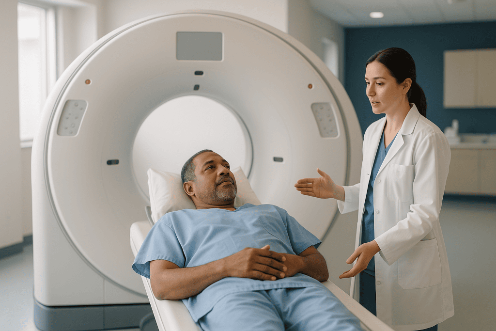
In a routine contrast-enhanced CT, the scanner waits a minute or more after the injection, by which point the contrast has spread out of the arteries into the veins, organs and soft tissues. That is ideal for spotting a tumor, an abscess or an inflamed bowel, because abnormal tissue takes up dye differently from healthy tissue. The arteries are visible, but they are no brighter than everything around them, so a narrowing is easy to miss.
A CT angiogram captures the arterial phase, the brief window, often only seconds long, when dye is concentrated in the arteries and has not yet reached the veins. To hit that window, the team uses a faster injection, often a small test dose or automated tracking to detect the moment the contrast arrives, and thinner slices. The result is arteries that glow against a dark background, which is what allows the radiologist to measure a channel to the millimeter.
The practical consequences for you are modest. The preparation, the arm cannula and the warm flush feel much the same. The report, however, will look very different. A CTA report describes vessel caliber, plaque, stenosis grade and anatomy of the arch and intracranial arteries. A standard contrast CT report may mention the carotids only in passing.
This is why a clinic that already has an old contrast CT of your neck on file may still request a dedicated CTA. The earlier scan was not wrong; it was simply asked a different question. The ct angiogram vs ct with contrast distinction, in short, is about what the scanner was looking for when it fired.
MRA vs CTA for carotid disease: where MRI fits in
Magnetic resonance imaging (MRI) uses a strong magnetic field and radio waves, not X-rays, to build pictures from the hydrogen atoms in water molecules. The NHS explains that a scan involves lying inside a large tube while the machine makes loud tapping noises, with the whole examination lasting anywhere from 15 to 90 minutes depending on how many sequences are needed.
For cerebrovascular disease, MRI contributes two things. The first is a view of the brain tissue itself. A sequence called diffusion-weighted imaging is particularly sensitive to fresh ischemic injury, often showing a small stroke within hours when a CT still looks normal. Finding such a spot in the territory supplied by a narrowed carotid links the two together and changes how urgently the narrowing is treated.
The second contribution is magnetic resonance angiography (MRA), a set of sequences that picture the arteries. Time-of-flight MRA relies on the signal from moving blood and needs no injection at all. Contrast-enhanced MRA uses a gadolinium-based agent, a different type of dye from the iodine used in CT, to sharpen the vessel picture, especially from the arch upward.
Set against CTA, MRA avoids radiation and iodine, which matters for younger people, those with iodine contrast reactions, and those with reduced kidney function (though gadolinium carries its own kidney-related cautions). Its weaknesses are also real. Turbulent flow at a tight narrowing can drop the signal and make the stenosis look worse than it is. Motion blurs the picture. Pacemakers, some implants and metal fragments may rule the scan out entirely, and some people find the enclosed space difficult.
In many pathways, MRI of the brain is ordered for the tissue question while either CTA or ultrasound answers the artery question. Which vascular test accompanies it depends on local expertise, the person’s kidneys and how quickly an answer is needed.
Carotid Doppler vs CT angiography: how each measures a narrowing
The two scans estimate the same number, the percentage of narrowing, by completely different routes. Knowing this explains why their results sometimes disagree.
CTA measures geometry. The radiologist finds the narrowest point, measures its diameter, then measures a normal segment of the internal carotid farther up and expresses the first as a fraction of the second. This mirrors the method used in the large surgical trials of the 1990s, so the thresholds that guide treatment were built around numbers of this kind. The measurement is reproducible but depends on image quality, on where the normal reference segment is chosen and on how well calcium is distinguished from contrast when the two sit side by side.
Duplex ultrasound measures physiology. The sonographer records velocities and compares them with validated ranges that correspond to bands of narrowing. Velocity rises steadily as the channel tightens, which makes the method sensitive for moderate and severe disease. But velocity depends on more than the narrowing. A tight stenosis on the opposite side can raise velocities in the artery being examined. A heart with weak output can lower them. Blood pressure, heart rhythm and the angle of the probe all nudge the numbers.
Both methods, then, carry known sources of error, and they tend to err in different directions. This is the practical case for using them together when an intervention is being considered: concordant results from two independent methods are far more reassuring than one result alone. When the two disagree substantially, teams typically repeat one of them, add MRA, or in selected cases turn to catheter angiography, the invasive reference standard in which contrast is injected directly into the artery through a thin tube. That last step is now uncommon for diagnosis alone, reserved for situations where the non-invasive tests have left a genuinely unanswered question.
Is a carotid ultrasound the same as a CT scan?
People searching for carotid ultrasound vs ct scan are often trying to work out whether they have been sent for a repeat of something they have already had. They have not. The two tests use different physics, different rooms and different staff, and they rarely substitute for one another.
Ultrasound is sound. A probe on the skin sends pulses into the neck and listens for echoes, building a live, moving picture that the sonographer can adjust in real time. There is no radiation, no injection and no tunnel. The person performing it, a vascular sonographer or technologist, is a skilled operator, and the quality of the result depends heavily on that skill. It is a focused test: it sees the carotid and vertebral arteries in the neck, and that is essentially all.
CT is X-rays. A rotating beam and detector capture hundreds of thin slices in seconds, which a computer reconstructs into three-dimensional images. The operator sets up the scan but the images are standardized and far less dependent on the hands of the person present. A CTA sees the whole vascular tree from the chest to the top of the head, plus the brain, bone and soft tissues along the way. The cost of that breadth is a dose of ionizing radiation and the need for iodine contrast.
Which comes first? Most stroke and vascular pathways favor ultrasound as the initial screen, precisely because it is safe enough to repeat and good enough to rule out significant disease in many people. CTA tends to enter when ultrasound shows a narrowing that might need treatment, when the ultrasound was technically limited, or when the clinical picture points to disease beyond the neck. The order can vary with local resources, and in an emergency setting a CTA may be done straight away because the scanner is already being used for the brain.
Side by side: carotid Doppler, CT angiography and MRI compared
The table below gathers the practical differences in one place. It is a guide to what each scan is good at, not a ranking; the right test depends on the question being asked, the person’s health and what is available locally.
| Feature | Carotid Doppler (duplex ultrasound) | CT angiography (CTA) | MRI with MR angiography (MRA) |
|---|---|---|---|
| What it uses | Sound waves | X-rays plus iodine contrast | Magnetic field and radio waves; gadolinium contrast optional |
| What it measures | Blood flow velocity and vessel wall in the neck | Vessel anatomy from aortic arch to brain | Brain tissue plus vessel anatomy |
| Radiation | None | Yes | None |
| Injection | None | Iodine contrast via arm vein | None for time-of-flight MRA; gadolinium for contrast-enhanced MRA |
| Sees inside the skull | No | Yes | Yes |
| Shows calcium clearly | Partly; can shadow the picture | Yes, very clearly | Poorly |
| Shows fresh brain injury | No | Limited in early hours | Yes, highly sensitive |
| Typical duration | About 30 minutes (Mayo Clinic) | Scanning takes minutes; appointment longer (Mayo Clinic) | 15 to 90 minutes (NHS) |
| Main cautions | Operator-dependent; limited by bone, calcium, neck anatomy | Kidney function, iodine contrast reactions, radiation | Pacemakers and some implants, claustrophobia, motion; may overestimate tight narrowing |
| Usual role | First-line screen and follow-up | Confirmation and treatment planning; emergency stroke imaging | Brain tissue assessment; vessel imaging when CTA is unsuitable |
Two patterns stand out. Ultrasound and MRI share the advantage of no radiation, which is why they dominate repeat surveillance. CTA and MRI share the ability to look inside the skull, which ultrasound simply cannot do. The scan that is right for a one-off emergency assessment may be the wrong one for checking the same artery every year, and vice versa.
Who is usually scanned first, and who is usually asked to wait
Urgency, more than anything else, decides who gets which scan and when.
People who have had a transient ischemic attack (TIA), a brief episode of stroke-like symptoms that resolves, or a minor stroke sit at the front of the queue. The reason is risk: the chance of a full stroke is highest in the days immediately after such an event, and if a tight carotid narrowing is responsible, the surgical trials showed the greatest benefit when treatment followed quickly. The NHS notes that carotid endarterectomy, the operation to clear the plaque, is ideally carried out within 2 weeks of symptoms when the artery is narrowed by 50% or more. Imaging must therefore happen within hours to days, and many pathways run ultrasound or CTA as part of the same-day or next-day assessment, with CTA often chosen in the emergency department because the brain is being scanned anyway.
A second group is people without symptoms in whom a narrowing has been found by chance, perhaps because a clinician heard a whooshing sound, called a bruit, over the neck, or because the carotid appeared on a scan done for another reason. Here the pace is measured. Ultrasound usually confirms and grades the finding, and the conversation turns to risk-factor control and whether any procedure would help, a question on which guidelines are cautious and the evidence is still evolving.
Then there are people who are asked to wait or who are steered toward a different test. Someone with significantly reduced kidney function may be directed away from CTA toward ultrasound and non-contrast MRA. A person with a pacemaker that is not MRI-conditional will not have MRI. Someone who has had a serious reaction to iodine contrast will have that noted before any CTA is booked. None of this is a denial of care; it is matching the tool to the person. The treating team weighs all of it, and the final choice is theirs to make with you.
What actually happens on the day of each scan
Knowing the sequence of a scan removes most of the anxiety around it, so here is each one in plain order.
For the carotid Doppler, you can usually eat and drink normally. You will be asked to remove necklaces and open or remove clothing around the neck. Lying on your back with a small pillow under your shoulders, you turn your head away from the side being examined. The gel is cool, the probe presses firmly but not painfully, and you may be asked to breathe out slowly or hold still while a measurement is taken. You can go about your day straight afterward.
For CT angiography, the preparation varies by department. Some ask you not to eat for a few hours beforehand; most will check your kidney function with a blood test and ask about previous contrast reactions, allergies and whether you could be pregnant. A small cannula is placed in an arm vein. You lie on the table, arms above your head or by your sides as instructed, and the table moves through the ring while a voice tells you when to hold your breath. The contrast injection brings a sudden warmth that spreads through the body and sometimes a sense that you have passed urine, though you have not. The scanning itself is over quickly. Afterward you are typically observed briefly and encouraged to drink fluids, which helps the kidneys clear the dye.
For MRI, you complete a safety questionnaire about metal in the body, change into a gown and remove anything magnetic. You lie on a sliding table, often with a padded frame around the head that contains the receiving coils, and are given earplugs or headphones for the noise. The NHS describes individual sequences lasting from a few seconds to a few minutes each, with the whole appointment running 15 to 90 minutes. Staying still is the single most useful thing you can do. If gadolinium is used, it is injected part-way through via a cannula. There is a call button in your hand throughout.
What happens if they find a blockage during a CT angiogram?
Finding a narrowing on CTA is common; it is often why the scan was ordered. What happens next depends on three things: how severe the narrowing is, whether it is on the side that matches your symptoms, and whether there is a complete blockage rather than a tight narrowing.
If the scan is being done in an emergency because of an acute stroke, a blockage in a large artery may trigger a rapid conversation about treatments that reopen the vessel, which is one reason emergency departments scan the arteries alongside the brain. That is a time-critical pathway with its own criteria, and the stroke team leads it.
Outside the emergency setting, the radiologist reports the degree of narrowing, describes the plaque and notes any disease elsewhere. The report goes to your treating clinician, who places it alongside your symptoms, your ultrasound if you had one, and your overall health. Discordance between tests is checked rather than ignored. A moderate or severe narrowing on the symptomatic side opens a discussion about carotid endarterectomy or stenting, procedures that aim to reduce the risk of future stroke; the NHS sets out the 50% narrowing threshold at which surgery is usually considered for people with symptoms. Lower grades of narrowing, or narrowing without symptoms, generally lead to intensive management of risk factors with medicines and lifestyle, plus planned follow-up imaging.
A complete blockage, counterintuitively, is not usually operated on. Once an internal carotid has fully closed, the brain on that side is being supplied through other routes, and reopening the vessel carries risks that the trials did not show to be worthwhile. Attention shifts to protecting those alternative routes and controlling risk factors.
Whatever the finding, it should come with a conversation rather than a letter alone. If the result reaches you before anyone has explained it, that is a reasonable thing to ask about.
The days and weeks after the scan: what the pathway usually looks like
The scan is the beginning of a sequence, not the end of one, and the pace of that sequence tracks the urgency of the original symptoms.
After the scan itself, the practical aftercare is light. Ultrasound needs none. After CTA, people are generally advised to drink well for the rest of the day; those taking certain diabetes medicines may be given specific instructions about pausing them around the contrast, which the prescribing clinician decides. After MRI with gadolinium, there is likewise little to do beyond watching the cannula site. Any delayed reaction, such as a rash or swelling appearing hours later, should be reported.
The report typically reaches your clinician within days, faster in urgent pathways where a radiologist may read the study the same day. For someone who has had a TIA or minor stroke, the next weeks are busy by design. The NHS describes the window for carotid surgery in symptomatic people as ideally within 2 weeks of symptoms, so a tight narrowing on the relevant side can lead to a surgical consultation within days of the scan. In parallel, medicines that reduce clotting and lower cholesterol and blood pressure are usually started or adjusted, and most people are advised not to drive for a period that their clinician will specify.
For someone with a narrowing found incidentally, the timeline stretches out. A follow-up ultrasound at an interval chosen by the team, often measured in months to a year, checks whether the plaque is stable. Blood pressure, cholesterol, blood glucose and smoking become the daily work, because these are what drive plaque growth.
Either way, keep a written list of which scans you have had and what each showed. Carotid disease is followed over years, and a clear personal record saves a great deal of repetition.
Risks, contrast, radiation and the honest trade-offs
Each scan carries risks that are small but worth understanding, because they explain why clinicians do not simply order the most detailed test for everyone.
Ultrasound has no known physical risk at diagnostic intensities. Its real risk is informational: a limited or misread study can either miss a narrowing or overstate one. That is why findings that would change treatment are usually confirmed by a second method.
CTA carries two specific concerns. Iodine contrast can, uncommonly, provoke an allergic-type reaction ranging from hives to, rarely, a severe response; departments screen for previous reactions and have treatment on hand. Contrast can also strain kidneys that are already impaired, which is why kidney function is checked beforehand and fluids are encouraged afterward. The radiation dose from a head and neck CTA is modest in absolute terms, but it is not zero, and repeated scans accumulate. For a one-off assessment in an older adult the balance clearly favors scanning; for annual surveillance in a younger person, ultrasound or MRA usually makes more sense.
MRI’s risks are mostly about the magnet. Ferromagnetic objects, inside or outside the body, can move or heat, so screening is strict. Gadolinium agents are generally well tolerated but have been linked to a rare serious condition in people with severe kidney disease and to small deposits in tissues whose significance is still being studied; newer agents are considered more stable. Claustrophobia is a genuine barrier for some, and open or wide-bore scanners, or simply a sedative chosen by the clinician, can help.
Finally, there is the risk shared by all three: finding something unexpected. A CTA of the neck will image the thyroid, the upper lungs and the spine along the way, and incidental findings there sometimes lead to further tests. Most prove harmless. It is still worth knowing before you lie down that a scan can raise questions as well as answer them.
What people often get wrong about carotid scans
Several beliefs about these scans circulate widely enough to deserve a direct correction.
The first is that the more expensive-looking scan must be the better one. Detail and usefulness are not the same thing. For deciding whether a narrowing is present and roughly how severe it is, ultrasound performs well and can be repeated without harm. CTA earns its place when the question is anatomical, not when it is merely available.
The second is that a clear carotid scan means no stroke risk. Carotid narrowing is one cause of ischemic stroke among several. Irregular heart rhythms that fling clots from the heart, disease in the small arteries deep in the brain, and problems with the vertebral arteries at the back of the neck can all produce the same symptoms with pristine carotids. A normal duplex is reassuring about one pathway, not about all of them.
The third is that a percentage on the report is an exact measurement. It is an estimate, derived by different methods on different scans, each with a margin of error. Two reports giving slightly different numbers are not a sign that someone made a mistake.
The fourth is that a complete blockage is the most dangerous finding and must be opened. As discussed earlier, a fully occluded carotid is generally managed medically rather than surgically, because the brain has already adapted and the operation would not help.
The fifth is that symptoms which went away do not count. They count enormously. A TIA is treated as a warning precisely because the stroke risk that follows is front-loaded into the first days and weeks. Imaging done promptly after a transient episode is some of the most consequential imaging in all of medicine.
And the last: that a scan can be requested like a menu item. Which test, and when, is a clinical judgment that depends on your history, your kidneys, your implants and your symptoms. Asking why a particular scan was chosen is always fair; expecting a specific one on demand usually is not.
Questions to ask your care team
A short list of questions, asked before the scan or when the result arrives, tends to produce clearer answers than a long one asked in a rush. These are the ones that matter most.
- Which scan am I having first, and what specific question is it meant to answer?
- If this scan shows a narrowing, will I need a second scan to confirm it before any treatment is discussed?
- Does this scan involve contrast dye or radiation, and has my kidney function been checked?
- Have you recorded my previous reactions to contrast, my implants and any metal in my body?
- How soon will the result be reviewed, and who will explain it to me?
- If the narrowing is on the same side as my symptoms, what would the next step be, and how quickly would it happen?
- If the narrowing is on the opposite side, or I have had no symptoms, how will you decide between monitoring and treatment?
- What should I do about my current medicines on the day of the scan?
- Am I safe to drive, and for how long should I avoid it?
- How often will the artery be re-imaged, and with which test?
Bring a pen or ask whether the answers can be written into your notes or an after-visit summary. The two most useful outcomes of this conversation are a clear understanding of why this scan rather than another, and a named person who will walk you through the result. Everything else follows from those.
If anything about the plan is unclear, say so before the scan rather than after. Teams would far rather answer a question at the start than untangle a misunderstanding once a report is in hand.
When to call your doctor
Carotid disease matters because of what it can do to the brain, so the warning signs to watch for are the signs of stroke. The CDC’s F.A.S.T. checklist captures the ones that need an emergency call, not a clinic appointment: sudden drooping of one side of the face, sudden weakness or numbness of an arm or leg, especially on one side, and sudden difficulty speaking or understanding speech. Sudden loss of vision in one eye or part of the visual field, a sudden severe headache unlike any before, and sudden loss of balance or coordination belong on the same list. Call emergency services immediately, even if the symptoms fade within minutes, because a brief episode is the strongest predictor of a larger one.
Separately, contact the department or your doctor promptly if, after a contrast scan, you develop a rash, itching, facial or throat swelling, wheeze or difficulty breathing; these can appear within minutes or, less often, hours later. Marked pain, redness or swelling at the cannula site, or a noticeable drop in how much urine you are passing in the day or two after contrast, also warrant a call.
If you are awaiting a scan after a transient episode and new symptoms occur, do not wait for the appointment; seek emergency care. And if a result has been issued but nobody has explained it, ring the clinic. An unexplained report is a gap in your care that you are entitled to have closed.
Everything in this article describes general evidence and typical pathways. The decisions about which scan, when, and what follows belong with the team looking after you, who know the details of your health that no article can.
Frequently asked questions
Is a CT with contrast the same as a CT angiogram?
No. Both use a CT scanner and iodine contrast, but a CT angiogram is timed to capture the seconds when dye is concentrated in the arteries, making vessel narrowing measurable. A routine contrast CT scans later, once dye has spread into veins and organs, which suits tumors and infections but shows arteries poorly. The experience feels similar; the report answers a different question.
Is a carotid ultrasound the same as a CT scan?
No. A carotid ultrasound uses sound waves from a handheld probe, with no radiation or injection, and images only the neck arteries while measuring blood flow speed. A CT scan uses X-rays, and a CT angiogram adds iodine contrast to map arteries from the chest to the brain. They are often used together, with ultrasound typically first and CT angiography for confirmation or planning.
What is a CT brain and carotid angiogram, and what does it show?
It is a single CT appointment that scans the brain, often first without contrast, then captures the carotid and brain arteries while iodine contrast fills them. It shows established stroke or bleeding in the brain, the degree and location of any carotid narrowing, how calcified the plaque is, whether its surface is ulcerated, and whether arteries inside the skull or at the aortic arch are also diseased.
What happens if they find a blockage during a CT angiogram?
The radiologist reports the severity, side and nature of the narrowing, and your treating team reviews it alongside your symptoms and any ultrasound result. A significant narrowing on the symptomatic side opens a discussion about surgery or stenting; the NHS cites 50% narrowing as the usual threshold. A complete blockage is generally managed medically, since reopening a fully closed carotid has not been shown to help.
How is carotid Doppler different from CT angiography in measuring narrowing?
Doppler measures physiology: blood velocity rises as the channel tightens, and those speeds are matched to bands of narrowing. CT angiography measures geometry: the narrowest diameter is compared with a normal segment of the artery. Each method has its own sources of error, so when results disagree substantially teams usually repeat a test or add MR angiography rather than act on one number alone.
MRA vs CTA for carotid disease: which is better?
Neither is universally better; they suit different people and questions. CT angiography shows calcium and fine anatomy very clearly and is fast, but involves X-rays and iodine contrast. MR angiography avoids both and pairs naturally with brain MRI, but takes longer, is affected by movement and implants, and can overestimate tight narrowings. Kidney function, allergies and urgency usually decide the choice.
Why do I need a second scan if the ultrasound already found a narrowing?
Because a treatment decision, particularly surgery, rests on the accuracy of the grading, and ultrasound is operator-dependent and limited by bone and heavy calcium. A second, independent method such as CT angiography or MR angiography confirms the degree of narrowing and checks arteries the ultrasound cannot see. Agreement between two methods gives the team far more confidence than one result alone.
Does a carotid ultrasound hurt or need preparation?
It is painless and usually needs no preparation. You lie on your back, gel is applied to the neck, and a probe is moved along the artery with gentle pressure; Mayo Clinic describes the test as typically taking about 30 minutes. You may be asked to remove a necklace or open your collar. There are no needles, no radiation and no restrictions afterward.
What should I expect from the contrast injection during CT angiography?
A cannula is placed in an arm vein and the contrast is injected quickly as the scanner runs. Most people feel a warm flush spreading through the body, a metallic taste and sometimes a sensation of having passed urine, all of which fade within a minute or so. Staff screen for previous reactions and kidney function beforehand, and you are usually asked to drink fluids afterward.
Can a normal carotid scan rule out stroke risk?
No. It makes carotid narrowing an unlikely cause, but strokes also arise from clots formed in the heart, disease of the small arteries deep in the brain, and problems in the vertebral arteries at the back of the neck, none of which a carotid scan assesses. Your team will consider these other causes, often with heart rhythm monitoring and brain imaging, alongside the carotid result.
References
- NHS: Carotid endarterectomy
- NHS: MRI scan
- MedlinePlus: Carotid artery disease
- CDC: Signs and symptoms of stroke
This article is for general information only and is not a substitute for professional medical advice. Please consult a qualified doctor about your individual situation.

More from the Blog

Which Specialist Evaluates Neurodegenerative Diseases, and When Is a Referral Made?
A neurologist usually evaluates suspected neurodegenerative disease, often one who focuses on memory, movement or nerve-and-muscle disorders; geriatricians and old-age psychiatrists share this work…

Before Craniosynostosis Surgery: The CT Scan, Blood Typing and Anesthesia Review Explained
Preparing for craniosynostosis surgery usually involves three checks. A low-dose 3D CT scan confirms which skull suture has fused and maps the bones for…

When Does Numbness or Tingling in the Feet Call for a Peripheral Neuropathy Evaluation?
Numbness or tingling in the feet usually warrants a peripheral neuropathy evaluation when it lasts more than a few weeks, spreads upward, affects both…

Is It Expected or Urgent? Blood Pressure Swings and Fainting in Autonomic Neuropathy Care
In autonomic neuropathy, brief lightheadedness on standing that settles within seconds is a known feature and belongs in your next routine review. Call your…

Genetic Testing for Huntington’s Disease: What the HTT Result Means for Planning Care
Genetic testing for Huntington's disease counts the CAG repeats in the HTT gene from a blood sample. A result of 40 or more repeats…

Preparing for Autonomic Disorder Testing: Caffeine, Medicines and the Symptom Diary
Preparing for autonomic testing usually means skipping caffeine, alcohol and nicotine for the window your laboratory specifies, wearing loose clothing without skin lotions, bringing…